



ABSTRACT
HIV-1-specific CD8 T cells can influence HIV-1 disease progression during untreated HIV-1 infection, but the functional and phenotypic properties of HIV-1-specific CD8 T cells in individuals treated with suppressive antiretroviral therapy remain less well understood. Here we show that a subgroup of HIV-1-specific CD8 T cells with stem cell-like properties, termed T memory stem cells (TSCM cells), is enriched in patients receiving suppressive antiretroviral therapy compared with their levels in untreated progressors or controllers. In addition, a prolonged duration of antiretroviral therapy was associated with a progressive increase in the relative proportions of these stem cell-like HIV-1-specific CD8 T cells. Interestingly, the proportions of HIV-1-specific CD8 TSCM cells and total HIV-1-specific CD8 TSCM cells were associated with the CD4 T cell counts during treatment with antiretroviral therapy but not with CD4 T cell counts, viral loads, or immune activation parameters in untreated patients, including controllers. HIV-1-specific CD8 TSCM cells had increased abilities to secrete interleukin-2 in response to viral antigen, while secretion of gamma interferon (IFN-γ) was more limited in comparison to alternative HIV-1-specific CD8 T cell subsets; however, only proportions of IFN-γ-secreting HIV-1-specific CD8 TSCM cells were associated with CD4 T cell counts during antiretroviral therapy. Together, these data suggest that HIV-1-specific CD8 TSCM cells represent a long-lasting component of the cellular immune response to HIV-1 that persists in an antigen-independent fashion during antiretroviral therapy but seems unable to survive and expand under conditions of ongoing viral replication during untreated infection.
IMPORTANCE Memory CD8 T cells that imitate the functional properties of stem cells to maintain lifelong cellular immunity have been hypothesized for many years, but only recently have such cells, termed T memory stem cells (TSCM cells), been physically identified and isolated in humans, mice, and nonhuman primates. Here, we investigated whether cellular immune responses against HIV-1 include such T memory stem cells. Our data show that HIV-1-specific CD8 T memory stem cells are detectable during all stages of HIV-1 infection but occur most visibly at times of prolonged viral antigen suppression by antiretroviral combination therapy. These cells may therefore be particularly relevant for designing antiviral immune defense strategies against the residual reservoir of HIV-1-infected cells that persists despite treatment and leads to viral rebound upon treatment discontinuation.
INTRODUCTION
Cytotoxic T cell responses against HIV-1 are mounted early in the disease process and can be readily detected in the vast majority of untreated HIV-1-infected patients (1, 2). Evidence from a number of investigations, including animal models (3), immunogenetic associations (4, 5), human cohort studies (6, 7), and phylogenetic explorations of viral sequence evolution (8, 9), suggests that these cells can importantly modulate clinical HIV-1 disease progression, particularly in rare groups of patients who spontaneously control HIV-1 infection in the absence of treatment. In these patients, HIV-1-specific CD8 T cells typically exhibit a polyfunctional profile characterized by strong abilities to proliferate, secrete antiviral cytokines, and execute major histocompatibility complex (MHC) class I-restricted cytolysis through perforin and granzyme B (6, 10, 11). In contrast, HIV-1-specific CD8 T cells in persons with progressive untreated disease seem to have markedly weaker cytotoxic activities, upregulate markers of immune senescence and functional exhaustion, and exhibit a monofunctional effector cell profile that focuses on secretion of gamma interferon (IFN-γ) (12–14). The role of HIV-1-specific CD8 T cells in patients undergoing suppressive antiretroviral therapy (ART) is less well understood. These individuals represent the majority of HIV-1-infected patients in Western countries and in most cases do not exhibit clinical signs of immune deficiency but typically demonstrate abnormal levels of immune activation which can be associated with accelerated immune aging, higher cardiovascular risks, and specific metabolic abnormalities (15). Prior studies have shown that HIV-1-specific cytotoxic T cells can persist when active viral replication is pharmacologically suppressed, although their frequency typically declines (16–18). Whether HIV-1-specific CD8 T cells from such patients influence the levels of immune activation, antiviral immune defense, or the reservoir of HIV-1-infected cells that persists despite treatment is uncertain and represents an area of ongoing investigation.
Antigen-specific memory T cells can be classified according to a hierarchical developmental program during which immature, long-lasting memory cell populations transition toward more short-lived, effector memory cells (19). Experimental animal studies, as well as theoretical considerations, suggested that the most immature antigen-specific memory T cells consist of small cell populations that exhibit stem cell-like properties otherwise encountered in classic tissue-specific stem cells. Recent studies have phenotypically identified such cells within the CD4 and the CD8 memory T cell compartments in humans (20), mice (21), and nonhuman primates (22) and indicated that these cells, termed T memory stem cells (TSCM cells), have superior abilities to proliferate homeostatically, persist long term, and resist apoptosis. Simultaneously, T memory stem cells can give rise to more differentiated central memory T cells (TCM cells), effector memory T cells (TEM cells), and terminally differentiated effector memory T cells (TTDEM cells) through transitional proliferation and effectively reconstitute cellular immunity in immunocompromised hosts during serial transplantation experiments in animal models (23). A recent study also showed that TSCM cells are able to persist and to preserve their precursor potential for up to 12 years after lymphocyte infusion in immunodeficient patients (24) and play important roles for regenerating the memory T cell pool after hematopoietic stem cell transplantation (25, 26). So far, T memory stem cells recognizing influenza virus (Flu), cytomegalovirus (CMV), and tumor antigens have been identified (20), but it is likely that T memory stem cells contribute to cellular immune responses against virtually any type of antigen.
In the present study, we performed a detailed analysis of HIV-1-specific CD8 TSCM cell responses in patients with different rates of spontaneous HIV-1 disease progression and in persons with viral suppression by antiretroviral combination therapy. These studies demonstrate that HIV-1-specific CD8 TSCM cells accumulate under conditions of pharmacological antigen withdrawal, gradually increase with prolonged durations of antiretroviral therapy, and correlate with the levels of CD4 T cells in patients treated with antiretroviral agents. As such, HIV-specific CD8 TSCM cells may be responsible for maintaining long-term, antigen-independent cellular immune memory to HIV-1.
MATERIALS AND METHODS
Patients.
Cryopreserved peripheral blood mononuclear cells (PBMCs) from HIV-1-infected individuals and HIV-1-negative individuals were used for this study according to protocols approved by the Institutional Review Board of the Massachusetts General Hospital (MGH). All study subjects gave written consent to participate.
Lymphocyte separation.
Fresh blood was collected in tubes containing acid citrate dextrose. PBMCs were extracted from whole blood by Ficoll-Hypaque density gradient centrifugation and cryopreserved in liquid nitrogen.
HLA typing.
High- and intermediate-resolution HLA class I typing was performed by sequence-specific PCRs according to standard procedures.
Synthetic peptides and peptide-MHC class I multimer complexes.
HIV-1 peptides corresponding to previously described optimal epitopes were synthesized at the MGH Peptide Core Facility on an automated peptide synthesizer using the 9-fluorenylmethoxy carbonyl technology. Peptide pools containing CMV, Epstein-Barr virus (EBV), and Flu (CEF) peptides or overlapping 15- to 20-mer peptides spanning the entire clade B consensus sequence of the HIV-1 gag sequence were similarly produced. Peptide-MHC class I multimers were purchased from ProImmune.
Antibodies.
Monoclonal antibodies directed against CD8, CD3, CD56, CD19, CD14, CD4, CCR7, CD45RO, CD95, HLA-DR, CD38, annexin V, IFN-γ, and interleukin-2 (IL-2) were purchased from BioLegend or Becton Dickinson (BD).
Immune phenotyping.
Cryopreserved T cells were thawed, purified using an EasySep human T cell enrichment kit (Stem Cell Technologies), and stained with blue viability dye (Invitrogen) for 20 min at 4°C, followed by staining with appropriately titrated, HLA class I-matched peptide-MHC class I tetramer complexes and antibodies directed against CCR7 at 37°C for 15 min in Ca2+-free medium as previously described (27). Cells were then washed and stained at 4°C for 20 min with different combinations of antibodies against the following molecules: CD3, CD8, CD4, CD56, CD19, CD14, CD45RO, CD95, HLA-DR, CD38, and annexin V. Afterwards, cells were fixed in 2% paraformaldehyde solution, acquired on a 5-laser Fortessa flow cytometer (BD), and analyzed using FlowJo (version X) software (Tree Star). Analysis and presentation of cell distributions were performed using GraphPad Prism (version 6) and SPICE (version 5.32; http://exon.niaid.nih.gov/spice/) software (28).
Intracellular cytokine staining.
Cryopreserved blood mononuclear cells were stimulated overnight in complete medium (RPMI [Invitrogen], 10% fetal bovine serum [FBS; Invitrogen], 100 μg/ml penicillin, 100 units/ml streptomycin [BioConcept], HEPES) in the presence of brefeldin A (1 μg/ml; BioLegend), anti-CD28 (0.5 μg/ml; BD), and 2 μg/ml of synthetic peptide. Stimulation with anti-CD3/CD28 magnetic beads (Life Technologies) served as a positive control. After stimulation, the cells were stained for CCR7 at 37°C for 15 min, followed by incubation with blue viability dye (Invitrogen) at 4°C for 20 min. Subsequently, surface antibodies against CD8, CD3, CD4, CD56, CD19, CD14, CD95, and CD45RO were added, and cells were treated with a fixation and permeabilization solution (BioLegend) according to the manufacturer's protocol. Cells were stained for 20 min at room temperature with antibodies directed to IFN-γ and IL-2. Cells were then resuspended in 2% paraformaldehyde (Affymetrix), acquired on an LSR Fortessa flow cytometer (BD), and analyzed using FlowJo (version X) software (Tree Star). Analysis and presentation of the distributions were performed using GraphPad Prism (version 6) and SPICE (version 5.32; http://exon.niaid.nih.gov/spice/) (28) software. The number of lymphocyte-gated events ranged from 0.6 × 10⁶ to 3 × 10⁶. The background in the unstimulated controls never exceeded 0.03%. To be considered a positive response, the proportion of cytokine-secreting cells had to be greater than 0.03% after subtraction of the background (obtained with medium only) and to be over 5-fold higher than the background.
Statistics.
Results are given as means or medians with ranges. Statistical analysis was performed using the Kruskal-Wallis test (for unpaired comparisons) and Friedman's test (for paired observations), followed by post hoc analysis with Dunn's test for multiple comparisons. A P value of <0.05 was considered significant. A nonparametric Spearman's rank correlation test was performed to assess the association between measured variables. The associations of measured variables across multiple time points from the same group of patients were assessed by generalized estimating equation (GEE) analysis adjusted for repeated measures. Statistical comparison of T cell subset distributions was performed using χ2 tests in SPICE (version 5.32) (28). Statistical analyses and illustrations were performed using FlowJo (version X), SPICE (version 5.32), GraphPad Prism (version 6), and R software.
RESULTS
Elevated proportions of HIV-1-specific CD8 TSCM cells in ART-treated HIV-1-infected patients.
To analyze the role of HIV-1-specific CD8 TSCM cells during HIV-1 infection, we determined the proportion of these cells within total populations of epitope-specific CD8 T cells in patients with untreated progressive HIV-1 infection, untreated controllers (with viral loads below 1,000 copies/ml), and patients receiving suppressive antiretroviral therapy. The clinical and demographic characteristics of the study cohorts are summarized in Table 1. For this analysis, we used MHC class I multimers to identify HIV-1-specific CD8 T cells, in combination with surface phenotyping antibodies, allowing us to classify cells as CD45RO− CCR7+ CD95− naive cells (TNA cells), CD45RO+ CCR7+ central memory cells (TCM cells), CD45RO+ CCR7− effector memory cells (TEM cells), and CD45RO− CCR7− terminally differentiated effector memory cells (TTDEM cells). CD8 TSCM cells were identified as CD45RO− CCR7+ cells expressing CD95, as described earlier (Fig. 1A) (20). CD8 TSCM cells specific for CMV, EBV, or Flu were simultaneously analyzed in a similar fashion.
TABLE 1.
Clinical and demographic characteristics of the study cohorts
| Cohort | No. of males | Avg age (yr) | Viral load (log10 no. of RNA copies/ml) |
CD4 count (no. of cells/μl) |
Epitopes | ||||
|---|---|---|---|---|---|---|---|---|---|
| Mean | SD | Range | Mean | SD | Range | ||||
| Untreated progressors (n = 14) | 13 | 39 | 5.8 | 6.2 | 3–6.8 | 541 | 181 | 240–974 | A02-SL9 (SLYNTVATL), A02-NV9 (NLVPMVATV-CMV), B08-EI8 (EIYKRWII), B08-RLL8 (RAKFKQLL-EBV), B14-DA9 (DRFYKTLRA) |
| Untreated controllers (n = 16) | 11 | 50 | 2.1 | 2.3 | 1.3–2.8 | 1,015 | 322 | 552–1,746 | A02-SL9 (SLYNTVATL), A02-NV9 (NLVPMVATV-CMV), A03-QK10 (QVPLRPMTYK), A03-AK11 (ALVEICTEMEK), A11-IK10 (IYQEPFKNLK), B07-GL9 (GPGHKARVL), B08-EI8 (EIYKRWII), B27-KK10 (KRWIILGLNK), B57-TW10 (TSTLQEQIGW) |
| ART treated (n = 33) | 28 | 51 | 1.7 | 1.9 | 1.3–2.7 | 781 | 243 | 241–1,367 | A02-SL9 (SLYNTVATL), A02-NV9 (NLVPMVATV-CMV), A03-QK10 (QVPLRPMTYK), B07-GL9 (GPGHKARVL), B08-EI8 (EIYKRWII), B08-RLL8 (RAKFKQLL-EBV), B14-DA9 (DRFYKTLRA), B27-KK10 (KRWIILGLNK), B57-TW10 (TSTLQEQIGW) |
FIG 1.

Relative proportions and absolute counts of HIV-1-specific CD8 TSCM cells in HIV-1-infected patients. (A) Representative flow cytometry gating strategy used for the identification of total and multimer-specific CD8 TSCM cells. FSC-H, forward scatter height. (B) Pie charts summarizing the composition of HIV-1-specific CD8 T cell populations in 6 untreated progressors (UP; n = 10 responses), in 14 untreated controllers (UC; n = 27 responses), and in 20 ART-treated patients (ART; n = 28 responses). Data for CD8 T cell populations specific for CMV, EBV, or Flu (CEF) (n = 25 responses) in 16 HIV-1-positive patients are also shown. Statistical comparisons among pie charts were performed using the χ2 test in SPICE (version 5.32). (C) Relative proportions of the indicated CD8 T cell subsets within total HIV-1-specific or CEF-specific CD8 T cells. (D) Relative proportions of HIV-1-specific or CEF-specific CD8 TSCM cells in the indicated untreated HIV-1-infected patients (untreated progressors and untreated controllers) and in ART-treated patients. (E) Absolute counts of HIV-1-specific and CEF-specific CD8 T cell subsets in untreated HIV-1-infected patients (untreated progressors and untreated controllers) and in ART-treated patients. (F) Absolute counts of HIV-1-specific or CEF-specific CD8 TSCM cells in the indicated study cohorts. Statistical comparisons in panels C to F were performed using the Kruskal-Wallis test, followed by post hoc analysis with Dunn's test for multiple comparisons.
Overall, we observed that HIV-1-specific CD8 T cells were most frequently detected in untreated controllers, followed by untreated progressors; the smallest quantities of HIV-1-specific cytotoxic T lymphocytes (CTLs) were observed in ART-treated individuals (see Fig. S1A in the supplemental material). Interestingly, HIV-1-specific CD8 TSCM cells were readily detectable in all patient cohorts; however, the relative contributions of these cells to individual multimer-positive HIV-1-specific CD8 T cell populations were the largest in ART-treated persons (Fig. 1B to D). The proportions and the subset compositions of HIV-1-specific CD8 T cells were not associated with the restricting HLA class I alleles (see Fig. S1B to D in the supplemental material). The absolute counts of HIV-1-specific CD8 TSCM cells also tended to be the highest in patients receiving antiretroviral combination therapy (Fig. 1E and F). An opposite trend was seen for the relative proportions and absolute levels of HIV-1-specific effector memory CD8 T cells, which were the highest in untreated progressors, followed by untreated controllers and ART-treated patients (Fig. 1B, C, and E). No substantial differences between the proportions of HIV-1-specific central memory and terminally differentiated HIV-1-specific CD8 T cells from the different study cohorts were observed (Fig. 1B and C).
Notably, the proportions of CMV-, EBV-, and Flu-specific CD8 TSCM cells approximated those of HIV-1-specific CD8 TSCM cells in ART-treated patients, while they were significantly higher than the corresponding proportions of HIV-1-specific CD8 TSCM cells in untreated controllers or progressors (Fig. 1B to D). The proportions and absolute counts of CMV-, EBV-, or Flu-specific central memory or effector memory CD8 T cells were not different from the proportions and absolute counts of the respective populations of HIV-1-specific CD8 T cells in the different study groups (Fig. 1B, C, and E).
For comparative purposes, we also analyzed the proportions of total CD8 and CD4 TSCM cells in these study cohorts (see Fig. S2 and S3 in the supplemental material). Corresponding to our observations for HIV-1-specific CD8 T cells and consistent with the findings of prior studies (29), we noted that the relative proportions and absolute counts of total CD8 TSCM cells in ART-treated HIV-1-infected patients were significantly higher than those in the other two HIV-1-infected patient cohorts and reached approximately the same levels observed in HIV-1-negative individuals (see Fig. S2 in the supplemental material). In line with the findings of previously published studies (29), ART-treated patients had a higher proportion of CD8 TCM cells and a lower proportion of TEM cells than untreated patients with chronic progressive HIV-1 infection. Moreover, no differences in the proportions or the counts of CD4 TSCM cells were observed among the different study cohorts (see Fig. S3 in the supplemental material).
Gradual increase of HIV-1-specific CD8 TSCM cells during prolonged antiretroviral therapy.
Since CD8 TSCM cells have enhanced abilities to persist long term and do not seem to depend on continuous antigenic challenge for their survival (22, 24), they may become enriched during prolonged periods of antiretroviral therapy. To analyze this, we individually assessed the proportions of HIV-1-specific CD8 TSCM cells in ART-treated patients stratified according to their duration of antiretroviral therapy. These studies indicated that prolonged periods of antiretroviral therapy are associated with gradual increases in the relative proportions (Fig. 2A to C) and absolute counts (Fig. 2D and E) of HIV-1-specific CD8 TSCM cells; this was also true for total CD8 and CD4 TSCM cell populations (Fig. 3; see also Fig. S4 in the supplemental material). Of note, we observed a statistically significant positive correlation between the duration of suppressive antiretroviral therapy and the relative proportions or the absolute counts of HIV-1-specific CD8 TSCM cells (Fig. 4A; see also Fig. S5A in the supplemental material) and total CD8 TSCM cells (Fig. 4B; see also Fig. S5B in the supplemental material). The duration of antiretroviral therapy was also positively associated, although at a weaker level, with the proportions of HIV-1-specific and total central memory CD8 T cells (Fig. 4A and B). In contrast, the proportions of HIV-1-specific and total effector memory CD8 T cells and total terminally differentiated effector memory CD8 T cells were negatively associated with the duration of antiretroviral therapy (Fig. 4A and B). Notably, the proportions and absolute counts of total memory stem cell and central memory CD4 T cells were also positively associated with the duration of antiretroviral therapy, while the proportions of effector memory and terminally differentiated CD4 T cells were negatively associated with the duration of antiretroviral therapy (see Fig. S5C and D in the supplemental material). Together, these data support the notion that HIV-1-specific CD8 TSCM cells represent a very stable and durable form of the adaptive cellular immune response against HIV-1 that persists long term and becomes increasingly visible when viral antigen exposure is suppressed by antiretroviral therapy for prolonged periods of time.
FIG 2.

Relative proportion and absolute counts of HIV-1-specific CD8 TSCM cells in ART-treated HIV-1-infected patients. (A) Pie charts summarizing the composition of HIV-1-specific CD8 T cell subsets in 6 untreated progressors (UP; n = 10 responses) and in 28 patients treated with ART for 0.2 to 2 years (n = 11 responses), 3 to 7 years (n = 22 responses), or 8 to 14 years (n = 13 responses). Statistical comparisons among pie charts were performed using the χ2 test in SPICE (version 5.32). (B and C) Relative proportions of the indicated HIV-1-specific CD8 T cell subsets (B) and HIV-1-specific CD8 TSCM cells (C) in the indicated ART-treated study cohorts stratified according to the duration of antiretroviral treatment. (D) Absolute counts of HIV-1-specific CD8 T cell subsets in the indicated study cohorts stratified according to the duration of antiretroviral treatment. (E) Absolute counts of HIV-1-specific CD8 TSCM cells in the indicated study cohorts. Statistical comparisons in panels B to E were performed using the Kruskal-Wallis test, followed by post hoc analysis with Dunn's test for multiple comparisons. y, years.
FIG 3.

Relative proportions and absolute counts of total CD8 TSCM cells in ART-treated HIV-1-infected patients. (A) Pie charts reflecting the subset distribution of total CD8 T cells in 14 untreated HIV-1-infected patients and in 64 ART-treated patients stratified according to treatment duration. Statistical comparisons among pie charts were performed by the χ2 test in SPICE (version 5.32). *, statistically significant difference from the results for all the other cohorts (P ≤ 0.028). (B and C) Relative proportions of the indicated CD8 T cell subsets (B) and of CD8 TSCM cells (C) in the indicated study cohorts. (D and E) Absolute counts of the indicated CD8 T cell subsets (D) and of CD8 TSCM cells (E) in the indicated study cohorts. Statistical comparisons in panels B to E were performed using the Kruskal-Wallis test, followed by post hoc analysis with Dunn's test for multiple comparisons.
FIG 4.
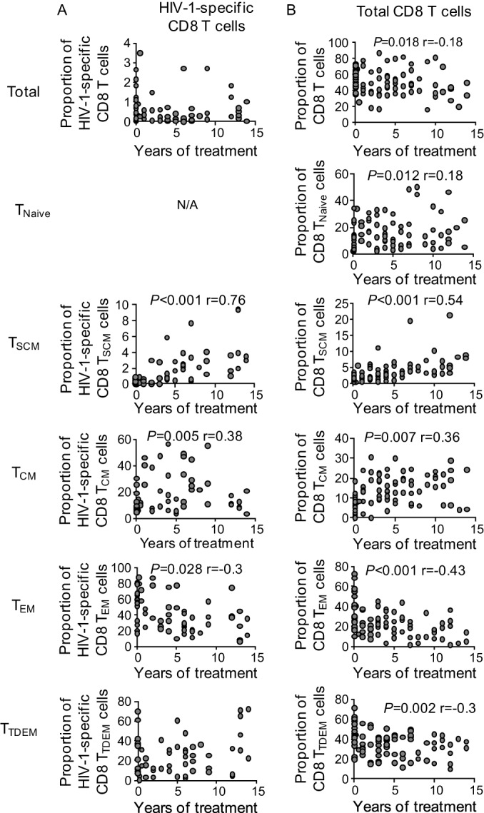
Correlations between the proportions of HIV-1-specific or total CD8 TSCM cells and duration of antiretroviral therapy. Data show associations between the indicated HIV-1-specific CD8 T cell subsets from 23 patients (A) (n = 59 responses) or total CD8 T cells from 45 patients (B) and the corresponding durations of antiretroviral therapy. Data from all study patients and all available time points were cumulatively analyzed using generalized estimating equation (GEE) analysis adjusted for repeated measures for the same patients. N/A, not applicable.
Associations between HIV-1-specific CD8 TSCM cells and clinical HIV-1 disease progression.
We subsequently analyzed the possible influence of HIV-1-specific CD8 TSCM cells on clinical parameters of HIV-1 disease progression during untreated infection. In a cross-sectional analysis, we observed that the proportions of total CD8 TSCM cells and CD8 TNA cells were positively associated with CD4 T cell counts (Fig. 5A), as opposed to the proportions of alternative total CD8 T cell subsets that were unrelated to CD4 T cell counts (data not shown). The counts of total HIV-1-specific CD8 T cells, HIV-1-specific CD8 TSCM cells (Fig. 5B), or any other HIV-1-specific CD8 T cell population (data not shown) also did not correlate with CD4 T cell counts. Notably, when patients treated with suppressive antiretroviral therapy were selectively considered, we observed that the CD4 T cell counts were positively associated with the relative proportions and the absolute counts of HIV-1-specific CD8 TSCM cells (Fig. 5C and D) and with the absolute counts of total CD8 TSCM cells (Fig. 5E and F).
FIG 5.

Associations between total and HIV-1-specific CD8 TSCM cells and markers of clinical HIV-1 disease progression. (A) Association between CD4 T cell counts (number of cells per microliter) and proportions of CD8 TNaive or TSCM cells in 14 untreated progressor (UP) and 16 untreated controller (UC) patients. (B) Association between CD4 T cell counts (number of cells per microliter) and proportions of HIV-1-specific CD8 T cells or HIV-1-specific CD8 TSCM cells in 6 untreated progressor (n = 10 responses) and 14 untreated controller (n = 23 responses) patients. (C and D) Association between CD4 T cell counts (number of cells per microliter) and proportions (C) or absolute counts (D) of HIV-1-specific CD8 TSCM cells in 28 ART-treated patients (n = 46 responses). (E and F) Association between proportions (E) and absolute counts (F) of total CD8 TSCM cells and the absolute CD4 T cell counts in 64 ART-treated patients. (G) Association between HIV-1 loads (number of RNA copies per milliliter) and the proportions of CD8 T cells or the indicated T cell subsets in 14 untreated progressor and 16 untreated controller patients. (H) Association between HIV-1 loads (number of RNA copies per milliliter) and the proportions of total HIV-1-specific CD8 T cells or the indicated HIV-1-specific T cell subsets in 6 untreated progressor (n = 11 responses) and 14 untreated controller (n = 26 responses) patients. Spearman's rank correlation coefficients (r values) are shown.
We subsequently assessed the associations between the different total and HIV-1-specific CD8 T cell subsets and the corresponding plasma viral loads in untreated HIV-1-infected patients. The proportions of total CD8 TSCM, CD8 TNA, and CD8 TCM cells were inversely correlated with HIV-1 loads, while the proportions of total CD8 TEM cells were positively associated with HIV-1 loads (Fig. 5G). However, there was no association between the viral loads and the proportions of either total HIV-1-specific CD8 T cells or HIV-1-specific CD8 TSCM, TCM, or TEM cells (Fig. 5H).
We also analyzed possible connections between HIV-1-specific CD8 TSCM cells and immune activation parameters, which can independently predict HIV-1 disease progression in untreated HIV-1-infected patients and are associated with immune dysfunction and accelerated immune senescence in patients treated with antiretroviral therapy. The proportions of total and HIV-1-specific HLA-DR/CD38-coexpressing CD8 T cells decreased during antiretroviral treatment (see Fig. S6A and B in the supplemental material) and were unrelated to the proportions of total and HIV-1-specific CD8 TSCM cells but were positively associated with the proportions of total and HIV-1-specific TTDEM CD8 T cells, consistent with immune activation as a driving force for CD8 T cell differentiation (data not shown). We also noted that within untreated HIV-1-infected patients, the proportions of total CD38/HLA-DR+ CD8 T cells, CD38/HLA-DR+ CD8 TSCM cells, and total CD38/HLA-DR+ HIV-1-specific CD8 T cells were inversely related to CD4 T cell counts (see Fig. S6C to E in the supplemental material). Inverse relationships between the proportions of total CD38/HLA-DR+ CD8 T cells and CD4 T cell counts were also observed when ART-treated patients were selectively considered (see Fig. S6F to H in the supplemental material).
Cytokine secretion of HIV-1-specific CD8 TSCM cells.
To analyze the functional properties of HIV-1-specific CD8 TSCM cells, we determined the IFN-γ and IL-2 production in these cells (Fig. 6A). Consistent with prior findings (7, 16, 30), the proportions of total IFN-γ-secreting HIV-1-specific CD8 T cells were reduced in ART-treated patients compared to those in untreated controllers (see Fig. S7A in the supplemental material), and the proportions of total IFN-γ- and IL-2-coproducing HIV-1-specific CD8 T cells appeared to be the lowest in untreated progressors (see Fig. S7B and C in the supplemental material). Overall, we observed that virus-specific CD8 TSCM cells made only very small contributions to the total CD8 T cell population secreting IFN-γ or IL-2 after stimulation with HIV-1 or alternative viral antigens (see Fig. S7D in the supplemental material), suggesting that these cells have more limited roles for direct antiviral immune defense. However, HIV-1-specific CD8 TSCM cells able to coproduce IFN-γ and IL-2 were enriched in ART-treated patients, with the proportions of cells in ART-treated patients exceeding the corresponding proportions of cells in untreated controllers (Fig. 6B). Throughout all patient groups, IFN-γ- and IL-2-coproducing HIV-1-specific CD8 T cells were preferentially detected in the more immature CD8 TSCM and TCM cell compartments (Fig. 6C). In contrast, IFN-γ-monosecreting cells were most dominantly observed in HIV-1-specific CD8 T cells with a TEM cell or terminally differentiated phenotype and less frequently in HIV-1-specific CD8 T cells with a TCM or TSCM cell phenotype (Fig. 6D). Notably, IFN-γ- and IL-2-cosecreting HIV-1-specific CD8 TSCM cells were almost completely absent in untreated HIV-1 progressors (data not shown) but accounted for a clearly detectable proportion of all cytokine-secreting HIV-1-specific CD8 TSCM cells in ART-treated persons and, to a lesser extent, in untreated controllers (Fig. 6C). The proportions of IFN-γ- and IL-2-cosecreting cells within CMV-, EBV-, or Flu-specific CD8 TSCM cells were also detectable at levels similar to those measured in ART-treated patients (Fig. 6C). The proportions of IFN-γ- and IL-2-cosecreting HIV-1-specific CD8 TSCM, TCM, or TTDEM cells were unrelated to CD4 T cell counts (Fig. 7A); however, the proportions of IFN-γ-monosecreting HIV-1-specific CD8 TSCM cells, but not the proportions of TCM or TTDEM cells, were positively associated with CD4 T cell counts (Fig. 7B). There was no association between the cytokine secretion profiles of HIV-1-specific CD8 TSCM cells and the duration of antiretroviral therapy (data not shown).
FIG 6.

Cytokine secretion properties of HIV-1-specific CD8 TSCM cells. (A) Flow cytometry gating strategy used for the identification of cytokine-secreting HIV-1-specific CD8 T cell subsets. (B) Pie charts reflecting the subset distribution of IFN-γ/IL-2 coproducing or IFN-γ-producing virus-specific CD8 T cells in the indicated HIV-1-positive patients, 12 untreated controller HIV-1-infected patients (n = 25 responses), and 14 ART-treated patients (n = 22 responses). Statistical comparisons among the pie charts were performed by the χ2 test in SPICE (version 5.32). (C and D) Proportions of IFN-γ/IL-2 coproducing (C) or IFN-γ-producing (D) HIV-1-specific (n = 47 responses) and CEF-specific (n = 16 responses) CD8 T cell subsets in the indicated patient populations. Statistical comparisons in panels C and D were performed using the Kruskal-Wallis test (for comparisons among distinct groups of patients) or Friedman's test (for paired comparisons among different cell subsets within each patient group), followed by post hoc analysis with Dunn's test for multiple comparisons. *, P ≤ 0.05; **, P ≤ 0.01; ***, P ≤ 0.001.
FIG 7.
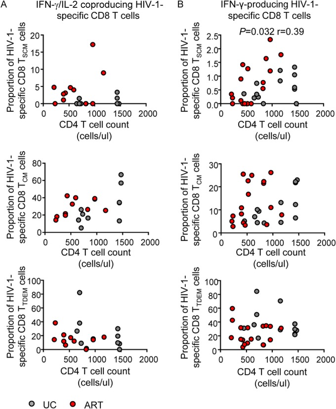
Associations between cytokine-producing HIV-1-specific CD8 TSCM cells and clinical HIV-1 disease progression. Data show associations between CD4 T cell counts (number of cells per microliter) and the proportion of cytokine-secreting HIV-1-specific CD8 TSCM, TCM, and TTDEM cells from untreated controllers (UC; n = 10 and n = 13 for IFN-γ/IL-2 coproducing and IFN-γ-secreting CD8 T cells, respectively) and from ART-treated patients (n = 11 and n = 17 for IL-2 and IFN-γ-secreting CD8 T cells, respectively). Spearman's rank correlation coefficients are shown.
DISCUSSION
Recent studies suggest that memory CD8 T cell development originates from a small population of highly immature and long-lived lymphocytes whose characteristics imitate many of the functional characteristics typically ascribed to tissue stem cells and that serve as precursor cells for more differentiated effector memory CD8 T cells (20). This view is supported by data from two long-term gene therapy clinical trials demonstrating that these T memory stem cells are able to persist and preserve their precursor potential for up to 12 years, consistent with the critical role of these cells for maintaining lifelong pathogen-specific immune defense (24). Moreover, CD4 TSCM cells have recently been shown to provide an important cellular source of HIV-1 long-term persistence, further supporting the role of TSCM cells as extremely long-lasting memory cells (31, 32). Although HIV-1-specific T cells can influence HIV-1 disease progression in untreated patients to some extent (33), numerous studies suggest that these cells are disturbed and dysfunctional during progressive disease (12, 13, 34–36). Here, we performed the first characterization of HIV-1-specific CD8 T memory stem cells in individuals with untreated progressive, untreated controlled, or ART-treated HIV-1 infection. Interestingly, we found that the proportions of HIV-1-specific TSCM cells are reduced during untreated infection with persistently high levels of viremia, as well as in individuals who spontaneously controlled HIV-1 replication; this suggests that the pool size and integrity of the HIV-1-specific CD8 TSCM cells are compromised in chronic HIV-1 infection, even when viral loads are very low or undetectable. In contrast, pharmacological inhibition of HIV-1 replication restored normal populations of HIV-1-specific CTLs, and the frequencies of CD8 TSCM cells gradually increased for the duration of antiretroviral therapy. Moreover, CD4 T cell counts during antiretroviral therapy, but not during untreated infection, were associated with the HIV-1-specific CD8 TSCM cell frequency. These observations suggest that HIV-1-specific CD8 TSCM cells are unable to survive and expand under conditions of chronic antigenic stimulation, even if it is occurring at very low levels. Continuous antigenic challenge may possibly inhibit self-renewal and homeostatic proliferation, which represent key features responsible for maintaining CD8 T memory stem cells.
While HIV-1-specific CD8 T cells from untreated HIV-1-infected individuals have been studied for many years, the function of HIV-1-specific CD8 T cells in patients undergoing suppressive antiretroviral therapy is less well understood. Multiple studies suggested that the frequency of HIV-1-specific CD8 T cells is reduced when active viral replication is pharmacologically suppressed; however, this process seems to be primarily related to the elimination of effector memory and terminally differentiated CD8 T cells, which, upon pharmacological antigen withdrawal, appear to undergo a process similar to the contraction phase of T cells that occurs after natural antigenic clearance in alternative infections (37). In contrast, long-lasting HIV-1-specific CD8 T cells, such as T memory stem cells and central memory CD8 T cells, seem to be restored and to some extent expanded under these conditions (38). In line with these findings, previous data showed that the suppression of viral replication is associated with dynamic changes in the T cell receptor (TCR) repertoire of HIV-1-specific CD8 T cells, likely reflecting changes in the CD8 T cell subset composition (16). How immature HIV-1-specific CD8 T memory stem cells seem to be able to survive long term in an antigen-independent fashion is an important area of future investigation. The ability of a relatively high proportion of these cells to secrete IL-2 may allow these cells to proliferate independently of CD4 T helper cells (30, 39). Indeed, IL-2-secreting HIV-1-specific CD8 TSCM cells accounted for a clearly detectable proportion of all cytokine-secreting CD8 TSCM cells within ART-treated persons. However, given that IL-2 may activate TSCM cells and support transitional proliferation rather than homeostatic proliferation into more differentiated T cell subsets, additional mechanisms are likely to govern self-renewal and long-term persistence as well. For example, it is tempting to speculate that T memory stem cells may reactivate stem cell-like molecular programs for regulating long-term maintenance of this specific cell compartment, and initial studies indeed suggest that stem cell pathways, such as the β-catenin signaling cascade, may regulate the behavior and fate of T memory stem cells (21). Whether T memory stem cells expanded during antiretroviral treatment can still contribute to antiviral immune defense in a clinically significant way is an important area of evaluation in future studies. Selective depletion of these cells in models of simian immunodeficiency virus infection in nonhuman primates treated with suppressive antiretroviral therapy would be highly informative in this regard.
Interestingly, our work showed that the proportions of HIV-1-specific CD8 TSCM cells did not differ substantially between patients with high-level viral replication and those with spontaneous viral control (40, 41), despite the fact that total CD8 TSCM cells were negatively associated with viral loads both in our work and in a recent study (29). Moreover, the frequency of HIV-1-specific CD8 TSCM cells was not associated with CD4 T cell counts or viral loads during untreated infection, and there was no association between the proportions of HIV-1-specific CD8 TSCM cells and protective HLA class I alleles. These results suggest that HIV-1-specific CD8 TSCM cells do not directly participate in antiviral immune defense and do not represent a correlate of immune protection during natural untreated infection. However, such a view does not exclude the possibility that alternative, more subtle characteristics of HIV-1-specific CD8 TSCM cells, such as TCR affinity to the peptide-MHC class I complex or their functional ability to repopulate effector memory populations, may distinguish HIV-1-specific CD8 TSCM cells in HIV-1 controllers from those in progressors.
The identification of antigen-specific TSCM cells may offer novel opportunities for manipulating T cell immunity through therapeutic vaccination or adoptive immunotherapy and holds promise that the clinical induction of antigen-specific T memory stem cells would provide a long-lasting cellular source that can continuously repopulate effector cell populations and increase antigen-specific immune surveillance long term. On the basis of the findings obtained in this study, it is less likely that HIV-1-specific CD8 T memory stem cells could be designed to enhance immune activity against HIV-1 during untreated infection. It is, however, possible that the relative enrichment of T memory stem cells in ART-treated patients could be exploited to increase antiviral cellular immunity during treatment with agents that reverse HIV-1 latency and to reduce the residual reservoir of HIV-1-infected cells. Prior studies have indeed suggested that HIV-1-specific CD8 T cells may be able to contribute to the elimination of HIV-1-infected CD4 T cells in which viral gene expression has been pharmacologically induced (42–44). The long-term persistence, the antigen independence, and the multipotency of HIV-1-specific CD8 TSCM cells raise the possibility that these cells can be manipulated to provide a source for cytotoxic effector cells during treatment with latency-reversing agents and in this way may be able to support elimination of the residual reservoir of HIV-1-infected cells in individuals treated with suppressive antiretroviral therapy.
Supplementary Material
ACKNOWLEDGMENTS
This work was supported by the U.S. National Institutes of Health (grants AI078799, AI089339, HL121890, AI098484, HL126554, AI116228, and AI087452 to X.G.Y. and grants AI098487 and AI106468 to M.L.). PBMC sample collection was supported by the Bill and Melinda Gates Foundation (OPP 1066973), the Mark and Lisa Swartz Foundation, the Ragon Institute of MGH, MIT, and Harvard, and the International HIV Controller Consortium.
Footnotes
Supplemental material for this article may be found at http://dx.doi.org/10.1128/JVI.00789-15.
REFERENCES
- 1.Pantaleo G, Koup RA. 2004. Correlates of immune protection in HIV-1 infection: what we know, what we don't know, what we should know. Nat Med 10:806–810. doi: 10.1038/nm0804-806. [DOI] [PubMed] [Google Scholar]
- 2.Walker B, McMichael A. 2012. The T-cell response to HIV. Cold Spring Harbor Perspect Med 2:a007054. doi: 10.1101/cshperspect.a007054. [DOI] [PMC free article] [PubMed] [Google Scholar]
- 3.Schmitz JE, Kuroda MJ, Santra S, Sasseville VG, Simon MA, Lifton MA, Racz P, Tenner-Racz K, Dalesandro M, Scallon BJ, Ghrayeb J, Forman MA, Montefiori DC, Rieber EP, Letvin NL, Reimann KA. 1999. Control of viremia in simian immunodeficiency virus infection by CD8+ lymphocytes. Science 283:857–860. doi: 10.1126/science.283.5403.857. [DOI] [PubMed] [Google Scholar]
- 4.Pereyra F, Jia X, McLaren PJ, Telenti A, de Bakker PI, Walker BD, Ripke S, Brumme CJ, Pulit SL, Carrington M, Kadie CM, Carlson JM, Heckerman D, Graham RR, Plenge RM, Deeks SG, Gianniny L, Crawford G, Sullivan J, Gonzalez E, Davies L, Camargo A, Moore JM, Beattie N, Gupta S, Crenshaw A, Burtt NP, Guiducci C, Gupta N, Gao X, Qi Y, Yuki Y, Piechocka-Trocha A, Cutrell E, Rosenberg R, Moss KL, Lemay P, O'Leary J, Schaefer T, Verma P, Toth I, Block B, Baker B, Rothchild A, Lian J, Proudfoot J, Alvino DM, Vine S, Addo MM, Allen TM, et al. 2010. The major genetic determinants of HIV-1 control affect HLA class I peptide presentation. Science 330:1551–1557. doi: 10.1126/science.1195271. [DOI] [PMC free article] [PubMed] [Google Scholar]
- 5.Carrington M, Nelson GW, Martin MP, Kissner T, Vlahov D, Goedert JJ, Kaslow R, Buchbinder S, Hoots K, O'Brien SJ. 1999. HLA and HIV-1: heterozygote advantage and B*35-Cw*04 disadvantage. Science 283:1748–1752. doi: 10.1126/science.283.5408.1748. [DOI] [PubMed] [Google Scholar]
- 6.Migueles SA, Laborico AC, Shupert WL, Sabbaghian MS, Rabin R, Hallahan CW, Van Baarle D, Kostense S, Miedema F, McLaughlin M, Ehler L, Metcalf J, Liu S, Connors M. 2002. HIV-specific CD8+ T cell proliferation is coupled to perforin expression and is maintained in nonprogressors. Nat Immunol 3:1061–1068. doi: 10.1038/ni845. [DOI] [PubMed] [Google Scholar]
- 7.Betts MR, Nason MC, West SM, De Rosa SC, Migueles SA, Abraham J, Lederman MM, Benito JM, Goepfert PA, Connors M, Roederer M, Koup RA. 2006. HIV nonprogressors preferentially maintain highly functional HIV-specific CD8+ T cells. Blood 107:4781–4789. doi: 10.1182/blood-2005-12-4818. [DOI] [PMC free article] [PubMed] [Google Scholar]
- 8.Borrow P, Lewicki H, Wei X, Horwitz MS, Peffer N, Meyers H, Nelson JA, Gairin JE, Hahn BH, Oldstone MB, Shaw GM. 1997. Antiviral pressure exerted by HIV-1-specific cytotoxic T lymphocytes (CTLs) during primary infection demonstrated by rapid selection of CTL escape virus. Nat Med 3:205–211. doi: 10.1038/nm0297-205. [DOI] [PubMed] [Google Scholar]
- 9.Cao J, McNevin J, Malhotra U, McElrath MJ. 2003. Evolution of CD8+ T cell immunity and viral escape following acute HIV-1 infection. J Immunol 171:3837–3846. doi: 10.4049/jimmunol.171.7.3837. [DOI] [PubMed] [Google Scholar]
- 10.Hersperger AR, Pereyra F, Nason M, Demers K, Sheth P, Shin LY, Kovacs CM, Rodriguez B, Sieg SF, Teixeira-Johnson L, Gudonis D, Goepfert PA, Lederman MM, Frank I, Makedonas G, Kaul R, Walker BD, Betts MR. 2010. Perforin expression directly ex vivo by HIV-specific CD8 T-cells is a correlate of HIV elite control. PLoS Pathog 6:e1000917. doi: 10.1371/journal.ppat.1000917. [DOI] [PMC free article] [PubMed] [Google Scholar]
- 11.Migueles SA, Osborne CM, Royce C, Compton AA, Joshi RP, Weeks KA, Rood JE, Berkley AM, Sacha JB, Cogliano-Shutta NA, Lloyd M, Roby G, Kwan R, McLaughlin M, Stallings S, Rehm C, O'Shea MA, Mican J, Packard BZ, Komoriya A, Palmer S, Wiegand AP, Maldarelli F, Coffin JM, Mellors JW, Hallahan CW, Follman DA, Connors M. 2008. Lytic granule loading of CD8+ T cells is required for HIV-infected cell elimination associated with immune control. Immunity 29:1009–1021. doi: 10.1016/j.immuni.2008.10.010. [DOI] [PMC free article] [PubMed] [Google Scholar]
- 12.Lichterfeld M, Kaufmann DE, Yu XG, Mui SK, Addo MM, Johnston MN, Cohen D, Robbins GK, Pae E, Alter G, Wurcel A, Stone D, Rosenberg ES, Walker BD, Altfeld M. 2004. Loss of HIV-1-specific CD8+ T cell proliferation after acute HIV-1 infection and restoration by vaccine-induced HIV-1-specific CD4+ T cells. J Exp Med 200:701–712. doi: 10.1084/jem.20041270. [DOI] [PMC free article] [PubMed] [Google Scholar]
- 13.Trautmann L, Janbazian L, Chomont N, Said EA, Gimmig S, Bessette B, Boulassel MR, Delwart E, Sepulveda H, Balderas RS, Routy JP, Haddad EK, Sekaly RP. 2006. Upregulation of PD-1 expression on HIV-specific CD8+ T cells leads to reversible immune dysfunction. Nat Med 12:1198–1202. doi: 10.1038/nm1482. [DOI] [PubMed] [Google Scholar]
- 14.Lichterfeld M, Mou D, Cung TD, Williams KL, Waring MT, Huang J, Pereyra F, Trocha A, Freeman GJ, Rosenberg ES, Walker BD, Yu XG. 2008. Telomerase activity of HIV-1-specific CD8+ T cells: constitutive up-regulation in controllers and selective increase by blockade of PD ligand 1 in progressors. Blood 112:3679–3687. doi: 10.1182/blood-2008-01-135442. [DOI] [PMC free article] [PubMed] [Google Scholar]
- 15.Lederman MM, Funderburg NT, Sekaly RP, Klatt NR, Hunt PW. 2013. Residual immune dysregulation syndrome in treated HIV infection. Adv Immunol 119:51–83. doi: 10.1016/B978-0-12-407707-2.00002-3. [DOI] [PMC free article] [PubMed] [Google Scholar]
- 16.Janbazian L, Price DA, Canderan G, Filali-Mouhim A, Asher TE, Ambrozak DR, Scheinberg P, Boulassel MR, Routy JP, Koup RA, Douek DC, Sekaly RP, Trautmann L. 2012. Clonotype and repertoire changes drive the functional improvement of HIV-specific CD8 T cell populations under conditions of limited antigenic stimulation. J Immunol 188:1156–1167. doi: 10.4049/jimmunol.1102610. [DOI] [PMC free article] [PubMed] [Google Scholar]
- 17.Ndhlovu ZM, Proudfoot J, Cesa K, Alvino DM, McMullen A, Vine S, Stampouloglou E, Piechocka-Trocha A, Walker BD, Pereyra F. 2012. Elite controllers with low to absent effector CD8+ T cell responses maintain highly functional, broadly directed central memory responses. J Virol 86:6959–6969. doi: 10.1128/JVI.00531-12. [DOI] [PMC free article] [PubMed] [Google Scholar]
- 18.Papasavvas E, Foulkes A, Yin X, Joseph J, Ross B, Azzoni L, Kostman JR, Mounzer K, Shull J, Montaner LJ. 12 February 2015. PDC and functional HIV Gag p55-specific T cells before treatment interruption can inform set-point plasma HIV viral load after treatment interruption in chronically suppressed HIV-1 subjects. Immunology doi: 10.1111/imm.12452. [DOI] [PMC free article] [PubMed] [Google Scholar]
- 19.Farber DL, Yudanin NA, Restifo NP. 2014. Human memory T cells: generation, compartmentalization and homeostasis. Nat Rev Immunol 14:24–35. doi: 10.1038/nri3567. [DOI] [PMC free article] [PubMed] [Google Scholar]
- 20.Gattinoni L, Lugli E, Ji Y, Pos Z, Paulos CM, Quigley MF, Almeida JR, Gostick E, Yu Z, Carpenito C, Wang E, Douek DC, Price DA, June CH, Marincola FM, Roederer M, Restifo NP. 2011. A human memory T cell subset with stem cell-like properties. Nat Med 17:1290–1297. doi: 10.1038/nm.2446. [DOI] [PMC free article] [PubMed] [Google Scholar]
- 21.Gattinoni L, Zhong XS, Palmer DC, Ji Y, Hinrichs CS, Yu Z, Wrzesinski C, Boni A, Cassard L, Garvin LM, Paulos CM, Muranski P, Restifo NP. 2009. Wnt signaling arrests effector T cell differentiation and generates CD8+ memory stem cells. Nat Med 15:808–813. doi: 10.1038/nm.1982. [DOI] [PMC free article] [PubMed] [Google Scholar]
- 22.Lugli E, Dominguez MH, Gattinoni L, Chattopadhyay PK, Bolton DL, Song K, Klatt NR, Brenchley JM, Vaccari M, Gostick E, Price DA, Waldmann TA, Restifo NP, Franchini G, Roederer M. 2013. Superior T memory stem cell persistence supports long-lived T cell memory. J Clin Invest 123:594–599. doi: 10.1172/JCI66327. [DOI] [PMC free article] [PubMed] [Google Scholar]
- 23.Cieri N, Camisa B, Cocchiarella F, Forcato M, Oliveira G, Provasi E, Bondanza A, Bordignon C, Peccatori J, Ciceri F, Lupo-Stanghellini MT, Mavilio F, Mondino A, Bicciato S, Recchia A, Bonini C. 2013. IL-7 and IL-15 instruct the generation of human memory stem T cells from naive precursors. Blood 121:573–584. doi: 10.1182/blood-2012-05-431718. [DOI] [PubMed] [Google Scholar]
- 24.Biasco L, Scala S, Basso Ricci L, Dionisio F, Baricordi C, Calabria A, Giannelli S, Cieri N, Barzaghi F, Pajno R, Al-Mousa H, Scarselli A, Cancrini C, Bordignon C, Roncarolo MG, Montini E, Bonini C, Aiuti A. 2015. In vivo tracking of T cells in humans unveils decade-long survival and activity of genetically modified T memory stem cells. Sci Transl Med 7:273ra213. doi: 10.1126/scitranslmed.3010314. [DOI] [PubMed] [Google Scholar]
- 25.Roberto A, Castagna L, Zanon V, Bramanti S, Crocchiolo R, McLaren JE, Gandolfi S, Tentorio P, Sarina B, Timofeeva I, Santoro A, Carlo-Stella C, Bruno B, Carniti C, Corradini P, Gostick E, Ladell K, Price DA, Roederer M, Mavilio D, Lugli E. 2015. Role of naive-derived T memory stem cells in T cell reconstitution following allogeneic transplantation. Blood 125:2855–2864. doi: 10.1182/blood-2014-11-608406. [DOI] [PMC free article] [PubMed] [Google Scholar]
- 26.Cieri N, Oliveira G, Greco R, Forcato M, Taccioli C, Cianciotti B, Valtolina V, Noviello M, Vago L, Bondanza A, Lunghi F, Marktel S, Bellio L, Bordignon C, Bicciato S, Peccatori J, Ciceri F, Bonini C. 2015. Generation of human memory stem T cells upon haploidentical T-replete hematopoietic stem cell transplantation. Blood 125:2865–2874. doi: 10.1182/blood-2014-11-608539. [DOI] [PubMed] [Google Scholar]
- 27.Cellerai C, Harari A, Stauss H, Yerly S, Geretti AM, Carroll A, Yee T, Ainsworth J, Williams I, Sweeney J, Freedman A, Johnson M, Pantaleo G, Kinloch-de Loes S. 2011. Early and prolonged antiretroviral therapy is associated with an HIV-1-specific T-cell profile comparable to that of long-term non-progressors. PLoS One 6:e18164. doi: 10.1371/journal.pone.0018164. [DOI] [PMC free article] [PubMed] [Google Scholar]
- 28.Roederer M, Nozzi JL, Nason MC. 2011. SPICE: exploration and analysis of post-cytometric complex multivariate datasets. Cytometry A 79:167–174. doi: 10.1002/cyto.a.21015. [DOI] [PMC free article] [PubMed] [Google Scholar]
- 29.Ribeiro SP, Milush JM, Cunha-Neto E, Kallas EG, Kalil J, Somsouk M, Hunt PW, Deeks SG, Nixon DF, SenGupta D. 2014. The CD8+ memory stem T cell (TSCM) subset is associated with improved prognosis in chronic HIV-1 infection. J Virol 88:13836–13844. doi: 10.1128/JVI.01948-14. [DOI] [PMC free article] [PubMed] [Google Scholar]
- 30.Zimmerli SC, Harari A, Cellerai C, Vallelian F, Bart PA, Pantaleo G. 2005. HIV-1-specific IFN-gamma/IL-2-secreting CD8 T cells support CD4-independent proliferation of HIV-1-specific CD8 T cells. Proc Natl Acad Sci U S A 102:7239–7244. doi: 10.1073/pnas.0502393102. [DOI] [PMC free article] [PubMed] [Google Scholar]
- 31.Buzon MJ, Sun H, Li C, Shaw A, Seiss K, Ouyang Z, Martin-Gayo E, Leng J, Henrich TJ, Li JZ, Pereyra F, Zurakowski R, Walker BD, Rosenberg ES, Yu XG, Lichterfeld M. 2014. HIV-1 persistence in CD4 T cells with stem cell-like properties. Nat Med 20:139–142. doi: 10.1038/nm.3445. [DOI] [PMC free article] [PubMed] [Google Scholar]
- 32.Jaafoura S, de Goer de Herve MG, Hernandez-Vargas EA, Hendel-Chavez H, Abdoh M, Mateo MC, Krzysiek R, Merad M, Seng R, Tardieu M, Delfraissy JF, Goujard C, Taoufik Y. 2014. Progressive contraction of the latent HIV reservoir around a core of less-differentiated CD4+ memory T cells. Nat Commun 5:5407. doi: 10.1038/ncomms6407. [DOI] [PMC free article] [PubMed] [Google Scholar]
- 33.Northfield JW, Loo CP, Barbour JD, Spotts G, Hecht FM, Klenerman P, Nixon DF, Michaelsson J. 2007. Human immunodeficiency virus type 1 (HIV-1)-specific CD8+ TEMRA cells in early infection are linked to control of HIV-1 viremia and predict the subsequent viral load set point. J Virol 81:5759–5765. doi: 10.1128/JVI.00045-07. [DOI] [PMC free article] [PubMed] [Google Scholar]
- 34.Yamamoto T, Price DA, Casazza JP, Ferrari G, Nason M, Chattopadhyay PK, Roederer M, Gostick E, Katsikis PD, Douek DC, Haubrich R, Petrovas C, Koup RA. 2011. Surface expression patterns of negative regulatory molecules identify determinants of virus-specific CD8+ T-cell exhaustion in HIV infection. Blood 117:4805–4815. doi: 10.1182/blood-2010-11-317297. [DOI] [PMC free article] [PubMed] [Google Scholar]
- 35.Vigano S, Bellutti Enders F, Miconnet I, Cellerai C, Savoye AL, Rozot V, Perreau M, Faouzi M, Ohmiti K, Cavassini M, Bart PA, Pantaleo G, Harari A. 2013. Rapid perturbation in viremia levels drives increases in functional avidity of HIV-specific CD8 T cells. PLoS Pathog 9:e1003423. doi: 10.1371/journal.ppat.1003423. [DOI] [PMC free article] [PubMed] [Google Scholar]
- 36.Peretz Y, He Z, Shi Y, Yassine-Diab B, Goulet JP, Bordi R, Filali-Mouhim A, Loubert JB, El-Far M, Dupuy FP, Boulassel MR, Tremblay C, Routy JP, Bernard N, Balderas R, Haddad EK, Sekaly RP. 2012. CD160 and PD-1 co-expression on HIV-specific CD8 T cells defines a subset with advanced dysfunction. PLoS Pathog 8:e1002840. doi: 10.1371/journal.ppat.1002840. [DOI] [PMC free article] [PubMed] [Google Scholar]
- 37.Lichterfeld M, Yu XG, Cohen D, Addo MM, Malenfant J, Perkins B, Pae E, Johnston MN, Strick D, Allen TM, Rosenberg ES, Korber B, Walker BD, Altfeld M. 2004. HIV-1 Nef is preferentially recognized by CD8 T cells in primary HIV-1 infection despite a relatively high degree of genetic diversity. AIDS 18:1383–1392. doi: 10.1097/01.aids.0000131329.51633.a3. [DOI] [PubMed] [Google Scholar]
- 38.Migueles SA, Weeks KA, Nou E, Berkley AM, Rood JE, Osborne CM, Hallahan CW, Cogliano-Shutta NA, Metcalf JA, McLaughlin M, Kwan R, Mican JM, Davey RT Jr, Connors M. 2009. Defective HIV-specific CD8+ T cell polyfunctionality, proliferation, and cytotoxicity are not restored by antiretroviral therapy. J Virol 83:11876–11889. doi: 10.1128/JVI.01153-09. [DOI] [PMC free article] [PubMed] [Google Scholar]
- 39.Feau S, Arens R, Togher S, Schoenberger SP. 2011. Autocrine IL-2 is required for secondary population expansion of CD8+ memory T cells. Nat Immunol 12:908–913. doi: 10.1038/ni.2079. [DOI] [PMC free article] [PubMed] [Google Scholar]
- 40.Walker BD, Yu XG. 2013. Unravelling the mechanisms of durable control of HIV-1. Nat Rev Immunol 13:487–498. doi: 10.1038/nri3478. [DOI] [PubMed] [Google Scholar]
- 41.Saez-Cirion A, Pancino G, Sinet M, Venet A, Lambotte O. 2007. HIV controllers: how do they tame the virus? Trends Immunol 28:532–540. doi: 10.1016/j.it.2007.09.002. [DOI] [PubMed] [Google Scholar]
- 42.Shan L, Deng K, Shroff NS, Durand CM, Rabi SA, Yang HC, Zhang H, Margolick JB, Blankson JN, Siliciano RF. 2012. Stimulation of HIV-1-specific cytolytic T lymphocytes facilitates elimination of latent viral reservoir after virus reactivation. Immunity 36:491–501. doi: 10.1016/j.immuni.2012.01.014. [DOI] [PMC free article] [PubMed] [Google Scholar]
- 43.Graf EH, Pace MJ, Peterson BA, Lynch LJ, Chukwulebe SB, Mexas AM, Shaheen F, Martin JN, Deeks SG, Connors M, Migueles SA, O'Doherty U. 2013. Gag-positive reservoir cells are susceptible to HIV-specific cytotoxic T lymphocyte mediated clearance in vitro and can be detected in vivo. PLoS One 8:e71879. doi: 10.1371/journal.pone.0071879. [DOI] [PMC free article] [PubMed] [Google Scholar]
- 44.Deng K, Pertea M, Rongvaux A, Wang L, Durand CM, Ghiaur G, Lai J, McHugh HL, Hao H, Zhang H, Margolick JB, Gurer C, Murphy AJ, Valenzuela DM, Yancopoulos GD, Deeks SG, Strowig T, Kumar P, Siliciano JD, Salzberg SL, Flavell RA, Shan L, Siliciano RF. 2015. Broad CTL response is required to clear latent HIV-1 due to dominance of escape mutations. Nature 517:381–385. doi: 10.1038/nature14053. [DOI] [PMC free article] [PubMed] [Google Scholar]
Associated Data
This section collects any data citations, data availability statements, or supplementary materials included in this article.