

ABSTRACT
Mutations in the polymerase genes are known to play a major role in avian influenza virus adaptation to mammalian hosts. Despite having avian origin PA and PB2, the 2009 pandemic H1N1 virus (pH1N1) can replicate well in mammalian respiratory tracts, suggesting that these proteins have acquired mutations for efficient growth in humans. We have previously shown that PA from the pH1N1 virus A/California/04/09 (Cal) strongly enhances activity of an otherwise avian polymerase complex derived from A/chicken/Nanchang/3-120/01 (Nan) in mammalian cells. However, this enhancement was observed at 37°C but not at the lower temperature of 34°C. An additional introduction of Cal PB2 enhanced activity at 34°C, suggesting the presence of unidentified residues in Cal PB2 that are required for efficient growth at low temperature. Here, we sought to determine the key PB2 residues which confer enhanced polymerase activity and virus growth in human cells at low temperature. Using a reporter gene assay, we identified novel mutations, PB2 V661A and V683T/A684S, which are involved in enhanced Cal polymerase activity at low temperature. The PB2 T271A mutation, which we previously reported, also contributed to enhanced activity. The growth of recombinant Cal containing PB2 with Nan residues 271T/661V/683V/684A was strongly reduced in human cells compared to wild-type virus at low temperature. Among the four residues, 271A and 684S are conserved in human and pH1N1 viruses but not in avian viruses, suggesting an important role in mammalian adaptation of pH1N1 virus.
IMPORTANCE The PB2 protein plays a key role in the host adaptation, cold sensitivity, and pathogenesis of influenza A virus. Despite containing an avian origin PB2 lacking the mammalian adaptive mutations 627K or 701N, pH1N1 influenza virus strains can replicate efficiently in the low temperature upper respiratory tract of mammals, suggesting the presence of unknown mutations in the pH1N1 PB2 protein responsible for its low temperature adaptation. Here, in addition to PB2 271A, which has been shown to increase polymerase activity, we identified novel PB2 residues 661A and 683T/684S in pH1N1 which confer enhanced polymerase activity and virus growth in mammalian cells especially at low temperature. Our findings suggest that the presence of these PB2 residues contributes to efficient replication of the pH1N1 virus in the upper respiratory tract, which resulted in efficient human-to-human transmission of this virus.
INTRODUCTION
Influenza pandemics occur when a new strain crosses the species barrier and acquires the ability to replicate and transmit efficiently in human hosts. The emergence of a new pandemic virus is driven by both adaptive mutation of individual viral genes and reassortment of viral genome segments. Evidence suggests that preferential binding of human or avian influenza virus hemagglutinin (HA) to α-2,6 or α-2,3 sialylated receptors, respectively, confers a restricted host range (1). It is also well established that mutations in polymerase proteins play a key role for avian virus adaptation to humans (2–7). Avian virus polymerases do not function well in mammalian cells (3), but specific mutations in avian virus polymerase genes allow for efficient viral replication in mammalian hosts. These key residues are mainly found to reside in PA and PB2 (2–4, 8, 9). In addition, efficient replication at lower temperatures is also considered a major factor that restricts viral host range (4). Human influenza viruses replicate in the human upper respiratory tract at close to 33 to 34°C, while avian viruses replicate in the avian intestinal tract at about 39 to 41°C (4, 10). Most avian influenza viruses exhibit a low temperature-sensitive phenotype, displaying reduced viral replication and protein production at lower temperatures, but more efficient replication at higher temperatures. Thus, the ability to replicate efficiently at low temperature is likely to be required for adaptation of influenza viruses to the human population.
Beginning in April 2009, the pandemic H1N1 (pH1N1) virus emerged in Mexico and rapidly spread worldwide, causing a global pandemic (11, 12). The pH1N1 virus is a swine-origin influenza virus strain with a unique genomic constellation derived from avian-, swine-, and human-origin genes (11, 12). Of note, this virus contains avian-like PA and PB2 polymerase gene segments but replicates well in mammalian cells (12, 13), suggesting that these virus segments have acquired mutations required for efficient growth in humans. Using an in vitro reporter gene assay, we previously determined the role of each polymerase component of the pH1N1 strain A/California/04/2009 (Cal) in mammalian host adaptation. We analyzed activity of polymerase complexes of the avian strain A/chicken/Nanchang/3-120/01 (Nan) containing components from Cal (8). A Nan polymerase complex containing Cal PA was highly active at 37 and 39°C but not at 34°C (8). Importantly, the additional introduction of Cal PB2 enhanced activity to a similar level to the Cal polymerase complex at 34°C, indicating that PA and PB2 are essential for the activity of Cal in mammalian cells at low temperature (8).
A PB2 residue at position 627 is a well-known determinant of cold sensitivity of the influenza virus polymerase complex (4, 5, 10). Avian influenza viruses most commonly possess a glutamine (E) at position 627 of PB2, while human viruses contain a lysine (K) at this position. An E627K substitution in PB2 confers the ability of an avian virus to replicate efficiently at low temperatures in vitro (10). It has also been reported that the PB2 E627K substitution in a highly pathogenic H5N1 avian influenza virus supports efficient virus replication in the upper respiratory tract of infected mice, where the temperature is low (14). Moreover, Steel et al. showed that the D701N mutation in PB2 was involved in virus replication in vitro at low temperature (15). Interestingly, pH1N1 virus can replicate well in the upper respiratory tract of infected animals (13), despite containing an avian-like PB2 lacking E627K or D701N substitutions. We previously found that PB2 residue 271A, which is conserved among human isolates, enhanced avian polymerase activity and virus replication in mammalian cells (3). However, introduction of an avian type A271T residue in Cal PB2 did not completely abolish the activity of the Cal polymerase complex at low temperature (3), suggesting the presence of additional residues that contribute to efficient polymerase activity and virus growth in mammalian cells at low temperature.
In this study, we further addressed the Cal PB2 residues required for enhanced polymerase activity and virus growth in human cells at low temperature. We constructed Cal/Nan PB2 chimeras to determine specific regions of Cal PB2 that confer enhanced activity at low temperature. Using a reporter gene assay, we found that the Cal PB2 C-terminal region, containing amino acids 645 to 759, contains key residues that are essential for high polymerase activity at low temperature. Stepwise mutation analysis revealed that mutation at residues V661A, V683T/A684S, and T271A enhance avian virus polymerase activity. We also found that these four residues are required for viral protein production and multistep viral growth in the context of Cal infection, especially at low temperature. Taken together, we identified novel PB2 residues, 661A and 683T/684S, which confer enhanced polymerase activity and viral growth in mammalian cells at low temperature.
MATERIALS AND METHODS
Virus strains and cell culture.
Influenza A virus A/California/04/2009 (H1N1) was provided by R. Webster and R. Webby (St. Jude Children's Research Hospital, Memphis, TN). Calu-3 cells were kindly provided by D. Topham (University of Rochester). MDCK, 293T, and Calu-3 cells were maintained in Dulbecco modified Eagle medium (DMEM; Corning) supplemented with 8% fetal bovine serum (Life Technologies), 1× GlutaMAX (Life Technologies), and gentamicin (Life Technologies).
Plasmids.
Construction of pCAGGS and pPolI vectors containing A/chicken/Nanchang/3-120/01 (Nan) and Cal genes were described previously (3, 16, 17). Chimera PB2 genes were created using compatible restriction sites in Nan and Cal PB2 and then cloned into pCAGGS. Mutations in the Nan or Cal PB2 gene were created by site-directed mutagenesis. Mutations in the PB2 segment were confirmed by sequence analysis. Primer sequences are available upon request. pPolI-NP-Luc was provided by T. Wolff (Robert-Koch Institute, Berlin, Germany), and pRL-SV40 (Promega) was provided by L. Martínez-Sobrido (University of Rochester).
Virus rescue.
Recombinant Cal viruses were rescued using the 12-plasmid rescue system (18). Briefly, 293T and MDCK cocultures in a six-well plate were transfected using Lipofectamine 2000 (Invitrogen) with 0.1 μg each of pPolI plasmids encoding the eight segments from Cal together with 0.4 μg of each pCAGGS plasmid encoding Cal PA, PB1, PB2, or NP genes as described previously (19). For the rescue of CalPB2-271T, CalPB2-3mut (PB2-661V/683V/684A), or CalPB2-4mut (PB2-271T/661V/683V/684A), we replaced pPolI-CalPB2 with pPolI-CalPB2-271T, pPolI-CalPB2-3mut, or pPolI-CalPB2-4mut, respectively. Rescued viruses were plaque purified, and stock virus was propagated in MDCK cells. Mutations in the PB2 segment were confirmed by sequence analysis. Virus titers were determined by immunofluorescence analysis of nucleoprotein expression in Calu-3 cells.
Transfection-based reporter gene assay.
Polymerase activity assays in 293T cells were performed as described previously (3, 8). Briefly, 293T cells in a 12-well plate were transfected with 0.4 μg each of pCAGGS-NP, -PA, -PB1, and -PB2 and 0.1 μg each of pPolI-NP-Luc and pRL-SV40 using Lipofectamine 2000 (Invitrogen) at 34 or 37°C for 24 h. Luciferase production was measured using the dual-luciferase reporter assay system (Promega). Polymerase activity was normalized to Renilla luciferase expression as a transfection control. An initial experiment using polymerase complex containing Cal PB2 wild type (wt) showed that the luciferase activity at 24 h posttransfection (hpt) was higher than that at 12 hpt but lower than that at 36 or 48 hpt, confirming that the activity measured in our experimental setting at 24 hpt is not saturated (data not shown).
Virus protein production.
Confluent Calu-3 cells in a 24-well plate were infected with each Cal virus at a multiplicity of infection (MOI) of 0.2 for 1 h and incubated in DMEM containing 0.15% bovine serum albumin at 34 or 37°C. At 12 h postinfection (hpi), total cell lysates were subjected to Western blot analysis to detect HA, M1, and β-actin proteins as described below.
Virus growth kinetics.
Confluent Calu-3 cells in a 12-well plate were infected with each Cal virus at an MOI of 0.025 for 1 h and incubated in DMEM containing 0.15% bovine serum albumin at 34 or 37°C. At 2, 12, 24, 48, and 72 hpi, 10% of the culture supernatant was collected and titrated on MDCK cells. Virus titers were calculated by the method of Reed and Muench (20) and expressed as the 50% tissue culture infective doses/ml.
Western blot analysis.
Calu-3 or 293T cells were lysed with Triton X-100 lysis buffer (20 mM HEPES [pH 7.5], 1.5 mM MgCl2, 500 mM NaCl, 0.2 mM EDTA, 1% Triton X-100, 20% glycerol) or passive lysis buffer (dual-luciferase reporter assay system; Promega), respectively. Cell lysates were separated by SDS-PAGE (4 to 12% gel; Life Technologies), and proteins were then transferred onto a polyvinylidene difluoride membrane (Millipore). The blot was blocked in 2% dry milk and then incubated with primary antibodies as follows: mouse anti-Influenza A virus PB2 monoclonal antibody (MAb; 1:1,000, NR-4541; BEI Resources), mouse anti-β-actin MAb (1:10,000; clone AC-15 [Sigma] or clone 8H10D10 [Cell Signaling]), mouse anti-influenza A virus HA MAb (1:4,000, NR42019; BEI Resources) or mouse anti-influenza A virus M1 MAb (1:4,000, GA2B; Thermo Scientific). The membrane was then incubated with HRP-conjugated goat anti-mouse IgG (1:10,000, Bio-Rad). Target proteins were visualized using SuperSignal West Femto maximum sensitivity substrate (Thermo Scientific). Images were obtained using a ChemiDoc XRS system (Bio-Rad) and analyzed using Quantity One 1-D analysis software (Bio-Rad).
Statistical analysis.
Statistical analysis was performed using GraphPad Prism 5.0 software. Comparisons between two groups were performed using a two-tailed Student t test. P values of <0.05 were considered statistically significant.
RESULTS
The C-terminal regions of Cal PB2 confer enhanced polymerase activity at low temperature.
Our previous study suggests that Cal PB2 enhances the activity of Nan polymerase complex containing Cal PA at 34°C (8). Cal and Nan PB2 vary at 31 of their 759 amino acid residues (Table 1). To determine the region of Cal PB2 responsible for enhanced activity at low temperature, we constructed and characterized five Cal/Nan PB2 chimera cDNAs (Fig. 1A). These chimeric PB2 proteins were expressed at similar levels compared to wt Cal and Nan PB2 in 293T cells (Fig. 1B). Their activities in complexes with Nan NP, PB1, and Cal PA were evaluated by reporter gene assay. All of the PB2 proteins, including that of wt Nan, function well at 37°C in a Nan complex containing Cal PA (Fig. 1C, bottom). However, at 34°C, the activity with wt Nan PB2 was nearly 100-fold less than that of the complex containing Cal PB2 (P < 0.05, Fig. 1C). Among the chimeras, C1 showed almost the same activity as Cal (P > 0.05), and C2 was similar to Nan (P > 0.05), suggesting that Cal residues within the region 248 to 759 are responsible for enhanced activity at 34°C. Chimeras C3 and C4 showed activity between wt Cal and Nan (P < 0.05 versus that of wt Cal or Nan), suggesting that multiple residues in the region 248 to 759 are involved in low temperature activity. Chimera C5, which contains residues 644 to 759 in addition to residues 1 to 248 from Nan, showed much lower activity than C1 (P < 0.05), suggesting that the PB2 C-terminal region has a strong impact on its function in mammalian cells at low temperature (Fig. 1C).
TABLE 1.
Amino acid differences between Cal and Nan PB2
| Virus | Residue in PB2 at position: |
||||||||||||||||||||||||||||||
|---|---|---|---|---|---|---|---|---|---|---|---|---|---|---|---|---|---|---|---|---|---|---|---|---|---|---|---|---|---|---|---|
| 9 | 44 | 54 | 65 | 87 | 109 | 147 | 221 | 225 | 271 | 278 | 292 | 315 | 353 | 355 | 356 | 379 | 408 | 453 | 478 | 495 | 508 | 559 | 588 | 591 | 645 | 661 | 676 | 683 | 684 | 711 | |
| Cal | Asp | Ala | Arg | Asp | Asp | Val | Thr | Ala | Gly | Ala | Ala | Val | Ile | Lys | Arg | Val | Arg | Asp | Ser | Ile | Val | Arg | Ile | Thr | Arg | Leu | Ala | Thr | Thr | Ser | Asn |
| Nan | Glu | Ser | Lys | Glu | Asn | Ile | Val | Ser | Ser | Thr | Leu | Ile | Leu | Arg | Lys | Ile | Lys | Glu | Pro | Val | Thr | Leu | Thr | Ala | Leu | Met | Val | Ser | Val | Ala | Ser |
FIG 1.
The C-terminal region of Cal PB2 contributes to low temperature polymerase activity. (A) Schematic diagram of five Cal/Nan PB2 chimeras. (B) Expression of PB2 proteins in transfected 293T cells was determined by Western blotting using an anti-PB2 antibody, and similar loading was confirmed using an anti-actin antibody. (C) Activity of polymerase complexes composed of Nan NP, PB1, Cal PA and the indicated PB2, determined in 293T cells at 34 and 37°C. As a negative control, a sample with an empty vector in place of PB2 was included. The results were scaled to the value of the complex containing wt Cal PB2 at 34°C and are shown as the means plus standard deviations from three independent experiments.
PB2 residues at positions 271, 661, and 683/684 contribute to enhanced polymerase activity at low temperature.
We further analyzed the activity of PB2 containing site-specific mutations. In residues 645 to 759, PB2 contains six amino acid differences between Nan and Cal (Table 1). Among these, we mutated four residues at positions 661, 676, 683, and 684 which are surface exposed and in close proximity to each other. We also mutated the residue at 271, which we previously have shown plays a role in enhanced activity in mammalian cells (3). We created 15 Nan PB2 mutants containing these mutations individually and in various combinations (Fig. 2A). In the case of 683/684, we mutated both together due to their close proximity. These PB2 mutants were expressed with Nan NP and PB1, Cal PA, and a luciferase reporter gene in 293T cells, and their activity was determined at 34 or 37°C. At 37°C, all of the mutant PB2 proteins were highly active due the presence of Cal PA in the complex. However, at 34°C, striking differences were observed between the mutants (Fig. 2A). Among the mutants containing a single mutation, 271A (mut 1; 5.9-fold, P < 0.05) increased activity the most, followed by 683T/684S (mut 4; 2.0-fold, P < 0.05) and 661A (mut 2; 1.2-fold, P > 0.05), compared to the activity of Nan PB2 at 34°C. The results of the double mutants (mut 5 to mut 10) indicate that the activity of PB2 containing 271A was further enhanced by the addition of 661A (mut 5; 2.5-fold, P < 0.05), and 683T/684S (mut 7; 4.8-fold, P < 0.05), but not by 676T (mut 6; 1.1-fold, P > 0.05). The addition of both 661A and 683T/684S to 271A (mut 12) further increased activity by 12.7-fold (P < 0.05). mut 12 was 74.5-fold more active than Nan PB2 (P < 0.05) and almost as active as wt Cal PB2 (P > 0.05). All of the mutant PB2 proteins were expressed well to similar levels (Fig. 2B). To directly assess whether these mutations in PB2 protein affect polymerase activity of the Cal polymerase complex, we introduced the Nan residues (271T, 661V, or 683V/684A) into the Cal PB2 gene and constructed plasmids expressing either Cal PB2-271T, PB2-3mut (661V and 683V/684A), or PB2-4mut (271T, 661V, and 683V/684A). We then compared their activities in the Cal polymerase complex in 293T cells at 34 or 37°C. At 34°C, Cal PB2-271T or PB2-3mut significantly reduced the polymerase activity of Cal polymerase complex by 80.7 or 90.8%, respectively, compared to wt Cal PB2 (P < 0.05, Fig. 3A). Cal PB2-4mut further reduced the activity by 97.4% at 34°C (P < 0.05, Fig. 3A). Although similar patterns were seen at 37°C, the reduced polymerase activity due to mutation of these residues was much more noticeable at 34°C than that at 37°C (Fig. 3A). In 293T cells, all PB2 mutants were expressed at least as well as wt Cal PB2 at both temperatures (Fig. 3B). Taken together, these results indicate that the amino acids at positions 271, 661, and 683/684 in PB2 are responsible for enhanced polymerase activity in mammalian cells especially at low temperature.
FIG 2.

Polymerase activity of complex containing Nan PB2 mutants. The indicated Nan PB2 mutants were expressed with Cal PA, Nan NP, and PB1, together with the reporter gene. The luciferase activity was measured at 24 hpt. The results were scaled to the value of the complex containing wt Cal PB2 at 34°C and are shown as means plus the standard deviations from three independent experiments. (B) Expression of wt and mutant PB2 and actin determined by Western blotting, as in Fig. 1B.
FIG 3.
Polymerase activity of Cal complex containing PB2 mutations. (A) The indicated PB2 cDNAs were cotransfected with Cal NP, PB1, and PA, together with the reporter gene in 293T cells at 34 or 37°C for 24 h. The luciferase activity was then measured. The activity of Cal polymerase complex containing wt Cal PB2 was set to 100%. The results are shown as means plus the standard deviations from three independent experiments. Asterisks indicate statistically significant differences (*, P < 0.05; Student t test) (B) Expression of PB2 and actin determined by Western blotting, as in Fig. 1B.
PB2 residues at positions 271, 661, and 683/684 are involved in viral protein production and growth in infected cells.
To assess the impact of mutating these PB2 residues in the context of viral infection, we rescued recombinant Cal viruses containing Cal PB2 with Nan residues 271T, 3mut (661V and 683V/684A), or 4mut (271T, 661V, and 683V/684A). All viruses were successfully rescued. First, we compared multistep virus growth in human airway epithelial Calu-3 cells following Cal wt or PB2 mutant infection. As anticipated, the growth of the CalPB2-4mut virus was strongly reduced compared to Cal wt (Fig. 4). The difference in virus titers at 34°C was more distinct than that detected at 37°C. At 24 hpi, the virus titer of CalPB2-4mut was 24.5-fold lower than that of Cal wt at 34°C, but only 5.4-fold less at 37°C. The virus titers of CalPB2-271T and CalPB2-3mut viruses showed an intermediate growth phenotype between Cal wt and CalPB2-4mut. These data indicate that the PB2 amino acids at positions 271, 661, and 683/684 play a key role in virus growth in Calu-3 cells, especially at low temperature. Next, we compared virus protein production in Calu-3 cells. Consistent with the results of the reporter gene assays and virus growth curves, CalPB2-4mut virus produced reduced levels of viral proteins at 34°C (Fig. 5). In contrast, the reduction of viral protein in CalPB2-4mut virus-infected cells, while present, was not as extreme at 37°C. These data demonstrate that the residues at positions 271, 661, and 683/684 in PB2 contribute to virus production in infected cells, especially at low temperature.
FIG 4.
Virus growth kinetics in Calu-3 cells infected with Cal wt and PB2 mutants. Cells were infected with Cal wt or the indicated PB2 mutants at an MOI of 0.025 and cultured at 34°C (A) or 37°C (B). After the indicated time points, 10% of the supernatant was collected and titrated in MDCK cells. The results are shown as means plus the standard deviations (n = 3).
FIG 5.
Virus protein synthesis in Calu-3 cells infected with Cal wt and PB2 mutants. Cells were infected with Cal wt or the indicated PB2 mutants at an MOI of 0.2 and cultured at 34 or 37°C. After 12 hpi, HA and M1 protein expression in total cell lysates was determined by Western blotting with specific MAbs. Actin protein expression was also detected as a loading control. The data shown are representative of two independent experiments.
Amino acid sequence variation of position 271, 661, and 683/684 in PB2 among influenza A virus lineages.
To analyze the presence of PB2 residues 271A, 661A, and 683T/684S among various influenza A virus lineages, we performed single nucleotide polymorphism analysis of PB2 proteins of avian (5,829 sequences), human (all subtypes of human viruses except for pH1N1 viruses, 3,700 sequences), swine (1,410 sequences), and pH1N1 viruses (2,215 sequences) isolated from North America available in the Influenza Research Database by 18 March 2015 (21). Among the residues we tested in vitro, the 271A mutation most strongly enhanced the activity, followed by the 683T/684S and 661A mutations. Consistent with our previous report (3), PB2 271A is highly conserved among human (99.5%), swine (90.6%), and pH1N1 viruses (99.9%), whereas 271T is most common in avian viruses (98.7%), suggesting that the T-to-A substitution at position 271 in PB2 plays a key role in adaptation of avian viruses and the pH1N1 virus containing an avian-origin PB2 to mammals. PB2 661A is well conserved among pH1N1 viruses (99.5%) and swine (89.6%) isolates. PB2 661A is also conserved among avian viruses (98.4%), while 661T is common in human viruses (94.5%), suggesting that although the presence of 661A enhances low-temperature polymerase activity, the residue is not required for maintenance in human populations (22). PB2 683T is commonly conserved in avian (98.9%), human (99.3%), swine (99.2%), and pH1N1 virus (99.8%). However, 684S is common in human (75.4%) and pH1N1 viruses (100%), while 684A is conserved among avian (99.0%) and swine viruses (95.9%). Because 684S is conserved among pH1N1, but not in swine isolates, we further analyzed amino acid sequence variation at position 684 in triple-reassortant swine influenza viruses containing avian origin PB2 isolated from North America between 1998 and 2008 (H1N1, H1N2, and H3N2 subtypes, 253 sequences), which are considered to be precursor viruses of the pH1N1 virus. Interestingly, 684A is well conserved among these viruses (99.2%), suggesting the possibility that an alanine-to-serine mutation at PB2 684 contributed to the emergence of the 2009 pH1N1 virus.
DISCUSSION
The PB2 protein has been shown to play an important role in determination of host range, cold sensitivity, and pathogenesis of influenza A virus (2, 4, 7, 10, 23). Several amino acid substitutions, including PB2 627K and 701N, have been reported to enhance activity of avian polymerase complexes in mammalian cells and are associated with host adaptation of avian influenza viruses, including highly pathogenic H5N1 or H7N9 avian influenza viruses, to mammals (5, 7, 23, 24). The pH1N1 virus can efficiently replicate in mammals despite its avian origin PB2 lacking 627K or 701N (13). Of note, the introduction of 627K or 701N into the pH1N1 virus had minimal impact on virus replication and pathogenicity in mice (25). It suggests that the mutation patterns of PB2 protein responsible for the adaptation into mammals vary between pH1N1 and other influenza viruses. Thus far, PB2 271A and 591R have been identified to enhance pH1N1 polymerase activity in mammalian cells (3, 26, 27). In addition, mutations in PA were also shown to contribute to mammalian host adaptation (8). We previously showed that an avian polymerase complex containing pH1N1 PA enhanced activity in mammalian cells only at 37°C and not at the low temperature 34°C. Additional incorporation of pH1N1 PB2 was required for enhanced activity in mammalian cells at 34°C, showing that mutations in PB2 contribute to low temperature activity (8). In the present study, we identified additional PB2 residues 661 and 683/684 which are required for enhanced polymerase activity and virus growth of the pH1N1 virus in mammalian cells, especially at low temperature.
Our reporter assay data using Cal/Nan PB2 chimeric cDNAs suggested that the 645-759 C-terminal region, in the absence of 627K or 701N, contains critical residues which confer increased polymerase activity in mammalian cells at low temperature (Fig. 1C). Interestingly, the C2 chimera containing Cal PB2 residues 1 to 247 fused to Nan PB2 residues 248 to 759 exhibited the lowest polymerase activity among the five chimeras (Fig. 1C), suggesting that residues within residues 248 to 644 also contribute to enhanced polymerase activity at low temperature. We have previously shown that residue 271A, which is well conserved among human influenza viruses, plays a role in pH1N1 polymerase activity in mammalian cells at low temperature (3). Indeed, introduction of 271A in Nan PB2 significantly enhanced the polymerase activity at 34°C (Fig. 2A), although it is still low compared to Cal PB2. Stepwise mutation analysis revealed that the introduction of 271A, 661A, and 683T/684S combination mutations to Nan PB2 enhanced activity to the level of the complex containing Cal PB2 (Fig. 2A), showing that these are the minimum residues required for enhanced activity at low temperature. Consistent with these data, the Cal polymerase complex or recombinant virus containing Cal PB2 with Nan residues 271T, 661V, and 683V/684A exhibited reduced polymerase activity and virus growth in mammalian cells especially at low temperature (Fig., 3, 4, and 5). Our sequence analysis showed that residues 271A and 684S are conserved in human and pH1N1 viruses, but not in avian viruses. Interestingly, 271A is very common in H1N1, H1N2, and H3N2 triple-reassortant swine influenza viruses (97.6%). However, these swine viruses did not contain 684S, which may indicate that the additional introduction of the 684S mutation contributed the emergence of pH1N1 from the swine virus.
Recently, the three-dimensional crystal structure of the trimeric polymerase complex (PA, PB1, and PB2) has been reported (28, 29). Interestingly, residues 661 and 683/684 are located at the surface of the PA-PB1-PB2 complex close to residue 701 (Fig. 6) (6, 30). These residues localize to a different area compared to residues 627 or 271 (Fig. 6). The majority of PB2 residues that affect polymerase activity in a host specific manner localize at the surface of the polymerase complex. Some previous studies suggest that the interaction of PB2 with host proteins, which act as either inhibitors or enhancers of the polymerase activity, plays a key role in avian virus adaptation to mammalian hosts (31, 32). If this is the case, it is highly likely that interactions with multiple cellular factors are involved in polymerase activity because of the different location of 271 compared to 661 and 683/684. It is possible that these residues may allow the pH1N1 polymerase to escape from host antiviral proteins or bind to host enhancing proteins that interact with the polymerase with low affinity. Identification of the host proteins involved will be required to better understand the host adaptation mechanism of influenza A viruses.
FIG 6.
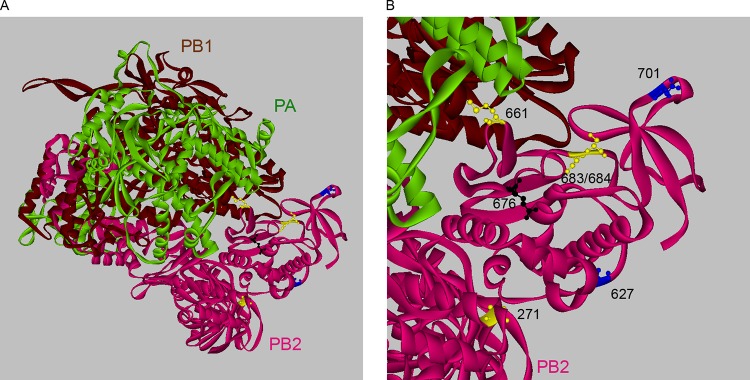
Residues 271, 661, and 683/684 are located on the surface of the polymerase complex. (A) Overall structure of the influenza A virus PA-PB1-PB2 complex (Protein Data Bank number 4WSB) (29). (B) Locations of residues 271, 661, and 683/684 in the complex. Residues 661 and 683/684 (yellow), which contribute to enhanced polymerase activity and virus replication, are located in close proximity to each other and to residue 701 (blue) on the surface of the molecule but far from residues 271 (yellow) or 627 (blue). Residue 676, which does not affect polymerase activity, is shown in black. The image was created using Discovery Studio ViewerPro (Accelyrs).
ACKNOWLEDGMENTS
This study was supported by the New York Influenza Center of Excellence, a member of the NIAID CEIRS network, under NIH contract HHSN266200700008C.
We thank David J. Topham, Luis Martinez-Sobrido, Richard Webby, Robert Webster, and Thorsten Wolff for reagents.
REFERENCES
- 1.Imai M, Kawaoka Y. 2012. The role of receptor binding specificity in interspecies transmission of influenza viruses. Curr Opin Virol 2:160–167. doi: 10.1016/j.coviro.2012.03.003. [DOI] [PMC free article] [PubMed] [Google Scholar]
- 2.Subbarao EK, London W, Murphy BR. 1993. A single amino acid in the PB2 gene of influenza A virus is a determinant of host range. J Virol 67:1761–1764. [DOI] [PMC free article] [PubMed] [Google Scholar]
- 3.Bussey KA, Bousse TL, Desmet EA, Kim B, Takimoto T. 2010. PB2 residue 271 plays a key role in enhanced polymerase activity of influenza A viruses in mammalian host cells. J Virol 84:4395–4406. doi: 10.1128/JVI.02642-09. [DOI] [PMC free article] [PubMed] [Google Scholar]
- 4.Naffakh N, Tomoiu A, Rameix-Welti MA, van der Werf S. 2008. Host restriction of avian influenza viruses at the level of the ribonucleoproteins. Annu Rev Microbiol 62:403–424. doi: 10.1146/annurev.micro.62.081307.162746. [DOI] [PubMed] [Google Scholar]
- 5.Manz B, Schwemmle M, Brunotte L. 2013. Adaptation of avian influenza A virus polymerase in mammals to overcome the host species barrier. J Virol 87:7200–7209. doi: 10.1128/JVI.00980-13. [DOI] [PMC free article] [PubMed] [Google Scholar]
- 6.Gabriel G, Dauber B, Wolff T, Planz O, Klenk HD, Stech J. 2005. The viral polymerase mediates adaptation of an avian influenza virus to a mammalian host. Proc Natl Acad Sci U S A 102:18590–18595. doi: 10.1073/pnas.0507415102. [DOI] [PMC free article] [PubMed] [Google Scholar]
- 7.Cauldwell AV, Long JS, Moncorge O, Barclay WS. 2014. Viral determinants of influenza A virus host range. J Gen Virol 95:1193–1210. doi: 10.1099/vir.0.062836-0. [DOI] [PubMed] [Google Scholar]
- 8.Bussey KA, Desmet EA, Mattiacio JL, Hamilton A, Bradel-Tretheway B, Bussey HE, Kim B, Dewhurst S, Takimoto T. 2011. PA residues in the 2009 H1N1 pandemic influenza virus enhance avian influenza virus polymerase activity in mammalian cells. J Virol 85:7020–7028. doi: 10.1128/JVI.00522-11. [DOI] [PMC free article] [PubMed] [Google Scholar]
- 9.Mehle A, Dugan VG, Taubenberger JK, Doudna JA. 2012. Reassortment and mutation of the avian influenza virus polymerase PA subunit overcome species barriers. J Virol 86:1750–1757. doi: 10.1128/JVI.06203-11. [DOI] [PMC free article] [PubMed] [Google Scholar]
- 10.Massin P, van der Werf S, Naffakh N. 2001. Residue 627 of PB2 is a determinant of cold sensitivity in RNA replication of avian influenza viruses. J Virol 75:5398–5404. doi: 10.1128/JVI.75.11.5398-5404.2001. [DOI] [PMC free article] [PubMed] [Google Scholar]
- 11.Garten RJ, Davis CT, Russell CA, Shu B, Lindstrom S, Balish A, Sessions WM, Xu X, Skepner E, Deyde V, Okomo-Adhiambo M, Gubareva L, Barnes J, Smith CB, Emery SL, Hillman MJ, Rivailler P, Smagala J, de Graaf M, Burke DF, Fouchier RA, Pappas C, Alpuche-Aranda CM, Lopez-Gatell H, Olivera H, Lopez I, Myers CA, Faix D, Blair PJ, Yu C, Keene KM, Dotson PD Jr, Boxrud D, Sambol AR, Abid SH, St George K, Bannerman T, Moore AL, Stringer DJ, Blevins P, Demmler-Harrison GJ, Ginsberg M, Kriner P, Waterman S, Smole S, Guevara HF, Belongia EA, Clark PA, Beatrice ST, Donis R, Katz J, Finelli L, Bridges CB, Shaw M, Jernigan DB, Uyeki TM, Smith DJ, Klimov AI, Cox NJ. 2009. Antigenic and genetic characteristics of swine-origin 2009 A(H1N1) influenza viruses circulating in humans. Science 325:197–201. doi: 10.1126/science.1176225. [DOI] [PMC free article] [PubMed] [Google Scholar]
- 12.Neumann G, Noda T, Kawaoka Y. 2009. Emergence and pandemic potential of swine-origin H1N1 influenza virus. Nature 459:931–939. doi: 10.1038/nature08157. [DOI] [PMC free article] [PubMed] [Google Scholar]
- 13.Itoh Y, Shinya K, Kiso M, Watanabe T, Sakoda Y, Hatta M, Muramoto Y, Tamura D, Sakai-Tagawa Y, Noda T, Sakabe S, Imai M, Hatta Y, Watanabe S, Li C, Yamada S, Fujii K, Murakami S, Imai H, Kakugawa S, Ito M, Takano R, Iwatsuki-Horimoto K, Shimojima M, Horimoto T, Goto H, Takahashi K, Makino A, Ishigaki H, Nakayama M, Okamatsu M, Takahashi K, Warshauer D, Shult PA, Saito R, Suzuki H, Furuta Y, Yamashita M, Mitamura K, Nakano K, Nakamura M, Brockman-Schneider R, Mitamura H, Yamazaki M, Sugaya N, Suresh M, Ozawa M, Neumann G, Gern J, Kida H, Ogasawara K, Kawaoka Y. 2009. In vitro and in vivo characterization of new swine-origin H1N1 influenza viruses. Nature 460:1021–1025. doi: 10.1038/nature08260. [DOI] [PMC free article] [PubMed] [Google Scholar]
- 14.Hatta M, Hatta Y, Kim JH, Watanabe S, Shinya K, Nguyen T, Lien PS, Le QM, Kawaoka Y. 2007. Growth of H5N1 influenza A viruses in the upper respiratory tracts of mice. PLoS Pathog 3:1374–1379. doi: 10.1371/journal.ppat.0030133. [DOI] [PMC free article] [PubMed] [Google Scholar]
- 15.Steel J, Lowen AC, Mubareka S, Palese P. 2009. Transmission of influenza virus in a mammalian host is increased by PB2 amino acids 627K or 627E/701N. PLoS Pathog 5:e1000252. doi: 10.1371/journal.ppat.1000252. [DOI] [PMC free article] [PubMed] [Google Scholar]
- 16.Desmet EA, Bussey KA, Stone R, Takimoto T. 2013. Identification of the N-terminal domain of the influenza virus PA responsible for the suppression of host protein synthesis. J Virol 87:3108–3118. doi: 10.1128/JVI.02826-12. [DOI] [PMC free article] [PubMed] [Google Scholar]
- 17.Bialas KM, Desmet EA, Takimoto T. 2012. Specific residues in the 2009 H1N1 swine-origin influenza matrix protein influence virion morphology and efficiency of viral spread in vitro. PLoS One 7:e50595. doi: 10.1371/journal.pone.0050595. [DOI] [PMC free article] [PubMed] [Google Scholar]
- 18.Neumann G, Watanabe T, Ito H, Watanabe S, Goto H, Gao P, Hughes M, Perez DR, Donis R, Hoffmann E, Hobom G, Kawaoka Y. 1999. Generation of influenza A viruses entirely from cloned cDNAs. Proc Natl Acad Sci U S A 96:9345–9350. doi: 10.1073/pnas.96.16.9345. [DOI] [PMC free article] [PubMed] [Google Scholar]
- 19.Hayashi T, MacDonald LA, Takimoto T. 2015. Influenza A virus protein PA-X contributes to viral growth and suppression of the host antiviral and immune responses. J Virol 89:6442–6452. doi: 10.1128/JVI.00319-15. [DOI] [PMC free article] [PubMed] [Google Scholar]
- 20.Reed L, Muench H. 1938. A simple method of estimating fifty per cent endpoints. Am J Epidemiol 27:493–497. [Google Scholar]
- 21.Squires RB, Noronha J, Hunt V, Garcia-Sastre A, Macken C, Baumgarth N, Suarez D, Pickett BE, Zhang Y, Larsen CN, Ramsey A, Zhou L, Zaremba S, Kumar S, Deitrich J, Klem E, Scheuermann RH. 2012. Influenza research database: an integrated bioinformatics resource for influenza research and surveillance. Influenza Other Respir Viruses 6:404–416. doi: 10.1111/j.1750-2659.2011.00331.x. [DOI] [PMC free article] [PubMed] [Google Scholar]
- 22.Miotto O, Heiny A, Tan TW, August JT, Brusic V. 2008. Identification of human-to-human transmissibility factors in PB2 proteins of influenza A by large-scale mutual information analysis. BMC Bioinform 9(Suppl 1):S18. doi: 10.1186/1471-2105-9-S1-S18. [DOI] [PMC free article] [PubMed] [Google Scholar]
- 23.Gabriel G, Czudai-Matwich V, Klenk HD. 2013. Adaptive mutations in the H5N1 polymerase complex. Virus Res 178:53–62. doi: 10.1016/j.virusres.2013.05.010. [DOI] [PubMed] [Google Scholar]
- 24.Mok CK, Lee HH, Lestra M, Nicholls JM, Chan MC, Sia SF, Zhu H, Poon LL, Guan Y, Peiris JS. 2014. Amino acid substitutions in polymerase basic protein 2 gene contribute to the pathogenicity of the novel A/H7N9 influenza virus in mammalian hosts. J Virol 88:3568–3576. doi: 10.1128/JVI.02740-13. [DOI] [PMC free article] [PubMed] [Google Scholar]
- 25.Herfst S, Chutinimitkul S, Ye J, de Wit E, Munster VJ, Schrauwen EJ, Bestebroer TM, Jonges M, Meijer A, Koopmans M, Rimmelzwaan GF, Osterhaus AD, Perez DR, Fouchier RA. 2010. Introduction of virulence markers in PB2 of pandemic swine-origin influenza virus does not result in enhanced virulence or transmission. J Virol 84:3752–3758. doi: 10.1128/JVI.02634-09. [DOI] [PMC free article] [PubMed] [Google Scholar]
- 26.Mehle A, Doudna JA. 2009. Adaptive strategies of the influenza virus polymerase for replication in humans. Proc Natl Acad Sci U S A 106:21312–21316. doi: 10.1073/pnas.0911915106. [DOI] [PMC free article] [PubMed] [Google Scholar]
- 27.Yamada S, Hatta M, Staker BL, Watanabe S, Imai M, Shinya K, Sakai-Tagawa Y, Ito M, Ozawa M, Watanabe T, Sakabe S, Li C, Kim JH, Myler PJ, Phan I, Raymond A, Smith E, Stacy R, Nidom CA, Lank SM, Wiseman RW, Bimber BN, O'Connor DH, Neumann G, Stewart LJ, Kawaoka Y. 2010. Biological and structural characterization of a host-adapting amino acid in influenza virus. PLoS Pathog 6:e1001034. doi: 10.1371/journal.ppat.1001034. [DOI] [PMC free article] [PubMed] [Google Scholar]
- 28.Reich S, Guilligay D, Pflug A, Malet H, Berger I, Crepin T, Hart D, Lunardi T, Nanao M, Ruigrok RW, Cusack S. 2014. Structural insight into cap-snatching and RNA synthesis by influenza polymerase. Nature 516:361–366. doi: 10.1038/nature14009. [DOI] [PubMed] [Google Scholar]
- 29.Pflug A, Guilligay D, Reich S, Cusack S. 2014. Structure of influenza A polymerase bound to the viral RNA promoter. Nature 516:355–360. doi: 10.1038/nature14008. [DOI] [PubMed] [Google Scholar]
- 30.Li Z, Chen H, Jiao P, Deng G, Tian G, Li Y, Hoffmann E, Webster RG, Matsuoka Y, Yu K. 2005. Molecular basis of replication of duck H5N1 influenza viruses in a mammalian mouse model. J Virol 79:12058–12064. doi: 10.1128/JVI.79.18.12058-12064.2005. [DOI] [PMC free article] [PubMed] [Google Scholar]
- 31.Mehle A, Doudna JA. 2008. An inhibitory activity in human cells restricts the function of an avian-like influenza virus polymerase. Cell Host Microbe 4:111–122. doi: 10.1016/j.chom.2008.06.007. [DOI] [PMC free article] [PubMed] [Google Scholar]
- 32.Moncorge O, Mura M, Barclay WS. 2010. Evidence for avian and human host cell factors that affect the activity of influenza virus polymerase. J Virol 84:9978–9986. doi: 10.1128/JVI.01134-10. [DOI] [PMC free article] [PubMed] [Google Scholar]