Abstract
The emerging porcine epidemic diarrhea virus (PEDV) requires trypsin supplementation to activate its S protein for membrane fusion and virus propagation in cell culture. By substitution of a single amino acid in the S protein, we created a recombinant PEDV with an artificial furin protease cleavage site N terminal of the putative fusion peptide (PEDV-SFCS). PEDV-SFCS exhibited trypsin-independent cell-cell fusion and was able to replicate in culture cells independently of trypsin, though to low titer.
TEXT
Entry of the enveloped coronaviruses (CoVs) is mediated by the spike (S) glycoprotein. The S protein can be functionally divided into two domains, S1 and S2, which enable the consecutive stages of receptor binding and membrane fusion, respectively. Like other class I viral fusion proteins, the CoV S proteins rely on proteolytic cleavage by host proteases for fusion priming/activation; cleavage enables the release of the fusion peptide and its insertion into the target cell membrane in a controlled manner. To this end, most CoVs exploit endogenous cellular proteases, such as furin, plasma membrane proteases, and endolysosomal proteases, that cleave the S protein during virus exit from the infected cell or during its entry at or in the target cell (1, 2). Cleavage of CoV S proteins has been shown to occur at the junction of receptor binding domain S1 and membrane fusion domain S2 and just upstream of the putative fusion peptide (S2′ site) in the S2 domain, with the latter cleavage supposed to be the most critical for fusion, as it liberates the fusion peptide (1, 2).
Porcine epidemic diarrhea virus (PEDV) is an emerging coronavirus that causes acute diarrheal disease in swine in Asia and since 2013 in the Americas. In contrast to other CoVs, PEDV requires the presence of an exogenous protease, trypsin, in the cell culture medium to activate the S protein for membrane fusion and for virus propagation in cell culture (3). This trypsin dependency in vitro is consistent with the tissue tropism of PEDV in vivo, where PEDV is confined to the trypsin-rich small intestine. Trypsin activates the PEDV S protein for membrane fusion after the binding of the virion to the host cell (4). Genetic and mutational analyses have pointed to a conserved arginine just upstream of the fusion peptide (S2′ site) in membrane fusion domain S2 as the trypsin cleavage site for fusion activation, yet direct evidence of functional cleavage at this position is lacking (4).
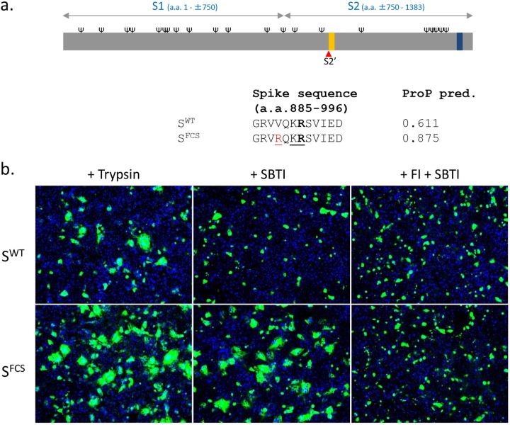
To assess the cleavage position required for PEDV S protein fusion activation and to test whether activation by exogenous trypsin can be bypassed by an endogenous host protease, we introduced an artificial cleavage recognition site for the furin protease into the PEDV spike protein (Fig. 1). The proprotein convertase furin is a type I membrane protein that is ubiquitously expressed in eukaryotic cells. Furin is located primarily in the trans-Golgi network but also occurs at the cell surface and as an extracellular, truncated soluble form (5). It cleaves cellular and viral proteins C terminal of a substrate recognition motif containing basic amino acids, with R-X-X-R and R-X-K/R-R (R, arginine; K, lysine; X, any amino acid) representing the minimal and highly favored motifs, respectively. We constructed a mutant PEDV S protein (strain CV777) with a valine-to-arginine single-residue substitution at amino acid position 888 (V888R), thereby creating a furin cleavage sequence site (VQKR→RQKR) N terminal of the predicted fusion peptide (6). Bioinformatic analysis predicted that this position can be cleaved by furin with a prediction score of 0.875, relative to 0.611 for the wild-type S (SWT) protein (Fig. 1a).
FIG 1.
The PEDV S protein with an artificial furin cleavage site at the S2′ position (SFCS) mediates trypsin-independent cell-cell fusion. (a) Schematic representation of the PEDV S protein (drawn to scale). Indicated are the positions of receptor binding domain S1 and membrane fusion domain S2, predicted N-glycosylation sites (Ψ; NetNGlyc server), the predicted S2′ cleavage site (red triangle), the fusion peptide (orange bar), and the transmembrane domain (black bar; TMHMM server). (Lower panel) Amino acid (a.a.) sequence at the putative S2′ cleavage site (R891, indicated in bold) for the wild-type CV777 S protein (SWT) and for a mutant S protein (SFCS) with a valine-to-arginine substitution at amino acid position 888 (colored in red), creating a furin cleavage site sequence (underlined) [R-X-(K/R)-R, where X is any amino acid]. Furin scores for the S2′ position predicted (pred.) by the ProP 1.0 server are indicated for both S variants. (b) Vero cells were transfected with expression plasmids encoding wild-type PEDV S (SWT) or SFCS and cultured in the absence or presence of a furin inhibitor (FI; 40 μM), a soybean trypsin inhibitor (SBTI; 40 μg/ml), or trypsin (15 μg/ml), as indicated. At 40 h posttransfection, cells were fixed and permeabilized. Nuclei (blue) were stained with DAPI (4′,6-diamidino-2-phenylindole), and PEDV S (green) expression was visualized by its C-terminally appended FLAG tag using anti-Flag monoclonal antibody (MAb) (Sigma). The transfection experiments were repeated two times, and representative images are shown.
First, we assessed whether the introduction of the furin cleavage site in S (SFCS) enables trypsin-independent cell-cell fusion. Therefore, we transiently expressed the SFCS protein and the SWT protein, each provided with a C-terminally appended Flag tag, in Vero cells. At 6 h posttransfection, the culture medium was replaced by fresh medium supplemented either with a furin inhibitor or with a soybean trypsin inhibitor, the latter to ensure that trypsin activity was completely absent. At 47 h posttransfection, cells were treated with trypsin for 1 h or left untreated. In the presence of trypsin, both the SWT and SFCS spike proteins were able to efficiently form syncytia (Fig. 1b). As expected, the SWT spike protein was unable to mediate cell-cell fusion when trypsin was omitted from the cell culture medium. However, the formation of multiple, large syncytia were seen after expression of the SFCS spike protein in the absence of trypsin activity. Trypsin-independent syncytium formation by SFCS may be inhibited by the inclusion of furin inhibitor in the culture medium (Fig. 1b). These data indicate that the creation, through a single amino acid substitution, of a furin cleavage site at the S2′ position renders the PEDV S protein prone to activation of its membrane fusion capacity by endogenous furin.
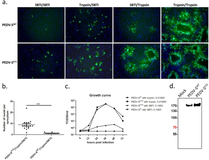
To assess whether the furin cleavage site at the S2′ position of the S protein enables the virus to infect cultured cells independently of trypsin, we introduced the mutation encoding the SV888R substitution into the viral genome. We generated recombinant viruses encoding either the wild-type CV777 S protein (PEDV-SWT) or the SFCS mutant (PEDV-SFCS) using our recently established PEDV reverse-genetics system (7). To facilitate the analyses, the nonessential ORF3 gene in the genomes of both recombinant viruses was replaced by the green fluorescent protein (GFP) gene. Recombinant PEDV-SWT and PEDV-SFCS were successfully rescued in the presence of trypsin, and the relevant S gene region of their genomes was confirmed by sequencing. Next, we inoculated Vero cells in parallel with PEDV-SWT and PEDV-SFCS in the absence and presence of trypsin for 2 h. The infection was subsequently continued in the absence or presence of trypsin for another 10 h, after which infected (i.e., GFP-positive) cells were visualized by fluorescence microscopy. In the absence of trypsin, hardly any infection was seen on Vero cells for PEDV-SWT, whereas clear infection was observed for the PEDV-SFCS mutant (Fig. 2a). Both viruses efficiently infected Vero cells in the presence of trypsin. Syncytia were abundantly observed when trypsin was absent after virus inoculation for the PEDV-SFCS mutant but not for PEDV-SWT (Fig. 2a and b). These results confirm our earlier observation that the furin recognition site creating mutation SV888R confers trypsin-independent cell-cell fusion.
FIG 2.
Recombinant PEDV encoding S of the PEDV CV777 strain with an artificial furin cleavage site at the S2′ site (PEDV-SFCS) mediates trypsin-independent cell-cell fusion. (a) Cells were infected with GFP-expressing recombinant PEDVs carrying wild-type CV777 S (PEDV-SWT) or mutated CV777 S (PEDV-SFCS) in the presence of trypsin (15 μg/ml) or soybean trypsin inhibitor (SBTI; 40 μg/ml), as indicated. At 2 h postinfection, cells were washed and further cultured in the presence of soybean trypsin inhibitor (SBTI; 40 μg/ml) or trypsin (15 μg/ml), as indicated. Cells were fixed at 12 h p.i. Nuclei were stained with DAPI (blue), and images of PEDV-infected cells (GFP positive, green) were acquired. Experiments were repeated two times, and representative images are shown. (b) Vero cells were infected with PEDV-SWT and PEDV-SFCS in the presence of trypsin for 2 h and further cultured in the presence of soybean trypsin inhibitor (as indicated above). At 12 h p.i., the numbers of nuclei per focus of infection were counted. Statistical significance was assessed by unpaired one-tailed Student’s t test; **, P < 0.01. (c) Vero cells were infected with recombinant PEDV-SWT and PEDV-SV888R (MOI 0.01) in the absence or presence of trypsin. The infectivity in the culture medium was monitored by taking small samples from the medium at various time points postinfection and titrating them on Vero cells in the presence of trypsin. TCID50, 50% tissue culture infective dose. (d) Vero cells were mock infected or infected with PEDV-SWT and PEDV-SFCS in the presence of the trypsin inhibitor for 2 h (MOI, 10) and further cultured in the absence of trypsin, as described for panel a. At 24 h p.i., the cell culture supernatants of these cultures were pelleted through a 20% sucrose cushion. Pellets were dissolved in Laemmli sample buffer and subjected to Western blotting. Virion-incorporated SWT and SFCS proteins were detected by their C-terminally appended FLAG tag using an anti-Flag MAb (Sigma). Sizes of marker proteins are indicated at the left in kilodaltons.
To assess whether the SV888R mutation would also enable PEDV to propagate in the absence of trypsin, the growth kinetics of PEDV-SWT and PEDV-SFCS on Vero cells were compared in the absence and presence of trypsin. Vero cells were inoculated in parallel with PEDV-SWT and PEDV-SFCS at a multiplicity of infection (MOI) of 0.01 or 0.1. Both viruses displayed similar growth kinetics and reached similar titers in the presence of trypsin (Fig. 2c). In the absence of trypsin, no infectious progeny was detected with either virus after inoculation at a low MOI (0.01 [data not shown]). At a 10-fold-higher MOI, PEDV-SFCS—but not PEDV-SWT—yielded low infectious virus titers starting from 12 h postinfection (p.i.). To check the cleavage status of the spike protein as it occurs on PEDV-SFCS virions, we performed Western blot analysis. The results indicate that the introduction of the furin cleavage site at the S2′ site did not result in detectable cleavage (Fig. 2e).
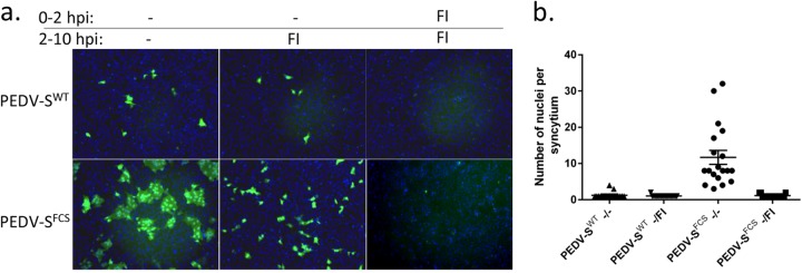
Huh-7 human hepatoma cells are known to express high levels of furin protease (8). We therefore analyzed whether these cells could support infection by PEDV-SFCS. Of note, infection of Huh-7 cells in the presence of trypsin could not be assessed since these cells are affected too strongly by trypsin at the concentrations required for propagation of PEDV in cell culture. Inoculation of Huh-7 cells with PEDV-SFCS resulted in trypsin-independent infection and the development of syncytia (Fig. 3a and b). Virus entry as well as cell-cell fusion could be inhibited when the furin inhibitor was present during these processes. Interestingly, some PEDV-SWT infection was also observed on Huh-7 cells, which could be inhibited by furin inhibitor, yet no syncytia were seen. Whether this infectivity correlates with the predicted suboptimal proprotease cleavage site at the S2′ position in the PEDV SWT protein (Fig. 1) remains to be seen.
FIG 3.

Furin inhibitor blocks trypsin-independent entry and formation of syncytia by PEDV-SFCS in human hepatoma (Huh-7) cells. (a) Cells were inoculated with PEDV-SWT or PEDV-SFCS at an MOI of 0.5 (the MOI was based on virus infectivity on Vero cells in the presence of trypsin). The furin inhibitor decanoyl-Arg-Val-Lys-Arg-chloromethylketone (FI; 40 μM) was present in the cell culture supernatant from −1 h to 2 h p.i. or from 2 h to 12 h p.i. To prevent trypsin-mediated entry, soybean trypsin inhibitor (SBTI; 40 μg/ml) was present in the culture supernatant throughout the experiment. Cells were fixed at 12 h p.i. Nuclei were stained with DAPI (blue), and images of PEDV-infected cells (GFP positive; green) were acquired. Experiments were repeated two times, and representative images are shown. (b) Huh-7 cells were infected with PEDV-SWT and PEDV-SFCS in the presence of trypsin inhibitor for 2 h and further cultured in the absence or presence of furin inhibitor, as described for panel a. At 12 h p.i., the numbers of nuclei per focus of infection were counted.
The proteolytic activation process of the CoV spike fusion protein has long been rather enigmatic. Whereas processing at the S1/S2 junction by furin was documented already in the early 1980s (9, 10), this cleavage occurs only in a subset of coronaviruses and does not liberate the putative fusion peptide at the N terminus of the membrane-anchored subunit, as it does in other class I viral fusion proteins. The second, more universal, as well as more appropriately located, S2′ cleavage site was identified only recently, and evidence for its general importance in CoV infection has since been accumulating (4, 11–13; for a recent review, see reference 1). Cleavage at the S2′ position is generally carried out by cellular proteases occurring at the plasma membrane or in the endo-/lysosomal system, depending on the particular target cell and S2′ sequence. In the case of PEDV, however, in which the S protein lacks a canonical furin cleavage site at the S1/S2 and the S2′ position, activation supposedly occurs by trypsin-like enzymes in the gut, and the virus hence requires supplementation of trypsin for propagation in vitro. Earlier, we mapped this trypsin requirement to the S2′ cleavage site in the PEDV S protein and demonstrated the critical importance of the characteristic arginine at this site for the viability of the virus and for the cell fusion capacity of the S protein (4).
In the present study, we aimed to demonstrate the requirement for cleavage at the S2′ site and to alleviate the trypsin dependence of PEDV infection in vitro. Thus, we show that introduction—through a single point mutation—of an artificial furin cleavage motif N terminal of the spike fusion peptide confers cell-cell fusion and PEDV entry in a trypsin-independent manner. Both processes were blocked by a furin-specific inhibitor, thereby confirming the functionality of furin cleavage at the S2′ position. The observations add further evidence that cleavage just upstream of the fusion peptide is a general and essential requirement for activation of CoV spike proteins for membrane fusion (11–14). Propagation of PEDV-SFCS in the absence of trypsin was less efficient than in its presence, which might be due to trypsin also being required for virus release (15). Moreover, besides cleavage N terminal of the fusion peptide, additional cleavage(s) may be required to increase the S protein's membrane fusion efficiency. Cleavage of the S protein at the S1/S2 junction of the coronaviruses Middle East respiratory syndrome (MERS) CoV, severe acute respiratory syndrome (SARS) CoV, and infectious-bronchitis virus (IBV) has been implicated to precede and promote cleavage at the S2′ position (11, 13, 14). For PEDV, the lack of efficient cleavage of virion-incorporated SFCS proteins indicates that the introduced furin cleavage site at the S2′ site is rather inaccessible for furin. Whether cleavage at the S1/S2 junction or binding to the receptor enhances the efficiency of cleavage at the S2′ cleavage site awaits further investigation.
While field strains of PEDV strictly require exogenous trypsin for propagation in vitro, serial passaging of the virus on cultured cells can lead to trypsin independency (3, 4). Vero cells are the most commonly used cells for PEDV studies because of their resistance to the high concentrations of trypsin required for PEDV infection. Hence, the protease requirement of PEDV, in addition to the specific virus receptor, may function as a critical tropism determinant in vitro as well as in vivo. As a consequence, the rational or evolutionary adaptation of CoVs to the use of ubiquitous, endogenous proteases like furin for the activation of their S proteins may expand their tropism in vitro and in vivo (1).
ACKNOWLEDGMENT
This study was supported by a grant from the Natural Science Foundation of China (31272572) provided to Qigai He.
REFERENCES
- 1.Millet JK, Whittaker GR. 22 November 2014. Host cell proteases: critical determinants of coronavirus tropism and pathogenesis. Virus Res doi: 10.1016/j.virusres.2014.11.021. [DOI] [PMC free article] [PubMed] [Google Scholar]
- 2.Heald-Sargent T, Gallagher T. 2012. Ready, set, fuse! The coronavirus spike protein and acquisition of fusion competence. Viruses 4:557–580. doi: 10.3390/v4040557. [DOI] [PMC free article] [PubMed] [Google Scholar]
- 3.Hofmann M, Wyler R. 1988. Propagation of the virus of porcine epidemic diarrhea in cell culture. J Clin Microbiol 26:2235–2239. [DOI] [PMC free article] [PubMed] [Google Scholar]
- 4.Wicht O, Li W, Willems L, Meuleman TJ, Wubbolts RW, van Kuppeveld FJ, Rottier PJ, Bosch BJ. 2014. Proteolytic activation of the porcine epidemic diarrhea coronavirus spike fusion protein by trypsin in cell culture. J Virol 88:7952–7961. doi: 10.1128/JVI.00297-14. [DOI] [PMC free article] [PubMed] [Google Scholar]
- 5.Schafer W, Stroh A, Berghofer S, Seiler J, Vey M, Kruse ML, Kern HF, Klenk HD, Garten W. 1995. Two independent targeting signals in the cytoplasmic domain determine trans-Golgi network localization and endosomal trafficking of the proprotein convertase furin. EMBO J 14:2424–2435. [DOI] [PMC free article] [PubMed] [Google Scholar]
- 6.Madu IG, Roth SL, Belouzard S, Whittaker GR. 2009. Characterization of a highly conserved domain within the severe acute respiratory syndrome coronavirus spike protein S2 domain with characteristics of a viral fusion peptide. J Virol 83:7411–7421. doi: 10.1128/JVI.00079-09. [DOI] [PMC free article] [PubMed] [Google Scholar]
- 7.Li C, Li Z, Zou Y, Wicht O, van Kuppeveld FJ, Rottier PJ, Bosch BJ. 2013. Manipulation of the porcine epidemic diarrhea virus genome using targeted RNA recombination. PLoS One 8:e69997. doi: 10.1371/journal.pone.0069997. [DOI] [PMC free article] [PubMed] [Google Scholar]
- 8.Tay FP, Huang M, Wang L, Yamada Y, Liu DX. 2012. Characterization of cellular furin content as a potential factor determining the susceptibility of cultured human and animal cells to coronavirus infectious bronchitis virus infection. Virology 433:421–430. doi: 10.1016/j.virol.2012.08.037. [DOI] [PMC free article] [PubMed] [Google Scholar]
- 9.Frana MF, Behnke JN, Sturman LS, Holmes KV. 1985. Proteolytic cleavage of the E2 glycoprotein of murine coronavirus: host-dependent differences in proteolytic cleavage and cell fusion. J Virol 56:912–920. [DOI] [PMC free article] [PubMed] [Google Scholar]
- 10.Sturman LS, Ricard CS, Holmes KV. 1985. Proteolytic cleavage of the E2 glycoprotein of murine coronavirus: activation of cell-fusing activity of virions by trypsin and separation of two different 90K cleavage fragments. J Virol 56:904–911. [DOI] [PMC free article] [PubMed] [Google Scholar]
- 11.Belouzard S, Chu VC, Whittaker GR. 2009. Activation of the SARS coronavirus spike protein via sequential proteolytic cleavage at two distinct sites. Proc Natl Acad Sci U S A 106:5871–5876. doi: 10.1073/pnas.0809524106. [DOI] [PMC free article] [PubMed] [Google Scholar]
- 12.Burkard C, Verheije MH, Wicht O, van Kasteren SI, van Kuppeveld FJ, Haagmans BL, Pelkmans L, Rottier PJ, Bosch BJ, de Haan CA. 2014. Coronavirus cell entry occurs through the endo-/lysosomal pathway in a proteolysis-dependent manner. PLoS Pathog 10:e1004502. doi: 10.1371/journal.ppat.1004502. [DOI] [PMC free article] [PubMed] [Google Scholar]
- 13.Yamada Y, Liu DX. 2009. Proteolytic activation of the spike protein at a novel RRRR/S motif is implicated in furin-dependent entry, syncytium formation, and infectivity of coronavirus infectious bronchitis virus in cultured cells. J Virol 83:8744–8758. doi: 10.1128/JVI.00613-09. [DOI] [PMC free article] [PubMed] [Google Scholar]
- 14.Millet JK, Whittaker GR. 2014. Host cell entry of Middle East respiratory syndrome coronavirus after two-step, furin-mediated activation of the spike protein. Proc Natl Acad Sci U S A 111:15214–15219. doi: 10.1073/pnas.1407087111. [DOI] [PMC free article] [PubMed] [Google Scholar]
- 15.Shirato K, Matsuyama S, Ujike M, Taguchi F. 2011. Role of proteases in the release of porcine epidemic diarrhea virus from infected cells. J Virol 85:7872–7880. doi: 10.1128/JVI.00464-11. [DOI] [PMC free article] [PubMed] [Google Scholar]


