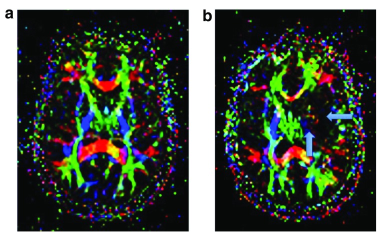
Figure 3a–b. Axial maps of fractional anisotropy (FA).
3a Normal FA shows the integrity and directionality of the white matter fibers (red: right-left, green: anterior-posterior, blue: craneo-caudal). 3b Altered (low) FA seen as loss of the normal colors of the left corticospinal tract in the internal capsule and of the left longitudinal fasciculus related to ischemic infarct of the territory of the left middle cerebral artery (arrows) in a patient with neuropsychiatric lupus and stroke. Figure origin: Department of Radiology, Hospital Clinic Barcelona.

