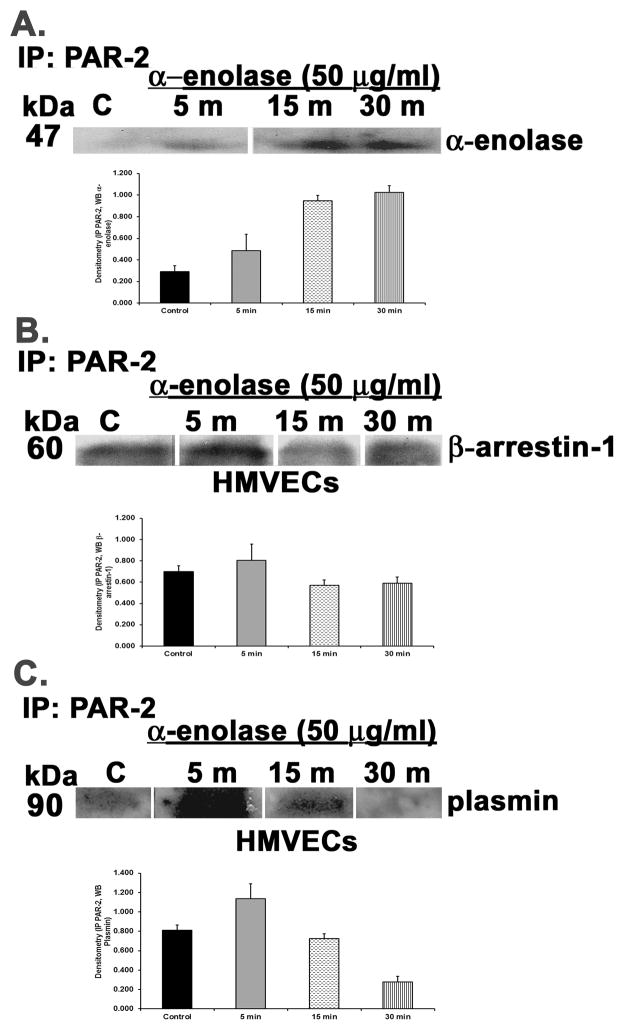
Figure 5. α-enolase causes occupancy, activation, and co-precipitation of the PAR-2 receptor on HMVECs with β-arrestin-1 and plasmin.
HMVECs were incubated with media or α-enolase [50 μg/ml] for 5–30 min, the cells were lysed, PAR-2 was immunoprecipitated, and the immunoprecipitate was blotted for α-enolase, β-arrestin-1 or plasmin. For each panel a bar graph is included which illustrates the mean±SEM of the densitometry of the bands of interest. Panel A demonstrates that α-enolase co-precipitated with the PAR-2 receptor beginning first at 5 min and continuing through 30 min. Panel B demonstrates the treatment of HMVEC with α-enolase induced co-precipitation of β-arrestin-1 beginning at 1 min and persisting through 30 min. Panel C depicts α-enolase elicited co-precipitation of plasmin with the PAR-2 receptor beginning at 5 min and continuing through 15 min. This figure represents identical results from two separate experiments using cells from different donors.

