Abstract
Spontaneous iliac vein rupture (SIVR) is a rare entity, which usually occurs without a precipitating factor, but can be a life-threatening emergency often requiring an emergency operation. This is a case report of SIVR in a 62-year-old female who presented to the emergency room with left leg swelling. Workup with contrast-enhanced computed tomography revealed a left leg deep vein thrombosis with May-Thurner syndrome and a hematoma in the pelvic cavity without definite evidence of arterial bleeding. She was managed conservatively without surgical intervention, and also underwent inferior vena cava filter insertion and subsequent anticoagulation therapy for pulmonary thromboembolism. This case shows that SIVR can be successfully managed with close monitoring and conservative management, and anticoagulation may be safely applied despite the patient presenting with venous bleeding.
Keywords: Spontaneous rupture, Iliac vein, Hemoperitoneum
INTRODUCTION
Spontaneous iliac vein rupture (SIVR) is a rare entity with only three cases having been documented in Korea [1,2]. However, clinical suspicion and early diagnosis of SIVR is very important since it may be life-threatening, often requiring aggressive management of unstable vital signs and emergent operation.
CASE
A 62-year-old female presented to the emergency department with left leg swelling. She had no history of medical diseases other than hypertension, which was well controlled with telmisartan/hydrochlorothiazide and sarpogrelate for 2 years. In addition, she had no history of trauma, but had been diagnosed with spinal stenosis, for which she had undergone operation 3 times and was on medications (gabapentin, tramadol/acetaminophen and etizolam) for the past 15 years. She had also undergone open cholecystectomy, oophorectomy and uterine myomectomy 20 years ago. For the past 2–3 years, the patient was only able to walk about 50 m with the aid of canes or walkers due to deterioration of her lower back and leg pain, with most of her daily activities being restricted to housework.
The symptom of acute left leg swelling started 2 days ago, with aggravation of her symptoms on the day of arrival to the emergency department. On arrival, her vital signs were stable, with a blood pressure of 106/57 mmHg, heart rate of 105 beats/min, respiratory rate of 18 times/min and body temperature of 36°C. Her mental state was clear and she complained of a mild discomfort and distention of the left lower abdomen but did not show any signs of peritoneal irritation such as rebound tenderness or rigidity. Her initial laboratory test results were as follows: hemoglobin 7.5 g/dL, hematocrit 23.1%, creatinine 0.59 mg/dL, potassium 3.0 mmol/L, D-dimer 5.25 μL/mL, and other test results including liver function tests, coagulation tests and electrolytes were within normal range. Her electrocardiography showed non-specific reactions with sinus tachycardia.
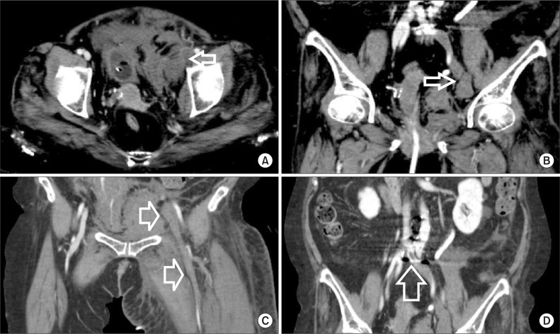
A contrast-enhanced computed tomography (CT) was then obtained (Fig. 1), revealing a hematoma in the pre-vesical area. Although there was no evidence of active contrast leakage on CT, rupture of the left external iliac vein (EIV) was highly suspicious (Fig. 1A). The presumed rupture site of the EIV was on the medial side, as shown by the location of a large hematoma (Fig. 1B). Deep vein thrombosis (DVT) of the left common iliac, external iliac, femoral and popliteal veins was also observed (Fig. 1C) and findings consistent with May-Thurner syndrome could be seen (Fig. 1D). She was immediately managed with hydration and red blood cell (RBC) transfusion and a decision to monitor her closely in the intensive care unit (ICU) was made instead of performing an emergency operation. Her hemoglobin and hematocrit levels rose to 8.8 g/dL and 26.2% respectively after initial resuscitation.
Fig. 1.
Initial abdominopelvic computed tomography showing (A) rupture of the left external iliac vein (EIV) with a hematomoa annotated by arrows. (B) A large hematomoa on the medial side of the left EIV annotated by arrows. (C) Wide-range deep vein thrombosis from left common iliac vein to left popliteal vein. (D) May-Thurner syndrome, overwhelmed iliac vein was annotated.
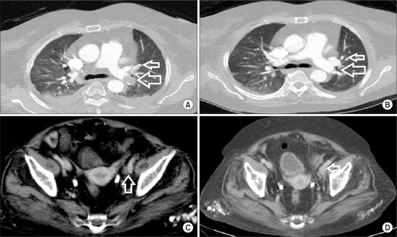
The next day, a follow-up CT was performed, in which the hematoma was still present in the retroperitoneal cavity but without any evidence of active bleeding. The extent of DVT had not changed either, and pulmonary thromboembolism (PTE) was found in both main pulmonary arteries (Fig. 2A). Therefore insertion of an inferior vena cava (IVC) filter was performed. RBC transfusion was continued due to a decrease in hemoglobin and hematocrit from 8.8 g/dL and 26.2% to 7.9 g/dL and 23.7%, respectively. From the third day onwards, follow up blood tests were normalized and vital signs were stable, and on the fourth hospital day, she was moved to the general ward.
Fig. 2.
Computed tomography finding showing pulmonary thromboembolism (A) at initial presentation with thrombus was annotated. (B) At 2 months follow-up. Thrombus was not observed. (C, D) Abdominopelvic computed tomography finding at 2 months follow-up showing a decreased hematoma around the medial side of the left external iliac vein was annotated by arrows.
On the fifth hospital day, a follow-up CT was performed again for further evaluation. PTE in the left superior lobar, inter-lobar, inferior lobar arteries and right superior lobar, basal segmental arteries was reported, and ill-defined consolidations in the left lower lobe were found, most likely due to lung infarction. DVT of the left common iliac, external iliac and femoral veins were remnant. She was started on low molecular weight heparin (LMWH) at therapeutic range, and was switched to warfarin on the seventh hospital day. There was no aggravation of imaging or laboratory abnormalities despite anticoagulation therapy. The IVC filter was retrieved on the twentieth hospital day. She was discharged after 25 days, and follow-up CT was performed two months after discharge in the outpatient clinic, which showed a decreased yet residual PTE in the left inter-lobar, lower lobar and basal segmental arteries (Fig. 2B), but the DVT had improved with gradual resolution of her leg edema. The hematoma in retroperitoneal space had decreased, but was still remnant around the medial side of the left EIV, which was the suspected site of EIV rupture (Fig. 2C, D).
DISCUSSION
Rupture of the iliac vein is rare and is most often related to trauma or iatrogenic injury during operation. A spontaneous rupture of the iliac vein is a rare disease, with less than forty-cases having been reported in the literature since the first reported case in 1961 by Hossne et al. [1,3]. The etiology of SIVR is not well defined, yet some proposed theories include mechanical, inflammatory and hormonal factors [1]. The interference of the left common iliac vein flow is the most representative example of a mechanical factor, such as May-Thurner syndrome [3] or Cockett’s syndrome [4]. Moreover, certain positions such as bending or straining (which may increase pressure to the left iliac vein between the right common iliac artery and the inguinal ligament), a previous pregnancy [5] or an intraabdominal mass may also contribute as a mechanical factor. Another study also reported that obstruction of flow or vibratory irritation may lead to vessel weakening [3]. Inflammatory factors such as thrombophlebitis or DVT may also affect the elasticity of the vessel wall [1,2]. On the other hand, estrogen in females may play a role to protect the vessels and promote compliance [1].
From the reported literature of 35 patients with SIVR between 1961 to 2004, the average age in women was 60.6±13.4 years [5] and 94% of cases were left-sided [5]. Also 79% of the cases were related to DVT or thrombophlebitis [5]. The main modality of treatment was bleeding control by direct suture or bypass reconstruction [6]. Thus surgical intervention was performed in the majority of cases, and the main cause for emergency operation was a lowered blood pressure due to bleeding [5]. Only two cases were treated non-operatively: one patient was treated conservatively with success, while another case of successful endovascular repair presented ultimately with a poor outcome due to prolonged anoxic injury [5].
In this case, the patient presented to the emergency room with acute left leg swelling and weakening. The cause of DVT was most likely due to the presence of May-Thurner syndrome but may also have resulted from long term decreased mobility owing to spinal stenosis. She was also overweight with a body mass index of 30.52 kg/m2, which may have contributed. These factors may have led to venous hypertension and a high abdominal pressure, which may have predisposed to SIVR. Prompt execution of a CT scan led to an accurate diagnosis, allowing for early initiation of resuscitation including hydration and RBC transfusion. The patient was initially observed closely in the ICU but always thoroughly ready for an emergency operation. This case was significant in that she successfully recovered with only conservative therapy by performing short-term blood tests and CT scans, without the need for surgical intervention. In most of the previously reported cases, an urgent exploratory operation was performed for suspected SIVR. However, conservative management has also been advocated in some previous studies and has been suggested to be safer than surgical procedures [1]. As in this case, conservative management can be an acceptable option in selected cases with stable initial vital signs, good response to hydration or transfusion, no specific findings on laboratory results and no evidence of active bleeding on imaging studies such as CT scans.
This patient also required treatment for PTE, which was achieved by LMWH, warfarin and IVC filter insertion. The use of anticoagulation in the situation of venous rupture is complicated and a difficult decision to make. There is no definite consensus and the risks/benefits of anticoagulation use should be weighed to decrease morbidity and mortality from both bleeding and respiratory collapse. Therefore the decision should be based on the clinical situation of the patient. In this case, an IVC filter was inserted first to minimize further propagation of thrombus to the pulmonary arteries. Short-term follow-up CT and blood tests were then performed to decide on the appropriate time for initiation of anticoagulation. Fortunately, by the fifth day, there were no further changes in blood tests or vital signs, and thus LMWH was initiated.
This study describes a case of SIVR complicated by the presence of DVT and PTE. This patient was managed successfully by non-operative means with intensive monitoring and timely initiation of anticoagulation. Conservative management of SIVR with DVT and PTE may be feasible and possibly safer than operative management in selected high operative risk patients.
Footnotes
Conflict of interest: None.
REFERENCES
- 1.Lin BC, Chen RJ, Fang JF, Lin KE, Wong YC. Spontaneous rupture of left external iliac vein: case report and review of the literature. J Vasc Surg. 1996;24:284–287. doi: 10.1016/S0741-5214(96)70106-4. [DOI] [PubMed] [Google Scholar]
- 2.Kim IH, Chon GR, Jo YS, Park SB, Han SD. Spontaneous left external iliac vein rupture. J Korean Surg Soc. 2011;81(Suppl 1):S82–S84. doi: 10.4174/jkss.2011.81.Suppl1.S82. [DOI] [PMC free article] [PubMed] [Google Scholar]
- 3.May R, Thurner J. The cause of the predominantly sinistral occurrence of thrombosis of the pelvic veins. Angiology. 1957;8:419–427. doi: 10.1177/000331975700800505. [DOI] [PubMed] [Google Scholar]
- 4.Steinberg JB, Jacocks MA. May-Thurner syndrome: a previously unreported variant. Ann Vasc Surg. 1993;7:577–581. doi: 10.1007/BF02000154. [DOI] [PubMed] [Google Scholar]
- 5.Tannous H, Nasrallah F, Marjani M. Spontaneous Iliac vein rupture: case report and comprehensive review of the literature. Ann Vasc Surg. 2006;20:258–262. doi: 10.1007/s10016-006-9003-5. [DOI] [PubMed] [Google Scholar]
- 6.Kwon TW, Yang SM, Kim DK, Kim GE. Spontaneous rupture of the left external iliac vein. Yonsei Med J. 2004;45:174–176. doi: 10.3349/ymj.2004.45.1.174. [DOI] [PubMed] [Google Scholar]


