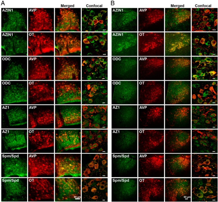
Figure 2.
Localization of AZIN1, ODC, AZ1, and spermine/spermidine with AVP and OT neurons of the SON (A) and PVN (B). A and B, Immunofluorescent colocalization of AZIN1, ODC, and AZ1 (all in green) with AVP (red) or OT (red) in the SON and PVN. Confocal images in the right panels show the merged subcellular localization of these proteins and spermine/spermidine with AVP or OT. Spm, spermine; Spd, spermidine. Scale bars for confocal images, 10 μm.

