Abstract
Osteomas are benign slow growing tumors of bone. Tumors are usually asymptomatic until they attain remarkable size and cause asymmetry or dysfunction. In view of few reported cases of giant osteoma of mandible, this article presents a case of giant osteoma of left mandible in a 53-year old male causing dyspnea due to compression of air way space.
Keywords: Osteoma, Giant osteoma, Mandible, Dyspnea
Introduction
Osteoma as a benign lesion and specific entity, was described by Jaffe in 1935 and since then hundreds of cases have been published [1]. It has been considered to be a neoplasm but reactive mechanism due to trauma or infection has been suggested in the pathogenesis of the lesion [2].
They are usually asymptomatic for many years and diagnosed incidentally on radiographs [3].
Osteomas are essentially restricted to craniofacial bones rarely affecting extragnathal skeleton, ranging in size from 2 to 30 mm. An osteoma with a diameter >30 mm or weighing >110 g is considered as giant osteoma [4].
Here, we report a case of mandibular giant osteoma causing facial disconfiguration and dyspnea with reviewing of the seven reported cases of mandibular giant osteomas [5–11] in the English literature.
Case Report
A 53-year old man was referred to the oral and maxillofacial surgery department, School of Dentistry, University of Beheshti (Tehran, Iran) in Jaunary 2014 with a history of left mandibular swelling since childhood. Despite the slow growth of the mass, patient stated the rapid growth of the lesion during last 6 months associated with discomfort and dyspnea in supine position and rotation of the head to the right side but he did not complain of pain and paraesthesia.
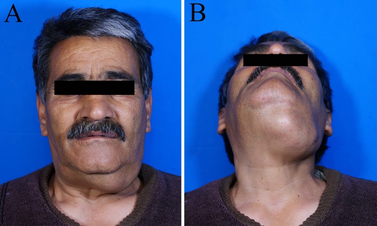
Obvious facial asymmetry was observed extraorally (Fig. 1). There was a well circumscribed bony hard mass in the submandibular area approximately 9.5 × 8 cm in size. On intraoral examination, expansion in the lower vestibule of edentulous ridge was seen. Expansion on the lingual cortical plate was more pronounced. The covering mucosa was intact.
Fig. 1.
Extraoral photographs show a swelling on left side of mandible
No history of previous medical history, trauma or infection was detected.
Computed tomography (CT) images showed a well circumscribed bony like hyperdense image with buccolingual expansion and lobulated surface on the left mandible extended to the midline with narrowing of the oropharynx airway (Fig. 2).
Fig. 2.

A 3D CT image, B axial bone algorithm view, C lateral neck radiograph. Images show well circumscribed bony like hyperdense mass with narrowing of oropharynx airway
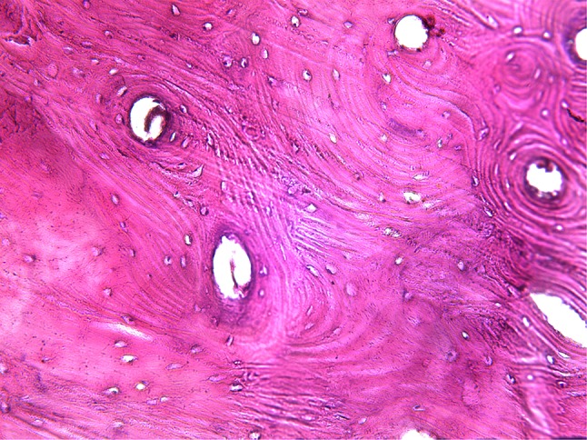
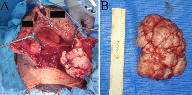
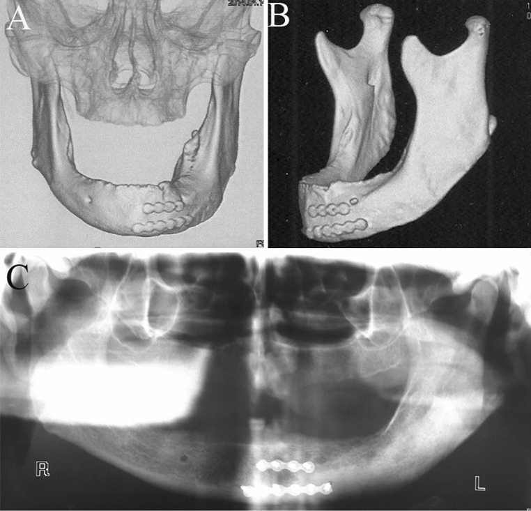
Clinical and radiographic evaluation showed no similar lesion in other parts of the jaws and no sign of Gardner’s syndrome was seen. Based on clinical and radiographic findings osteoma, central ossifying fibroma (COF) and osteoblastoma were considered in differential diagnosis. Incisional biopsy was done and histopathologic evaluation revealed compact mature bone with hematopoietic marrow tissue (Fig. 3). So, the diagnosis of osteoma was established. The patient underwent surgical procedure under general anesthesia. In present case the location of the mass was in the ramus and body of the mandible. The huge size of the mass did not provide the accessibility through intraoral approach. A submandibular approach and lip splitting was used to dissect the tissues sufficiently and reach to the entire lesion. Then para-midline mandibulotomy technique was used to retract the body of mandible, to expose the lingual side of the jaw and to preserve genioglossus and geniohyoid muscles (Fig. 4A). The mass was resected by a lindemann bur through its pedicle in inferior mandibular border and junction to the lingual cortex. The lesion was 10.5 × 6.5 × 5 cm in size and 725 grams in weight (Fig. 4B). The mandibular osteotomy cut was fixed with two miniplates. The incisions were sutured and a mini hemovac drain was placed in the surgery site for 48 h. The patient post-operatively received systemic antibiotics and was discharged after 3 days. Post-operative CT scans are shown in Fig. 5. After the surgery, patient was more relieved and the fullness sensation in his neck and jaw was eliminated. The facial deformity was corrected and the patient was able to eat and swallow with less discomfort. The patient’s breathing was without any difficulty and no sleep disturbance was reported after surgery.
Fig. 3.

Microscopic features consisting of mature lamellar bone with hematopoietic marrow (×200)
Fig. 4.

A Submandibular approach and lip splitting with paramidline mandibulotomy, B the surgical specimen measuring 10.5 cm in greatest diameter
Fig. 5.
Post-operative CT scans
Discussion
Osteoma is a relatively rare osteogenic benign neoplasm which affects 0.43–1 % of the population [2]. It may arise from periosteum (peripheral), endosteum (central) and even extraskeletal soft tissue. Peripheral osteoma affects mainly the frontal bone and mandible [9]. Central cases are also seen in mandible more than maxilla but the proportion of the central maxillary lesion is slightly higher than peripheral maxillary lesions [9, 12]. They are usually asymptomatic but 5 % of cases which are larger than 3 cm become symptomatic and cause complication.
Giant osteoma of mandible are rare with only seven cases reported in the English literature (Table 1). They occur most often in 5th and 6th decades of life with female predilection [5, 8–11]. The most common symptom of these tumors is facial asymmetry but dysphagia, snoring, dyspnea and limited mandibular movement have been reported in a few cases [5–7, 9]. Dyspnea was both remarkable and unusual finding of our case similar to Young et al. ’s [6] report.
Table 1.
Summary of reported cases of mandibular giant osteoma
| No | Source | Age/sex | Type | Symptom | Size (cm) | Side |
|---|---|---|---|---|---|---|
| 1 | Kerckhaert et al. | 53/F | C | Dysphagia | NA | R |
| 2 | Young et al. | 60/M | P | Dyspnea | 8.7 | L |
| 3 | Tarsitano et al. | 41/M | NA | Sleep apnoea | NA | R |
| 4 | Gawande et al. | 45/F | P | Swelling | 5 | R |
| 5 | Kalu et al. | 50/F | P | Dysphagia | NA | R |
| 6 | Bulut et al. | 37/F | p | Swelling | 3 | L |
| 7 | Kachewar et al. | 50/F | P | Swelling | 10.8 | R |
C central, P peripheral, NA not available
Previously described cases predominantly occurred in the posterior mandible just like present case [6–10]. However, this case was located in the left side, in spite of the other reports [5, 7–9, 11]. In contrast to Young et al. [6], Gawande et al. [8], and Kalu et al. [9] reports, the origin of this case is central, because of diffuse buccolingual expansion and attachment to cortical border of mandible.
The duration of the lesion similar to previous reported cases goes back to childhood period [5–8]. The greatest size with 10.8 cm in diameter was reported by Kachewar et al. [11].
The radiological appearance is a densely sclerotic, radiopaque sharply defined mass. Central osteomas are generally identified on routine radiographic examination. Fibrous dysplasia, COF, osteoblastoma must be considered in the differential diagnosis of central osteomas. In present case, osteoblastoma and COF were ruled out in differential diagnosis because of lack of pain and duration of the lesion since childhood period. Furthermore, COF may mature with time into completely radiopaque mass. Characteristically it is surrounded by a radiolucent rim.
Exostoses, peripheral ossifying fibroma, osteoid osteoma should be added in differential diagnosis of peripheral tumors.
Distinguishing between osteoma and exostosis because of the similar microscopic feature are difficult, but exostoses stop growing after puberty and are located in the attached gingiva.
It should be mentioned that CT is the best imaging choice for diagnosis, size identifying and anatomical location of the lesion [5].
Histologically, two types of osteomas can be distinguished: (1) compact and (2) cancellous.
The compact osteoma consists of dense, compact bone with few marrow spaces. The cancellous type comprises trabeculae of bone and fibrofatty marrow resembling mature bone.
Most cases reported in the jaws are histologically compact osteomas similar to the current case [6].
The treatment choice is surgery by considering several factors such as tumor location, extension and dimension. Recurrence is rare and there is no report of malignant transformation [5].
In conclusion, osteomas are slow growing asymptomatic masses seen predominantly in maxillofacial region. However giant tumors can cause problems like dyspnea, dysphagia, and facial asymmetry depending on the site of tumor. CT is the best imaging modality for designing treatment plan.
References
- 1.Gayathri G, Ravikumar R, Manjunath GA, Jyothi M. Osteoma of mandible. J Dent Sci Res. 2011;2(1):116–121. [Google Scholar]
- 2.Sanchez R, Gonzalez J, Arias gallo J, Carceller F. Giant osteoma of the ethmoid sinus with orbital extension: craniofacial approach and orbital reconstruction. Acta Otorhinolaryngol Ital. 2013;33(6):431–434. [PMC free article] [PubMed] [Google Scholar]
- 3.Johann A, Freitas J, Aguiar M, Araujo N, Mesquita R. Peripheral osteoma of the mandible: case report and review of the literature. J Craniomaxillofac Surg. 2005;33(4):276–281. doi: 10.1016/j.jcms.2005.02.002. [DOI] [PubMed] [Google Scholar]
- 4.Cheng K, Wang SH, Lin L. Giant osteoma of the ethmoid and frontal sinuses: clinical characteristics and review of the literature. Oncol Lett. 2013;5(5):1724–1730. doi: 10.3892/ol.2013.1239. [DOI] [PMC free article] [PubMed] [Google Scholar]
- 5.Kerckhaert A, Wolviusevanderwal K, Oosterhius J. A giant osteoma of the mandible: case report. J Craniomaxillafac Surg. 2005;33(4):282–285. doi: 10.1016/j.jcms.2005.03.001. [DOI] [PubMed] [Google Scholar]
- 6.Young S, Hyeon CH, Choi K. Giant osteoma of mandible causing breathing problem. Korean J Oral and Maxillofacial Radiology. 2006;36(4):217–220. [Google Scholar]
- 7.Tarsitano A, Marchetti C. Unusual presentation of obstructive sleep apnoea syndrome due to giant mandible osteoma. Acta Otorhinolaryngol Ital. 2013;33(1):63–66. [PMC free article] [PubMed] [Google Scholar]
- 8.Gawande P, Deshmukh V, Grade JB. A giant osteoma of the mandible. J Maxillofac Oral Surg. 2011 doi: 10.1007/s12663-010-0112-x. [DOI] [PMC free article] [PubMed] [Google Scholar]
- 9.Kalu U, Nashed M, Ayoub A. Huge peripheral osteoma of the mandible: a case report and review of the literature. Pathol Res Pract. 2007;203(3):185–188. doi: 10.1016/j.prp.2007.01.004. [DOI] [PubMed] [Google Scholar]
- 10.Bulut E, Acikgoz A, Ozan B, Gunhan O. Large peripheral osteoma of mandible. Int J Dent. 2010;2010:834761. doi: 10.1155/2010/834761. [DOI] [PMC free article] [PubMed] [Google Scholar]
- 11.Kachewar S, Bhadane S, Kukami D, Sankaye S. Giant peripheral osteoma of mandible. Int J Med Updat. 2012;7(1):66. [Google Scholar]
- 12.Kaplan I, Nicolaou Z, Hatuel D, Calderon S. Solitary central osteoma of the jaws: a diagnostic dilemma. Oral Surg Oral Med Oral Pathol Oral Radiol Endod. 2008;106(3):22–29. doi: 10.1016/j.tripleo.2008.04.013. [DOI] [PubMed] [Google Scholar]


