

Abstract
Objective
The aim of the study is to assess the clinical and radiological factors that increase the surgical difficulty in removal of mandibular impacted 3rd molar and design a new difficulty predictive index.
Methods
The data was collected from 100 patients with impacted mandibular 3rd molar who presented to Department of Oral and Maxillofacial Surgery, K.L.E’s Institute of Dental Sciences. Clinical and radiological parameters included in the New Index were noted. The tooth was then removed under local anesthesia and time taken for the removal was noted. The Pederson Index, New Index and time taken were co-related using kappa statistical analysis.
Results
The kappa agreement between Pederson Index and time taken was 66.50 % (0.2231) whereas between New Index and time was 89 % (0.7177) indicating that New Index is a better predictor of the difficulty.
Conclusion
The New Index is a reliable tool in predicting the difficulty in the removal of mandibular impacted third molar.
Keywords: Impaction, 3rd molar, Minor oral surgery
Introduction
Surgical removal of mandibular third molar is one of the most common procedures in any dental office. This is why, sometime in their career, every dental surgeon has faced difficulty in surgical removal of mandibular third molars. Both the patient and dentist must therefore have scientific evidence-based information concerning the estimated level of surgical difficulty of every case. The ability to predict the surgical difficulty of lower third molar extraction is essential when designing a treatment plan in that it helps to assess the competence of the dental practitioner for the particular operation, minimize complications, and optimize the preparation of the patient and assist in terms of the postoperative management of inflammation and pain [1–3]. We should know the difficulty in advance so as to plan the surgery, explain to the patient the expected difficulty and possible complications if any. It gives us an idea to schedule the appointment as required in difficult cases.
There are a number of previous studies to evaluate surgical difficulty in the extraction of impacted mandibular third molars [4–7]. However, most of these studies were based only on dental factors evaluated by radiologic assessment. Other authors believe it is difficult to estimate actual difficulty by radiologic methods only, and that it is only intraoperatively that actual difficulty can be estimated [8]. Some authors also believe that clinical variables such as age, gender, and weight of the patient are also very important [9].
An appropriate paradigm is needed to determine factors associated with surgical difficulty to treat patients adequately and provide students and residents with the tools necessary to predict the difficulty and the possible complications encountered during surgery. In this study we have assessed both clinical and radiologic variables that are responsible for increasing the difficulty encountered during the surgical extraction of mandibular third molars. We also propose a New Index based on both clinical and radiologic variables.
Materials and Methods
The required data for the study was collected from 100 human subjects with impacted mandibular 3rd molar teeth who presented to the Department of Oral and Maxillofacial Surgery, K.L.E’s V K Institute of Dental Sciences, Belgaum. The patients who were included in the study were all healthy adults in the age range of 18–45 years {males/females}; ASA class I and class II; if one or more episodes of infections such as pericoronitis, abcess has occurred; when there is caries in the 3rd molar with no hope of restoration or caries in the second molar which cannot be treated without removal; if periodontal disease caused by the position of the 3rd molar is also affecting the second molar.
Detailed case history of the patient was taken. All patients were ruled out for any medical conditions. Then a thorough examination of the third molar in question was done. Preoperative details were noted on the proforma (Table 1). The assessment included the Pederson’s Index, depth from point of elevation, the preoperative clinical grading chart, the root form, width of the root.
Table 1.
New Index proforma
| Score | |
|---|---|
| (A) Pederson’s Index | |
| 1. Angulation of the tooth | |
| Mesioangular | 1 |
| Horizontal | 2 |
| Vertical | 3 |
| Distoangular | 4 |
| 2. Depth | |
| Level A | 1 |
| Level B | 2 |
| Level C | 3 |
| 3. Ramus relationship | |
| Class I | 1 |
| Class II | 2 |
| Class III | 3 |
| (B) Depth from point of elevation | |
| 0–3 mm (slightly difficult) | 1 |
| 4–6 mm (moderately difficult) | 2 |
| >6 mm (very difficult) | 3 |
| (C) Pre-operative clinical assessment chart | |
| Mouth opening (adequate/reduced) | (1/2) |
| Tongue size (normal/large) | (1/2) |
| Angulation of external oblique ridge (obtuse/acute) | (1/2) |
| Cheek flexibility (flexible/non flexible) | (1/2) |
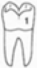
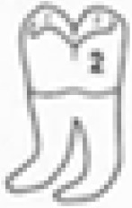
| (D) Width of root | |

|
|
| Thin (A ≥ B)—easy | 1 |
| Bulbous (B > A)—moderate | 2 |
| Thick (multiple roots B > A, B > thickness of all roots combined) | 3 |
| (E) Curvature of roots | |
 (straight roots) (straight roots) |
1 |
 (both roots distally curved) (both roots distally curved) |
2 |
 (distal root distally curved) (distal root distally curved) |
3 |
 (both roots curved towards each other) (both roots curved towards each other) |
4 |
 (mesial root distally curved) (mesial root distally curved) |
5 |
 (distal root mesially curved) (distal root mesially curved) |
6 |
 (mesial root mesially curved) (mesial root mesially curved) |
7 |
 (both roots mesially curved) (both roots mesially curved) |
8 |
 (both roots curved away from each other) (both roots curved away from each other) |
9 |
Total score = A + B + C + D + E
After thorough case history, clinical and radiographic evaluation, the patient is prepared for the surgery. The patient was positioned in the dental chair, baseline recordings of blood pressure was measured. Local anesthesia using 2 % lignocaine HCl with 1:200,000 adrenaline was administered to the operating site depending on the procedure to gain local analgesia. After the effect of local anesthesia was achieved, surgery was started under all aseptic precautions. Surgical procedure for the impacted third molar was performed. During the surgery, a verbal contact was maintained with the patient at all times. At any point of time when it was found that the anesthetic was not adequate, an incremental block was given later on during the procedure. A standard incision with no. 15 blade was made with the buccal vertical release incision. Approach was usually the ward’s incision. Soft tissue was reflected using a periosteal elevator. Bone removal was done for all the 100 impacted teeth using a straight carbide fissure bur (ss white carbide bur no. 702) and no. 8 round bur. Tooth division was done using the same bur. Elevation of distal tooth segment was followed by the mesial tooth segment. After removal of the tooth the alveolus was inspected for root fragments, loose bony fragments, attached dental follicle sac and curetted if necessary. Thorough inspection for fracture of the lingual plate or exposure of the inferior alveolar nerve was done. Thorough curettage of the socket was done. Closure was done using silk sutures. The time taken from the start of incision to the time the tooth was removed from the socket was noted for every case.
After all 100 cases, the study of quartiles was done on the proposed New score and the time taken and they were divided into three groups; slightly difficult, moderately difficult and very difficult, thus giving a New Index that helps in preoperatively predicting the difficulty in the surgical extraction of mandibular third molar. The Pederson Index score, the New Index score and the time taken are correlated using the kappa statistical analysis.
The protocol was reviewed by the appropriate institutional review board (IRB), was in compliance with the Helsinki declaration, and each subject in the project signed a detailed informed consent form.
Results
Note: for statistical analysis purposes, the total score was classified into “slightly difficult”, “moderately difficult”, and “difficult” groups on the basis of quartiles of the scores (i.e. ≤Q1 = slightly difficult; between Q1 and Q3 = moderately difficult; and ≥Q3 = difficult). The minimum score was 10 and maximum was 33 (Table 2).
Table 2.
Quartiles statistical analysis
| Quartiles | |||
|---|---|---|---|
| Q1 | Q2 | Q3 | |
| Pederson score | 4.0 | 5.0 | 6.0 |
| New Index score | 16.0 | 18.0 | 21.0 |
| Time taken | 15.5 | 22.0 | 32.0 |
Of the 100 cases; according to Pederson Index, slightly difficult included 34 patients, moderately difficult included 28 patients and difficult included 38 patients whereas our New Index included 28 patients in slightly difficult group, 45 in moderately difficult group and 27 in the difficult group as shown in (Table 3).
Table 3.
No. of patients in Pederson Index and New Index
| Pederson’s Index | New Index | |||
|---|---|---|---|---|
| Slightly difficult (N index) | Moderately difficult (N index) | Difficult (N index) | Total | |
| Slightly difficult (P index) | 13 | 15 | 6 | 34 |
| Moderately difficult (P index) | 7 | 16 | 5 | 28 |
| Difficult (P index) | 8 | 14 | 16 | 38 |
| Total | 28 | 45 | 27 | 100 |
Comparison between the Pederson Index and time taken shows a kappa agreement of 66.50 % (kappa value 0.2231) as shown in Tables 4 and 5 whereas between New Index and time taken shows a kappa agreement of 89 % (kappa value 0.71778) as shown in Tables 5 and 6.
Table 4.
Comparison of Pederson Index and time taken
| Pederson’s Index | Time taken | |||
|---|---|---|---|---|
| Slightly difficult (time taken) | Moderately difficult (time taken) | Difficult (time taken) | Total | |
| Slightly difficult (P index) | 10 | 20 | 4 | 34 |
| Moderately difficult (P index) | 12 | 12 | 4 | 28 |
| Difficult (P index) | 3 | 17 | 18 | 38 |
| Total | 25 | 49 | 26 | 100 |
Table 5.
Kappa agreement between Pederson Index and time taken
| Agreement | Expected agreement | Kappa | SE | Z value | p value | |
|---|---|---|---|---|---|---|
| Pederson Index | 66.50 % | 56.88 % | 0.2231 | 0.0749 | 2.98 | 0.0014 |
| New Index | 89.00 % | 61.02 % | 0.7178 | 0.0750 | 9.5700 | 0.00001 |
Table 6.
Comparison of New Index and time taken
| New Index | Time taken | |||
|---|---|---|---|---|
| Slightly difficult (time taken) | Moderately difficult (time taken) | Difficult (time taken) | Total | |
| Slightly difficult (New Index) | 20 | 6 | 2 | 28 |
| Moderately difficult (New Index) | 4 | 39 | 2 | 45 |
| Difficult (New Index) | 1 | 4 | 22 | 27 |
| Total | 25 | 49 | 26 | 100 |
Discussion
Preoperative assessment of surgical difficulty is fundamental to the planning of extraction of impacted third molars. The assessment is not only important to the dental surgeon who needs it to be able to decide whether or not to refer patients for specialist care, but it is also important in predicting the possible complications so that the patient can be informed [2]. Most researchers agree that postoperative complications are more commonly associated with more difficult extractions. With the range of difficult extractions from the studies being between 4.1 and 44.5 %, it is imperative that patients are, to the highest level of scientific certainty, informed of the possibility of complications after removal of their impacted mandibular third molars, based on a preoperative estimation of difficulty. Prediction of operative difficulty is therefore important for correct management (Table 7).
Table 7.
Kappa agreement between New Index and time taken
| Agreement | Expected agreement | Kappa | SE | Z value | p value |
|---|---|---|---|---|---|
| 89.00 % | 61.04 % | 0.7177 | 0.1062 | 6.7600 | 0.00001 |
MacGregor [10] made the first attempt to establish a model for assessing surgical difficulty [10]. This model served as the basis for subsequent studies. Previous assessment models are based on dental factors recorded on preoperative X-rays [1, 2, 11]. Three imaginary lines to determine the depth of the mandibular third molars in bone have been described earlier [12]. This method is taught to most undergraduate students, but is reported to be used little in practice [1]. Pell and Gregory described an alternative method, but it also has recently been found to be an unreliable method of determining surgical difficulty [13]. The Pederson scale is widely cited in oral and maxillofacial surgical texts as a useful way of predicting the difficulty of extraction of impacted lower third. However, this method has recently been found to be inadequate for the determination of surgical difficulty [14]. Edwards et al. [8] corroborated this by reporting that it is difficult to estimate actual surgical difficulty by radiologic assessment alone. Thus, a classification system based on clinical and radiographic results would be a useful tool [15].
In our study, mandibular third molars were studied in 100 patients in whom 100 third molars were extracted using transalveolar method. Of the 100 patients, 59 were males and 41 females. There was no significant difference between the number of male and female patients which is in contrast to the study by Carvalho et al. [16] who suggest that women seek third-molar surgery more frequently than men. According to Nakagawa et al. [17] the female gender is a risk factor because of the mandible’s lesser bone thickness. In the present study, however, gender was not a determinant of surgical difficulty. The mean age of the patients was 28.32 years.
A number of studies have used surgery time and surgical technique as determinants of difficulty [4, 11, 18, 19]. A study by Lago-Méndez et al. found both these factors to be reliable, statistically significant measures and the best way to predict surgical difficulty [19]. The surgery time is considered to be the gold standard in determining the surgical difficulty [4, 11, 15, 18, 19]. In our study, we have considered time taken for tooth removal (surgery time) to be the gold standard for determination of the surgical difficulty against which we have compared the conventional Pederson’s Index and our proposed New Index.
In our study, the kappa of agreement between the conventional Pederson’s Index and the surgery time is 66.50 % (p value = 0.2) in contrast to the kappa of agreement between our proposed New Index and the time taken for tooth removal which is 89.0 %. The results of our study indicates that there is no significant association between the conventional Pederson’s Index and the operation time. We have proposed a New Index in which factors like depth from point of elevation, mouth opening, tongue size, angulation of external oblique ridge, cheek flexibility, width of the root, curvature of roots were incorporated along with the Pederson’s Index. There is a significant association between this New Index and the operation time which reflects on the accuracy of the index.
In our opinion radiographic factors alone are insufficient predictors of the surgical difficulty encountered during the surgical removal of mandibular third molar. The Pederson Index originally aimed to grade the difficulty of surgical extraction; the intervals of classification have the same value and the points indicated difficulty. However the Pederson’s scores in almost all our patients was classified as moderately difficult which is in accordance with the study by Yuasa et al. [6]. The results of our study show that the New Index is better than the Pederson’s Index in terms of kappa agreement test. The difference between the New Index and Pederson’s Index is the inclusion of additional factors like depth from point of elevation, mouth opening, tongue size, angulation of external oblique ridge, cheek flexibility, width of the root, curvature of roots. This suggests that the additional clinical factors are equally important for the prediction of the surgical difficulty. Thus both clinical and radiographic factors when assessed together enables in accurate prediction of the surgical difficulty. Our New Index includes both clinical and radiologic factors, as a result of which there is a significant association between our index and the operation time.
In our opinion, this New Index is an accurate and valuable tool for the prediction of the surgical difficulty in the removal of mandibular third molar. This index is easy to calculate and can be used by general dental practitioners, residents and experienced oral and maxillofacial surgeons alike.
Conflict of interest
There is no conflict of interest.
Ethical standard
All human and animal studies have been approved by appropriate ethics committee and have therefore been performed in accordance with the ethical standards laid down in the 1964 declaration of Helsinki and its later amendments.
References
- 1.Renton T, Smeeton N, McGurk M. Factors predictive of difficulty of mandibular third molar surgery. Br Dent J. 2001;190:607. doi: 10.1038/sj.bdj.4801052. [DOI] [PubMed] [Google Scholar]
- 2.Akinwande JA. Mandibular third molar impaction—A comparison of two methods for predicting surgical difficulty. Niger Dent J. 1991;10:3. [Google Scholar]
- 3.Contar CM, de Oliveira P, Kanegusuku K, Berticelli RD, Azevedo-Alanis LR, Machado MA. Complications in third molar removal: a retrospective study of 588 patients. Med Oral Patol Oral Cir Bucal. 2010;15:e74–e78. doi: 10.4317/medoral.15.e74. [DOI] [PubMed] [Google Scholar]
- 4.Akadiri OA, Obiechina AE. Assessment of difficulty in third molar surgery—a systematic review. J Oral Maxillofac Surg. 2009;67:771–774. doi: 10.1016/j.joms.2008.08.010. [DOI] [PubMed] [Google Scholar]
- 5.Susarla SM, Dodson TB. Estimating third molar extraction difficulty: a comparison of subjective and objective factors. J Oral Maxillofac Surg. 2005;63:427–434. doi: 10.1016/j.joms.2004.12.003. [DOI] [PubMed] [Google Scholar]
- 6.Yuasa H, Kawai T, Suguira M. Classification of surgical difficulty in extracting impacted third molars. Br J Oral Maxillofac. 2002;40:26–31. doi: 10.1054/bjom.2001.0684. [DOI] [PubMed] [Google Scholar]
- 7.Santamaria J, Arteagatia MD. Radiologic variables of clinical significance in the extraction of impacted mandibular third molars. Oral Surg Oral Med Oral Pathol Oral Radiol Endod. 1997;84:469. doi: 10.1016/S1079-2104(97)90259-6. [DOI] [PubMed] [Google Scholar]
- 8.Edwards DJ, Brickley MR, Horton J, et al. Choice of anaesthetic and healthcare facility for third molar surgery. Br J Oral Maxillofac Surg. 1998;36:333. doi: 10.1016/S0266-4356(98)90643-X. [DOI] [PubMed] [Google Scholar]
- 9.Koerner KR. The removal of impacted third molars: principles and procedures. Dent Clin North Am. 1994;38:255. [PubMed] [Google Scholar]
- 10.MacGregor AJ. The radiological assessment of ectopic lower third molars. Ann R Coll Surg Engl. 1979;61:107. [PMC free article] [PubMed] [Google Scholar]
- 11.Gbotolorun OM, Arotiba GT, Ladeinde AL. Assessment of factors associated with surgical difficulty in impacted mandibular third molar extraction. J Oral Maxillofac Surg. 2007;65:1977–1979. doi: 10.1016/j.joms.2006.11.030. [DOI] [PubMed] [Google Scholar]
- 12.Howe GL. Minor oral surgery. 2. Bristol: John Wright and Sons; 1971. [Google Scholar]
- 13.Renton T, McGurk M. Evaluation of factors predictive of lingual nerve injury in third molar surgery. Br J Oral Maxillofac Surg. 2001;39:423–428. doi: 10.1054/bjom.2001.0682. [DOI] [PubMed] [Google Scholar]
- 14.Akadiri OA, Fasola AO, Arotiba JT. Evaluation of Pederson index as an instrument for predicting difficulty of third molar surgical extraction. Niger Postgrad Med J. 2009;16(2):105–108. [PubMed] [Google Scholar]
- 15.Farish SE, Bouloux GF. General technique of third molar removal. Oral Maxillofac Surg Clin North Am. 2007;19:23. doi: 10.1016/j.coms.2006.11.012. [DOI] [PubMed] [Google Scholar]
- 16.Carvalho RWF, do Egito Vasconcelos BC (2011) Assessment of factors associated with surgical difficulty during removal of impacted lower third molars. J Oral Maxillofac Surg 69(11):2714–2721. doi:10.1016/j.joms.2011.02.097 [DOI] [PubMed]
- 17.Nakagawa Y. Third molar position: reliability of panoramic radiography. J Oral Maxillofac Surg. 2007;65:1303–1308. doi: 10.1016/j.joms.2006.10.028. [DOI] [PubMed] [Google Scholar]
- 18.Muhonen, et al. Factors predisposing to postoperative complications related to wisdom tooth surgery among university students. J Am Coll Health. 1997;46:39–42. doi: 10.1080/07448489709595585. [DOI] [PubMed] [Google Scholar]
- 19.Lago-Méndez L, Diniz-Freitas M, Senra-Rivera C et al (2007) Relationships between surgical difficulty and postoperative pain in lower third molar extractions. J Oral Maxillofac Surg 65:979 [DOI] [PubMed]