Abstract
Purpose
The objective was to compare the pre- and postsurgical profile changes after surgical correction of prognathism and maxillary hypoplasia, as perceived by panels of orthodontists, maxillofacial surgeons, laypersons and patients and to identify photogrammetric changes that might be related to preferred ratings.
Materials and methods
Each panel consisted of six males and six females who rated sets of pre- and postsurgical lateral photographs of 20 female and 20 male patients using a five-point scale. Patients rated their own set of photographs. Pre- to postsurgical differences of photogrammetrically assessed landmarks were recorded as a surgical change.
Results
No significant differences in ratings between panels and patients could be detected. Significant correlation coefficients (r) were obtained between the ratings of all panel groups and between the ratings and changes in facial convexity (r = 0.351–0.542). Correlations with changes of the mentolabial angle were found to be significant for old orthodontists, male laypersons, and male patients (r = 0.332–0.609). Ratings of female and young laypersons were correlated with the horizontal changes in the lower face (r = 0.324–0.379).
Conclusion
Information gathered from this study will support the cooperation of the medical staff and might assist in treatment planning.
Keywords: Orthognathic surgery, Esthetics, Photogrammetry, Panel study, Facial convexity, Facial attractiveness
Introduction
Since today’s orthognathic surgery patients focus their expectations mostly on the postsurgical facial outcome, quality-of-life benefits are generally high if these patients perceive aesthetic improvement in facial features after surgery [1, 2]. However, the question arises as to whether the general public as the consumer of orthodontic services appreciates facial change brought about by orthognathic surgery in the same way as orthodontists or surgeons, since patients’ motives for treatment are not necessarily related to objectively determined needs.
What laypersons perceive as a beautiful, attractive or satisfying outcome may not agree with that of professionals whose judgment is based on experience and training [3, 4]. Several investigators have sought to answer this question by comparing ratings of facial attractiveness given by panels of experts that include orthodontists and surgeons and panels of laypersons. However, panel composition and assessment of data have been inconsistent and vary from study to study. On the one hand, some results suggest that preferences of facial attractiveness by laypersons, orthodontists, and surgeons are generally in agreement [5, 6]. On the other hand, laypersons might be expected to give lower improvement scores after surgery than do professionals and should be less critical in their evaluation of profiles displaying dysgnathia as well as normal reference profiles [7]. Professionals provide more reliable ratings on certain morphologic features, but layperson’s profile attractiveness ratings should be the best predictors of patients’ motivation for surgery [3, 4]. Hence, controversy remains in literature as to whether laypersons, professionals and patients agree in their perceptions of facial attractiveness. However, this information is urgently needed to counsel patients who seek orthognathic surgery and especially to communicate with patients on their treatment expectations. Therefore, the objective of this study was to compare the pre- and postsurgical changes of patients’ soft tissue profiles after surgical correction of prognathism and maxillary hypoplasia as perceived by maxillofacial surgeons, orthodontists, laypersons, and patients themselves, and to identify certain photogrammetric changes that might be related to the preferred ratings of panel members and patients.
Materials and Methods
Ethical Approval of the Study Protocol
Approval for the study was given by the ethics committee of the state medical association (Study No. 401). All the patients signed release forms permitting the use of their data and photographs for scientific purposes.
Subjects
Medical records were assembled from patients who were admitted, orthodontists, for surgery and consecutively underwent bimaxillary osteotomy for correction of prognathism and maxillary hypoplasia in 2012 and 2013. In a next step, patient files were selected using exclusion criteria to avoid any bias. These criteria were patient findings that exceeded routine orthognathic surgery planning, such as those with an anterior open bite greater than 1 cm, facial asymmetry with occlusal cants in the frontal plane, midline deviations and mandibular border asymmetry, matured cleft lip and palate, severe congenital facial or posttraumatic deformity, and obese patients [Body Mass Index (BMI) >30 kg/m2]. Of the remaining patient files, pre- and post-treatment files from 40 patients were randomly chosen after stratification for gender. Hence, the study sample included files of 20 females (mean age 21.8 ± SD deviation 5.5 years) and 20 males (mean age 22.5 ± 4.9 years).
Lateral Photogrammetry
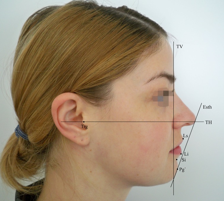
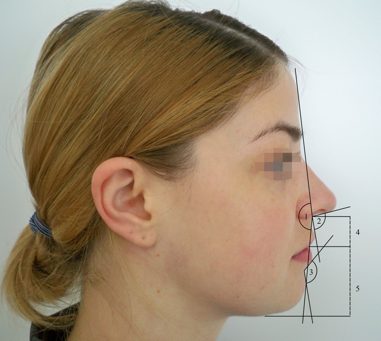
All subjects had received lateral photographs at first presentation 1 month before surgery (mean 1.4 ± 0.8 months) and at a follow-up appointment 6 months postsurgery (mean 6.4 ± 1.2 months). Lateral photographs were obtained using the following procedure: Subjects were asked to sit on a chair in front of a pale blue background, maintain a straight back, and look straight ahead with a relaxed facial expression and eyes fully open, lips gently closed, and not smiling (natural head position). The subjects’ faces were photographed in the right lateral view together with a metric scale in front of the midfacial vertical line (true vertical (TV)]. A high-resolution digital camera with flash (Nikon D3200, Nikon, Chiyoda, Tokyo, Japan) was firmly mounted on a photo stand 1 m in front of the subject. All photographs were taken at 2,048 × 1,536 pixels resolution and saved in JPEG file format. The images were then imported into a photographic software program (Adobe Photoshop version 7.0, Adobe Systems, San Jose, CA, USA) and were adjusted to life-size, so that 1 mm on the ruler represented 1 mm of actual scale. For two-dimensional photogrammetry, soft tissue landmarks, distances, and angles were traced with the tools of the software using a modified version of the soft tissue analysis described by Legan and Burstone [8] and Lew et al. [9]. Additionally, TV on nasion and true horizontal [TH, (perpendicular to TV through the tragus)] were constructed as reference lines for horizontal and vertical landmark movements. Pre- and postsurgical distances of each landmark towards the reference lines were measured and the differences were recorded as the vertical and horizontal surgical change, respectively. Furthermore, the corresponding soft tissue angles were constructed within the landmarks and measured in degrees. In accordance with landmark movements, differences between pre- and postsurgical angular measurements were also recorded as the surgically induced change (Figs. 1, 2).
Fig. 1.
Soft tissue landmarks and reference lines for tracing lateral photogrammes. Ls labrale superius, Li labrale inferius, Si labiomental sulcus, Pg′ soft tissue pogonion, Esth aesthetic line; Trg tragus point, TV true vertical, TH true horizontal
Fig. 2.
Soft tissue angles and distances for tracing lateral photogrammes. 1 facial convexity angle (FCA), 2 nasolabial angle (NLA), 3 mentolabial angle (MLA), 4 upper lip length (ULL), 5 lower lip length (LLL)
Booklets and Raters
Profile images were printed on glossy photo paper to an image size of 4 × 5 cm. Thirty-six survey booklets were made, each including the 40 sets of pre- and postsurgical patient profile photographs. Three sets of photos were shown per page. Six randomly selected sets (three female and three male profile sets) were duplicated and rated twice to test reproducibility. A rating scale was printed beneath each set of photos in the booklet to evaluate the differences in facial pre- and postsurgical aesthetics on a five-point scale on which −2 = markedly worsened, −1 = worsened, 0 = no change, +1 = improved, and +2 = markedly improved (Fig. 3). Raters were contacted by phone to determine their readiness for participation in the study. Each booklet included a questionnaire to provide gender, age, and profession. To obtain anonymity and avoid the possibility of tracing the booklets, no further personal data were sampled. Each rater was requested to read the given instructions before proceeding with the study. Raters were asked to evaluate the aesthetic change between pre- and postsurgical photographs in the most objective way, without being influenced by factors such as make-up, eye color, and hair style. The final booklet consisted of 18 pages and was sent to the raters together with a self-addressed envelope for a cost-free return.
Fig. 3.
Original set of lateral photographs (left side presurgical, right side postsurgical) with rating scale as it was printed in the booklet
The rating panels consisted of 12 orthodontists (six females, mean age 45.5 ± 7.6 years; six males, mean age 44.8 ± 8.9 years), 12 maxillofacial surgeons (six females, mean age 41.3 ± 4.7 years; six males, mean age 51.8 ± 10.4 years), and 12 laypersons (six females, mean age 37.3 ± 15.1 years; six males, mean age 37.7 ± 13.6 years). The laypersons were recruited from incidental contacts and had a relatively high socioeconomic background, but none of them was trained in dentistry or surgery. The data were collected and analyzed after all booklets were completed and returned. In addition, each orthognathic surgery patient rated his or her set of photographs given in the booklet with the same rating scale as did the panel members at the follow-up appointment 6 months postsurgery.
Statistics
Data were subjected to statistical analysis using the SPSS statistical software package, version 20.0 (IBM, SPSS, Chicago, IL). Normal distribution of datasets was confirmed using the Kolmogorov–Smirnov test. In statistically evaluating the reproducibility of the ratings on the five-point scale, the random error for a rater was calculated as , where SD is the standard deviation of the differences in the ratings of the duplicated photographs. The reliability of the final score was expressed as the intraclass correlation coefficient (ICC), as described by Kiekens et al. [10]. Variance of the random effects, Vb, was the between-subject variance, which reflected the variability of the five-point score between patients. The within-subject variance, Vw, reflected the variability of the panel members over the same patient. The ICC was then calculated as Vb/[Vb + Vw], which can be interpreted as the mean correlation of randomly selected pairs of single panel members. The ICC was 1 when all panel members agreed on all patients. If the panel members substantially disagreed on the same patient, the within-subject variance was large compared with the between-subjects variance, and the ICC was close to 0. When the five-point score was based on the average five-point scores of N randomly selected raters, the ICC for pairs of panels was ICC(N) = N × ICC(1)/[1 + (N − 1) × ICC(1)]. To assess significant differences between ratings of the panels, a two-way analysis of variance (ANOVA) was performed on the mean ratings. To assess the effect of age on ratings, age was dichotomized at the median age of the panel members and the patients. A median age of the panel members above 44 years was categorized as “old”; a median age below 44 as “young”; a median age of patients above 20 was categorized as “old,” and a median age below 20 as “young”. The paired t test was used to evaluate the intraclass effect of gender and age on all groups. Results were considered significant if the P value was <0.05 and highly significant if the P value was <0.01. Pearson’s correlation analysis was used to assess the degree and significance of correlations between the mean rating scores of all groups and photogrammetric changes. Reliability of photogrammetric measurements was determined by randomly selecting ten lateral photographs to have their tracings repeated by a second senior examiner. All respective values of method error calculation for the linear measurements ranged between 0.27 and 0.53 mm, and between 1.8° and 4.4° for angular measurements. There were no significant differences between the examiners’ measurements. All statistics were finally checked by biostatisticians at our institution.
Results
Random Error and Intraclass Correlation Coefficient (ICC)
The mean of the differences of scores between the original and duplicate photographs varied from −0.17 to 0.67 among each panel group. Statistical evaluation of the reproducibility of the ratings on the five-point scale revealed random errors for the measurements from 0.26 to 0.91 (mean 0.46 ± 0.19) for orthodontists, from 0.26 to 0.67 (0.39 ± 0.21) for maxillofacial surgeons, and from 0.26 to 0.76 (0.46 ± 0.17) for laypersons. No significant differences could be obtained between the three panel groups, female and male raters, and old and young raters (ANOVA, P = 0.943). The ICCs for the three panels, each consisting of one randomly selected rater, are displayed in Table 1. Generally, the ICC was higher in male raters among all panels, except the ICCs of female maxillofacial surgeons judging boys and female orthodontists judging girls. Since Vw was much smaller for male orthodontists judging boys, female maxillofacial surgeons judging boys, and male laypersons judging boys and girls, these panel raters agreed more over the same adolescent than did the other raters. Female orthodontists evaluating boys produced the lowest ICC and a remarkably high Vw. For the total number of 12 raters per panel, the ICCs were 0.95 for orthodontists, 0.95 for maxillofacial surgeons, and 0.97 for laypersons.
Table 1.
Variance among groups rating profile changes of girls and boys
| Girls | Boys | |||||
|---|---|---|---|---|---|---|
| Vb | Vw | ICC | Vb | Vw | ICC | |
| Orthodontists | ||||||
| Female | 0.61 | 0.67 | 0.48 | 0.85 | 1.37 | 0.38 |
| Male | 0.59 | 0.67 | 0.47 | 0.61 | 0.17 | 0.78 |
| Maxillofacial surgeons | ||||||
| Female | 0.64 | 0.67 | 0.49 | 0.89 | 0.17 | 0.84 |
| Male | 0.61 | 0.27 | 0.69 | 0.77 | 0.27 | 0.74 |
| Laypersons | ||||||
| Female | 0.96 | 0.57 | 0.63 | 1.05 | 1.21 | 0.47 |
| Male | 0.91 | 0.17 | 0.84 | 0.91 | 0.17 | 0.84 |
V b between-subject variance, V w within-subject variance, ICC intraclass correlation coefficient
Comparison Between Groups and Effect of Gender and Age
Highly significant correlations with r ranging from 0.610 to 0.811 were obtained between the overall ratings of all panels. Ratings of patients and panels revealed no significant correlations. However, no significant differences in rating were obtained between panel groups and patients’ group by two-way ANOVA (Table 2). In the orthodontists’ group exclusively, female orthodontists’ scores were significantly lower than scores given by male colleagues. Older maxillofacial surgeons gave significantly lower scores than did their younger counterparts. With respect to gender and age, no effect on scoring could be found in ratings by laypersons or patients.
Table 2.
Ratings of photographic profiles of patients (girls and boys) given by orthodontists, maxillofacial surgeons, laypersons, and patients themselves
| Ratings | |||
|---|---|---|---|
| Mean ± SD | t test | ANOVA | |
| Orthodontists | |||
| Female | 0.75 ± 0.95 | 0.002† | 0.288a |
| Male | 1.01 ± 0.85 | ||
| Old | 1.08 ± 0.84 | 0.864 | |
| Young | 0.85 ± 0.96 | ||
| Maxillofacial surgeons | |||
| Female | 0.93 ± 0.89 | 0.307 | |
| Male | 0.85 ± 0.80 | ||
| Old | 0.77 ± 0.87 | 0.006† | |
| Young | 0.98 ± 0.82 | ||
| Laypersons | |||
| Female | 0.79 ± 1.03 | 0.581 | |
| Male | 0.75 ± 0.95 | ||
| Old | 0.87 ± 0.92 | 0.126 | |
| Young | 0.72 ± 1.02 | ||
| Patients | |||
| Female | 0.75 ± 1.18 | 0.199 | 0.392b |
| Male | 1.20 ± 0.93 | ||
| Old | 1.04 ± 1.06 | 0.771 | |
| Young | 0.94 ± 1.09 | ||
SD standard deviation
†Highly significant at the level P < 0.01 (two-tailed)
aTested for orthodontists’, laypersons, and maxillofacial surgeons’ panels
bTested for panels and patients’ group
Pre- to Postoperative Changes of Photogrammetric Parameters and Correlation with Ratings
Pre- to postoperative photogrammetric comparisons revealed highly significant changes for all parameters except that of TH–Li, which displayed a weak significant change, and the nasolabial angle, upper lip length, and TH–Si presenting no significant changes (Table 3). Dividing the panels and the patients’ group into subgroups comprising gender and age revealed further significant correlations (Table 4). Between all female and male and between all young and old subgroups of the panels, significant, as well as highly significant correlations were obtained between the ratings and changes in facial convexity. Correlations between changes in the mentolabial angle and ratings were found to be significant for old orthodontists, male laypersons, and male patients. Ratings of female and young laypersons were significantly correlated with horizontal changes of the lower face represented by changes in TV–Li, TV–Si, and TV–Pg′. Within the latter photogrammetric parameter, significant correlations also existed between female orthodontists and young and old maxillofacial surgeons. Only weak significant correlations could be found between young orthodontists and changes in upper lip length and between young patients and changes in nasolabial angle.
Table 3.
Photogrammetric measurements and changes from pre- to postsurgery
| Parameter | Presurgery | Postsurgery | Changes | T test |
|---|---|---|---|---|
| Mean ± SD | Mean ± SD | Mean ± SD | P | |
| FCA | 176.27 ± 7.54 | 171.36 ± 4.53 | 4.91 ± 6.55 | <0.001† |
| NLA | 108.25 ± 11.48 | 106.38 ± 11.45 | 1.46 ± 8.05 | 0.157 |
| MLA | 150.99 ± 4.16 | 140.59 ± 8.32 | 10.45 ± 10.12 | <0.001† |
| ULL | 18.54 ± 3.16 | 17.49 ± 3.32 | 1.19 ± 3.50 | 0.083 |
| LLL | 48.95 ± 4.99 | 39.47 ± 6.95 | 9.54 ± 8.73 | <0.001† |
| Ls–Esth | −7.41 ± 2.26 | −5.14 ± 2.83 | −2.06 ± 2.28 | <0.001† |
| Li–Esth | −2.53 ± 2.19 | −3.99 ± 2.41 | 1.54 ± 2.21 | <0.001† |
| TV–Li | 7.84 ± 3.99 | 5.46 ± 3.74 | 2.24 ± 3.39 | <0.001† |
| TV–Si | 3.08 ± 4.41 | 0.42 ± 4.34 | 2.52 ± 3.80 | <0.001† |
| TV–Pg′ | 4.24 ± 3.92 | 1.09 ± 4.93 | 3.08 ± 4.69 | <0.001† |
| TH–Li | −36.69 ± 7.35 | −38.61 ± 7.33 | 1.93 ± 5.71 | 0.046* |
| TH–Si | −48.01 ± 8.62 | −48.74 ± 8.82 | 0.59 ± 8.09 | 0.586 |
| TH–Pg′ | −69.46 ± 6.27 | −66.07 ± 6.94 | −3.09 ± 6.78 | 0.004† |
Table 4.
Correlations between ratings and photogrammetric changes
| FCA | NLA | MLA | ULL | TV–Li | TV–Si | TV–Pg′ | |
|---|---|---|---|---|---|---|---|
| Orthodontists | |||||||
| Female | |||||||
| r | 0.462 | ns | ns | ns | ns | ns | 0.353 |
| P | 0.003† | 0.030* | |||||
| Male | |||||||
| r | 0.551 | ns | ns | ns | ns | ns | ns |
| P | < 0.001† | ||||||
| Young | |||||||
| r | 0. 542 | ns | ns | 0.327 | ns | ns | ns |
| P | < 0.001† | 0.045* | |||||
| Old | |||||||
| r | 0.507 | ns | 0.332 | ns | ns | ns | ns |
| P | < 0.001† | 0.042* | |||||
| Maxillofacial surgeons | |||||||
| Female | |||||||
| r | 0.404 | ns | ns | ns | ns | ns | ns |
| P | 0.012* | ||||||
| Male | |||||||
| r | 0.476 | ns | ns | ns | ns | ns | ns |
| P | 0.003† | ||||||
| Young | |||||||
| r | 0.426 | ns | ns | ns | ns | ns | 0.395 |
| P | 0.008† | 0.014* | |||||
| Old | |||||||
| r | 0.526 | ns | ns | ns | ns | ns | 0.332 |
| P | < 0.001† | 0.041* | |||||
| Laypersons | |||||||
| Female | |||||||
| r | 0.351 | ns | ns | ns | 0.335 | 0.324 | 0.346 |
| P | 0.031* | 0.040* | 0.047* | 0.033* | |||
| Male | |||||||
| r | 0.522 | ns | 0.361 | ns | ns | ns | ns |
| P | < 0.001† | 0.026* | |||||
| Young | |||||||
| r | 0.401 | ns | ns | ns | 0.345 | 0.333 | 0.379 |
| P | 0.013* | 0.034* | 0.041* | 0.019* | |||
| Old | |||||||
| r | 0.477 | ns | ns | ns | ns | ns | ns |
| P | 0.002† | ||||||
| Patients | |||||||
| Female | |||||||
| r | ns | ns | ns | ns | ns | ns | ns |
| P | |||||||
| Male | |||||||
| r | ns | ns | 0.609 | ns | ns | ns | ns |
| P | 0.006† | ||||||
| Young | |||||||
| r | ns | 0.457 | ns | ns | ns | ns | ns |
| P | 0.043* | ||||||
| Old | |||||||
| r | ns | ns | ns | ns | ns | ns | ns |
| P | |||||||
Abbreviations as given for Figs. 1 and 2. Only photogrammetric parameters whose changes correlate with scores of at least one subdivision of the panel or patient group are displayed
r Pearson’s correlation coefficient, ns not significant
* Significant at the level P < 0.05 (two-tailed); † highly significant at the level P < 0.01
Discussion
In this study, the ICCs fulfilled the requirement for an ICC to be equal to or above 0.80 for a panel size to be large enough to obtain reliable results in evaluating profile changes in adolescent faces by using photographs and a five-point scale [10]. The best agreement of panel members’ ratings of pre- to postoperative profile changes was obtained for male orthodontists judging boys, female maxillofacial surgeons judging boys, and male laypersons judging girls and boys. In contrast, the lowest agreement was found among female orthodontists judging boys. No significant differences in overall ratings were obtained between panels on the one hand and between the panels and patient group on the other hand. In accordance with the findings of other study groups [4, 6, 11], these results revealed that the ratings of pre- to postoperative changes towards higher facial attractiveness by orthodontists, maxillofacial surgeons, laypersons, and patients are generally in agreement, although significant correlations between ratings could be obtained only for the panels. In the recent literature, the influence of gender and age of panel members on their ratings is still disputable, and it varies among studies. Some studies indicated that the gender of panel members was not decisive for their ratings [12]. Other studies, however, indicated that female panel members are less critical than males [13] and female laypersons rate female faces as more attractive than do male laypersons [14]. In our study a significant effect of raters’ ages on rating scores was shown in the maxillofacial surgeon panel, in which older practitioners gave significantly lower scores than did the younger ones. With respect to gender, in the orthodontists’ group, female orthodontists rated facial changes significantly lower than did their male colleagues, suggesting that female orthodontists are more critical than male orthodontists. However, no impact of gender on scoring could be found in the laypersons’ panel.
Until now no study had explicitly addressed the differences between panels and patients in the correlations between ratings of profile changes and objective photogrammetric changes. Between all female, male, and young and old subgroups of the panels, highly significant and significant correlations were obtained between the changes of facial convexity and rating scores. Narrowing of the facial convexity angle positively influenced the ratings of the panels. That means in turn and in accordance to the findings of Fabré et al. [7], that the degree of facial concavity had a negatively predictive value for the panel evaluations. In this study as well, ratings by female and young laypersons, female orthodontists, and young and old maxillofacial surgeons were significantly correlated with the parameters indicating horizontal changes of the lower face. In the patients’ group, two significant correlations between ratings and photogrammetric changes were obtained. Ratings of males were correlated with changes in the mentolabial angle, while ratings of young patients with changes in the nasolabial angle. The latter correlation possesses only low validity, since this parameter was not significantly changed between pre- and postsurgical measurements. However, no correlation could be found with changes in facial convexity.
Although there is no difference in the overall ratings between groups, the results of this study suggest that laypersons’ assessments are more like those of orthodontists’ and maxillofacial surgeons’ panels than those of the patients’ group. Hence, findings in the laypersons’ panel cannot be transferred without concerns for patients. Facial convexity was the most distinctive photogrammetric parameter for the ratings of the panels, but not for the patients’ ratings. However, information gleaned from this study might elicit support from board members and medical staff insofar as assisting in treatment planning and recommendations to patients.
Conflict of interest
The authors declare they have no conflict of interest.
References
- 1.Espeland L, Høgevold HE, Stenvik A. A 3-year patient-centred follow-up of 516 consecutively treated orthognathic surgery patients. Eur J Orthod. 2008;30:24–30. doi: 10.1093/ejo/cjm081. [DOI] [PubMed] [Google Scholar]
- 2.Rustemeyer J, Gregersen J. Quality of life in orthognathic surgery patients: post-surgical improvements in aesthetics and self-confidence. J Craniomaxillofac Surg. 2012;40:400–404. doi: 10.1016/j.jcms.2011.07.009. [DOI] [PubMed] [Google Scholar]
- 3.Cochrane SM, Cunningham SJ, Hunt NP. A comparison of the perception of facial profile by the general public and 3 groups of clinicians. Int J Adult Orthod Orthog Surg. 1999;14:291–295. [PubMed] [Google Scholar]
- 4.Vargo JK, Gladwin M, Ngan P. Association between ratings of facial attractiveness and patients’ motivation for orthognathic surgery. Orthod Craniofac Res. 2003;6:63–71. doi: 10.1046/j.1439-0280.2003.2c097.x. [DOI] [PubMed] [Google Scholar]
- 5.Shelly AD, Southard TE, Southard KA, Casko JS, Jakobsen JR, Fridrich KL, Mergen JL. Evaluation of profile esthetic change with mandibular advancement surgery. Am J Orthod Dentofacial Orthop. 2000;117:630–637. doi: 10.1016/S0889-5406(00)70171-5. [DOI] [PubMed] [Google Scholar]
- 6.Maple JR, Vig KWL, Beck FM, Larsen PE, Shankere S. A comparison of providers’ and consumers’ perceptions of facial-profile attractiveness. Am J Orthod Dentofacial Orthop. 2005;128:690–696. doi: 10.1016/j.ajodo.2004.09.030. [DOI] [PubMed] [Google Scholar]
- 7.Fabré M, Mossaz C, Christou P, Kiliaridis S. Orthodontists’ and laypersons’ aesthetic assessment of Class III subjects referred for orthognathic surgery. Eur J Orthod. 2009;31:443–448. doi: 10.1093/ejo/cjp002. [DOI] [PubMed] [Google Scholar]
- 8.Legan HL, Cl Burstone. Soft tissue cephalometric analysis for orthognathic surgery. J Oral Surg. 1980;38:744–751. [PubMed] [Google Scholar]
- 9.Lew KK, Low FC, Yeo JF, Loh HS. Evaluation of soft tissue profile following intraoral ramus osteotomy in Chinese adults with mandibular prognathism. Int J Adult Orthodon Orthognath Surg. 1990;5:189–197. [PubMed] [Google Scholar]
- 10.Kiekens RMA, Maltha JC, van’t Hof MA, Straatman H, Kuijpers-Jagtman AM. Panel perception of change in facial aesthetics following orthodontic treatment in adolescents. Eur J Orthod. 2008;30:141–146. doi: 10.1093/ejo/cjm114. [DOI] [PubMed] [Google Scholar]
- 11.Kiekens RMA, van’t Hof MA, Straatman H, Kuijpers-Jagtman AM, Maltha JC. Influence of panel composition on aesthetic evaluation of adolescent faces. Eur J Orthod. 2007;29:95–99. doi: 10.1093/ejo/cjl060. [DOI] [PubMed] [Google Scholar]
- 12.Howells DJ, Shaw WC. The validity and reliability of ratings of dental and facial attractiveness for epidemiological use. Am J Orthod. 1985;88:402–408. doi: 10.1016/0002-9416(85)90067-3. [DOI] [PubMed] [Google Scholar]
- 13.Tedesco LA, Albino JE, Cunat JJ, Slakter MJ, Waltz KJ. A dentalfacial attractiveness scale. Part II. Consistency and perception. Am J Orthod. 1983;83:44–46. doi: 10.1016/0002-9416(83)90270-1. [DOI] [PubMed] [Google Scholar]
- 14.Cross JF, Cross J. Age, sex, race, and the perception of facial beauty. Dev Psychol. 1971;5:433–439. doi: 10.1037/h0031591. [DOI] [Google Scholar]


