Abstract
Objective. To investigate the level of expression of prostaglandin receptivity and uptake factors in eutopic and ectopic endometrium of women with endometriosis. Design. Prospective study. Setting. Human reproduction research laboratory. Patients. Seventy-eight patients with endometriosis and thirty healthy control subjects. Intervention(s). Endometrial and endometriotic tissue samples were obtained during laparoscopic surgery. Main Outcome Measure(s). Real-time polymerase chain reaction assay of mRNA encoding prostaglandin E2 receptors (EP1, EP2, EP3, and EP4), prostaglandin F2α receptor (FP), prostaglandin transporter (PGT), and multidrug resistance-associated protein 4 (MRP4); immunohistochemical localization of expressed proteins. Results. Marked increases in receptors EP3, EP4, and FP and transporters PGT and MRP4 in ectopic endometrial tissue were noted, without noticeable change associated with disease stage. An increase in EP3 expression and decreases in FP and PGT were observed in the eutopic endometrium of endometriosis patients in conjunction with the phases of the menstrual cycle. Conclusion(s). This study is the first to demonstrate a possible relationship between endometriosis and enhanced prostaglandin activity. In view of the wide range of prostaglandin functions, increasing cell receptivity and facilitating uptake in endometrial tissue could contribute to the initial steps of overgrowth and have an important role to play in the pathogenesis and symptoms of this disease.
1. Introduction
Endometriosis is a major health issue affecting nearly 10 percent of women of childbearing age. The main symptoms of this disease include chronic pelvic or abdominal pain, irregular bleeding, and in 40–50% of cases infertility [1]. The amount of pain experienced correlates poorly with disease stage. Endometriotic tissue may settle and proliferate in the fallopian tubes or the ovaries or enter the peritoneal cavity and deposit in ectopic sites. The causes and symptoms of endometriosis are multifactorial. Although knowledge of its underlying immunological and endocrine mechanisms is progressing, gray areas continue to obscure complete understanding of its pathology. Our studies were among the first to highlight dysfunctions in eutopic endometrium, including elevated levels of the monocyte chemoattractant factor MCP-1 [2]. In addition, we have shown that ectopic endometrial tissue by itself is capable of producing growth-promoting molecules such as vascular endothelial growth factor (VEGF) [3] as well as implantation-promoting integrins [4] while initiating peritoneal inflammation. This inflammation causes the release of mediators such as prostaglandins E2 (PGE2) and F2α (PGF2α), which play a role in reproductive functions such as ovulation, luteolysis, implantation, parturition, and lactation. Excessive release of prostaglandins may affect peritoneal function, causing pain [5] and disrupting processes such as oocyte maturation, ovulation, and fertilization [6, 7].
Prostaglandins are lipid compounds derived enzymatically from arachidonic acid. The reproductive system is the principal producer of PGE2 and PGF2α. The involvement of prostaglandins in pain [5, 8, 9], infertility [10–12], angiogenesis [13, 14], tissue remodeling [15], and cell proliferation [16] in association with various pathological conditions is well documented. We along with others have shown that an isoform of cyclooxygenase-2 (COX-2) is overexpressed in ectopic endometrial cells [17–20] and that PGE2, PGF2α, and other specific prostaglandins are present at abnormally high levels in uterine tissues of women suffering from menorrhagia, dysmenorrhoea, or endometriosis [21–23]. It is also well known that PGE2 and PGF2α are more concentrated in the peritoneal fluid of endometriosis patients [5, 10, 12, 24, 25]. Such evidence has led us to investigate a possible role of these prostaglandins in the pathogenesis of endometriosis. Our recent comparison of endometrium (eutopic and ectopic) from endometriosis patients to healthy eutopic endometrium showed overproduction of PGE2 and PGF2α, apparently promoted by increased expression of enzymes such as COX-2 with cPGES or AKR1C3 [26].
Regulation of production is not the only means by which the body modulates the action of PGE2 and PGF2α. Expression of prostaglandin receptors and transporters may also be regulated. Once released as messenger molecules, prostaglandins act locally on receptors in an autocrine or paracrine manner. The four main subtypes of PGE2 receptor are designated as EP1, EP2, EP3, and EP4, while the PGF2α receptor is called FP [27, 28]. The receptor subtype determines the nature of the physiological response. Reception either elicits the intracellular calcium-inositol triphosphate pathway or increases/decreases cyclic adenosine monophosphate (cAMP) activity. Engagement of some receptors may elicit both pathways, depending on cell type and receptor splice variety.
Prostaglandins were originally believed to exit from producer cells via passive diffusion because of their strongly lipophilic character. The discovery of the prostaglandin transporter protein PGT (SLCO2A1), which mediates prostaglandin uptake and release [29, 30], demonstrated that diffusion alone did not explain the penetration of prostaglandins through the cell membrane. Furthermore, a specific transporter, namely, multidrug resistance protein 4 (MRP4, ABCC4) of the ATP-binding cassette transporter superfamily, has been shown to mediate prostaglandin release [31]. Whether or not MRP4 is the only transporter that does this is still unclear.
Although it is clear that PGE2 and PGF2α play important roles in a number of female reproductive physiological processes as well as in endometriosis-associated infertility and pain [5, 10, 12, 32], current understanding of these roles remains incomplete. In the present study, we analyzed the expression of EP1, EP2, EP3, EP4, FP, PGT, and MRP4 in endometriosis patients in comparison to their expression in normal eutopic endometrium. We observed marked differences between eutopic and ectopic endometria in terms of prostaglandins receptivity and transport readiness.
2. Materials and Methods
2.1. Patients and Tissue Collection
The study received approval from the Human Research Ethics Committee at Saint-François d'Assise Hospital, and informed consent was obtained from all participants, who were recruited between February 2002 and March 2007. Endometriosis patients were aged 34.2 ± 3.6 years (n = 78) and were consulting for pelvic pain and/or infertility. They were diagnosed using laparoscopy and the disease stage (I–IV) was determined according to the Revised American Fertility Society classification system. Endometriotic tissue samples were collected from 28 of these patients. We also recruited healthy women aged 35.3 ± 3.8 (n = 30) scheduled for tubal ligation. These participants had no pelvic pathology or sign of endometrial hyperplasia or neoplasia and had not received any anti-inflammatory or hormonal medication for at least 3 months. Menstrual cycle dating was determined using the cycle history.
Endometrial and endometriotic biopsies were obtained during laparoscopy. Tissue was placed immediately at 4°C in sterile Hank's balanced salts solution (HBSS) (GIBCO Invitrogen Corp., Burlington, ON, Canada) containing 100 IU/mL penicillin, 100 μg/mL streptomycin, and 0.25 μg/mL amphotericin and transported to the laboratory. After washing in HBSS at 4°C, samples were frozen at −80°C in Eppendorf tubes for quantitative real-time PCR (qRT-PCR) or embedded in paraffin and stored at room temperature for immunohistochemical analysis.
2.2. Quantitative Real-Time PCR
Total RNA was extracted from endometrial tissue using the TRIzol reagent according to the manufacturer's instructions (Invitrogen Life Technologies, Inc., Grand Island, NY, USA) and reverse-transcribed in the presence of random hexamers. The qRT-PCR reaction was carried out in an ABI 7000 Thermal Cycler (Applied Biosystems, Foster City, CA, USA). The standard reaction mixture contained 2 μL of RT product, 0.5 μL of each primer (final concentration, 0.1 mM), 12.5 μL SYBR Green PCR Master Mix (Invitrogen Life technologies, Inc., Grand Island, NY, USA) consisting of Taq DNA polymerase reaction buffer, dNTP mix, SYBR green I, MgCl2, and Taq DNA polymerase. Following denaturing for 2 min at 95°C, the reactions were cycled 45 times with denaturing for 15 sec at 95°C and annealing for 60 sec at 60°C. The primer sequences are listed in Table 1. The primers were designed using Primer Express 2.0 (Applied Biosystems, Foster City, CA, USA) to span intron-exon boundaries to avoid amplification of genomic DNA and selected to have compatible Tm values (59–61°C). A relative quantification method was used. Expression of mRNA of EP1, EP2, EP3, EP4, FP, PGT, total MRP4, and MRP4 variant 1 was normalized to that of the gene GAPDH. After each run, melting curve analysis (55–95°C) was performed to verify the specificity of the PCR reaction. All samples were tested in duplicate and each run included a template control. Baseline curves, melting curves, melting points, crossing points, slopes, and errors were monitored for each gene.
Table 1.
List of primers used for qRT-PCR.
| Gene | Forward | Reverse |
|---|---|---|
| EP1 | 5′-ATGGTGGGCCAGCTTGTC-3′ | 5′-GCCACCAACACCAGCATTG-3′ |
| EP2 | 5′-TCCTTGCCTTTCACGATTT-3′ | 5′-AGAGCTTGGAGGTCCCATT-3′ |
| EP3 | 5′-TGGTCTCCGCTCCTGATAA-3′ | 5′-TGCATTCTTTCTGCTTCTCC-3′ |
| EP4 | 5′-TGCTCTTCTTCAGCCTGTCC-3′ | 5′-GAGCTACCGAGACCCATGTT-3′ |
| FP | 5′-TCTGGTCTGTGCCCACTTC-3′ | 5′-GACTCCAATACACCGCTCAAT-3′ |
| PGT | 5′-CTGGTGGATTTCATTAAACGG-3′ | 5′-GGCTGCTGAGGTGCCATAC-3′ |
| MRP4 total | 5′-AAAGTGCCAAAGTAATCCAGC-3′ | 5′-GTTCAAAGCCACAGAATCCA-3′ |
| MRP4 variant 1 | 5′-CGGGCATACAAAGCAGAA-3′ | 5′-GGACCCAAAGGCAACG-3′ |
2.3. Immunohistochemical Probe
Endometrium was fixed in 10% formalin (Fisher Scientific, New Jersey, USA) and then embedded in paraffin. Serial tissue sections 4 μm thick were rinsed in phosphate buffered saline (PBS) and treated with 3% hydrogen peroxide to block endogenous peroxidase activity. All antibodies were diluted in PBS containing 0.2% bovine serum albumin and 0.1% Tween 20. Sections were incubated for two hours at room temperature with the specific antibody (Cayman, Ann Arbor, USA). Rabbit polyclonal anti-human EP2 was diluted 1 : 800, and rabbit polyclonal antibodies directed against human EP1, EP3, EP4, FP, and PGT were all diluted 1 : 200. Rat monoclonal anti-human total MRP4 (Abcam, Cambridge, USA) was diluted 1 : 50. Sections were then held for 45 minutes at room temperature with peroxidase-conjugated goat anti-rabbit antibody (Jackson ImmunoResearch Laboratories, Inc., West Grove, USA) diluted 1 : 2000 or peroxidase-conjugated rabbit anti-rat antibody (Jackson ImmunoResearch Laboratories, Mississauga, Canada) diluted 1 : 500 and then for 40 minutes with the peroxidase substrate 3,3′-diaminobenzidine for 5 minutes at room temperature followed by rinsing in PBS, counterstaining with hematoxylin and mounting in Mowiol.
2.4. Statistical Analysis
Data that followed a normal (Gaussian) distribution were subjected to one-way analysis of variance (ANOVA) and Bonferroni's post hoc test for multiple comparisons, while data that were not normally distributed were analyzed using the Kruskal-Wallis test and Dunn's multiple comparison post hoctest for multiple comparisons. Comparison of the two groups was performed using the parametric unpaired t-test or the nonparametric Mann-Whitney test. All statistical analyses were performed using GraphPad Prism 5.0 Software (San Diego, CA, USA). Differences were considered to be statistically significant at P < 0.05.
3. Results
3.1. Expression of PGE2 and PGF2α Receptors and Transporters in Eutopic and Ectopic Endometria in the Diseased State
3.1.1. Prostaglandins E2 and F2α Receptors
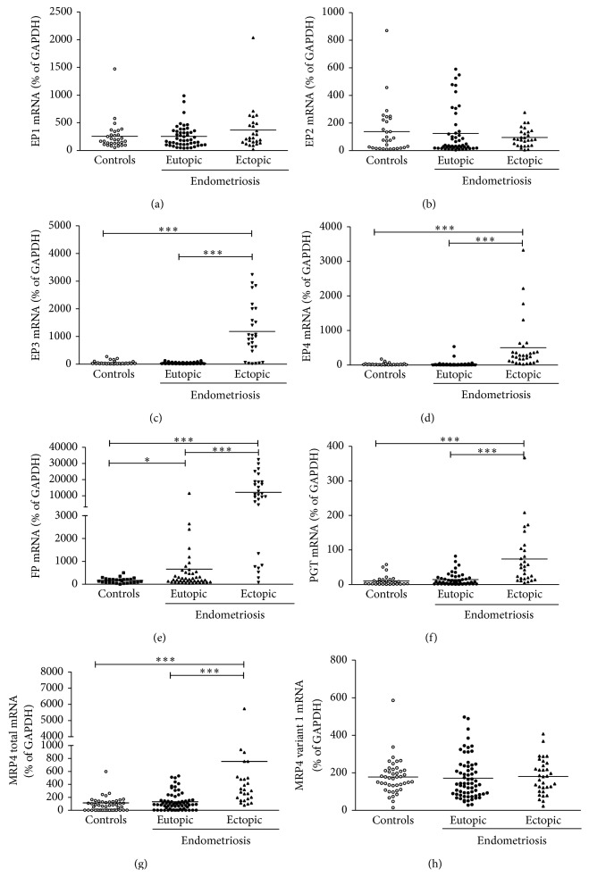
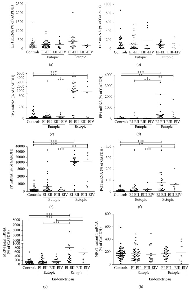
Neither EP1 nor EP2 was expressed differentially to any appreciable degree, whether the comparison was between endometriosis patients and healthy control patients, eutopic and ectopic endometrium in endometriosis patients (Figures 1(a) and 1(b)), endometriosis stages (Figures 2(a) and 2(b)), or the two phases of the menstrual cycle (Table 2).
Figure 1.
Expression of PGE2 and PGF2α receptors and prostaglandin transporters in endometrium, n = 30, 50, and 28, respectively, for healthy (control) subjects, eutopic tissue samples from endometriosis patients, and ectopic tissue samples. Total RNA was extracted and reverse-transcribed and mRNA was quantified by quantitative RT-PCR and normalized relative to GAPDH (internal control). (a) EP1, (b) EP2, (c) EP3, (d) EP4, (e) FP, (f) PGT, (g) total MRP4, and (h) MRP4 variant 1. The horizontal lines represent the mean for each set of data. * P < 0.05, ** P < 0.01, and *** P < 0.001.
Figure 2.
Expression of PGE2 and PGF2α receptors and prostaglandin transporters in endometrium at different stages of endometriosis, n = 30, 50, and 28, respectively, for healthy (control) subjects, eutopic tissue samples from patients with the disease, and ectopic tissue samples. Total RNA was extracted and reverse-transcribed and mRNA was quantified by quantitative RT-PCR and normalized relative to GAPDH (internal control). (a) EP1, (b) EP2, (c) EP3, (d) EP4, (e) FP, (f) PGT, (g) total MRP4, and (h) MRP4 variant 1. The horizontal lines represent the mean for each set of data. * P < 0.05, ** P < 0.01, and *** P < 0.001.
Table 2.
Effect of endometriosis and menstrual cycle phase on expression of prostaglandin receptors and transporters in endometrium, based on real-time PCR and normalized relative to GAPDH mRNA as mean % ± SD. Also, n = 30, 50, and 28, respectively, for healthy (control) subjects, eutopic tissue samples from endometriosis patients, and ectopic tissue samples.
| Gene |
Controls (mean ± SEM) |
Endometriosis | |
|---|---|---|---|
| Eutopic (mean ± SEM) |
Ectopic (mean ± SEM) |
||
| EP1 | |||
| Proliferative phase | 317,2 ± 100,5 | 307,0 ± 42,13 | 307,3 ± 54,11 |
| Secretory phase | 209,5 ± 35,33 | 200,2 ± 37,57 | 411,3 ± 128,9 |
|
| |||
| EP2 | |||
| Proliferative phase | 57,90 ± 17,46 | 66,60 ± 22,24 | 71,74 ± 14,89 |
| Secretory phase | 198,2 ± 51,72† | 179,5 ± 38,82† | 112,6 ± 17,9 |
|
| |||
| EP3 | |||
| Proliferative phase | 28,55 ± 6,652 | 29,76 ± 3,444 | 1021 ± 249,3∗∗∗+++ |
| Secretory phase | 73,94 ± 20,06† | 48,35 ± 6,463†† | 1310 ± 276,1∗∗∗+++ |
|
| |||
| EP4 | |||
| Proliferative phase | 34,38 ± 12,67 | 46,82 ± 22,68 | 380,2 ± 136,5∗∗∗+ |
| Secretory phase | 21,94 ± 6,654 | 10,47 ± 1,546 | 583,4 ± 205,2∗∗∗+++ |
|
| |||
| FP | |||
| Proliferative phase | 237,5 ± 31,18 | 1059 ± 497,2 | 9213 ± 1947∗∗∗+++ |
| Secretory phase | 97,27 ± 18,26†† | 229,6 ± 61,22†† | 14268 ± 2742∗∗∗+++ |
|
| |||
| PGT | |||
| Proliferative phase | 16,04 ± 4,058 | 21,74 ± 3,682 | 56,42 ± 14,73∗∗+ |
| Secretory phase | 5,964 ± 3,326†† | 6,760 ± 2,736††† | 88,69 ± 25,28∗∗∗+++ |
|
| |||
| MRP4 total | |||
| Proliferative phase | 142,9 ± 63,47 | 97,05 ± 15,51 | 591,5 ± 227∗∗+ |
| Secretory phase | 80,93 ± 16,17 | 154,7 ± 25,77 | 931,2 ± 386,5∗∗∗+++ |
|
| |||
| MRP4 variant 1 | |||
| Proliferative phase | 160,9 ± 15,28 | 157,8 ± 18,82 | 194,2 ± 24,08 |
| Secretory phase | 196,2 ± 24,35 | 178,8 ± 18,99 | 164,7 ± 20,93 |
Note: ** P < 0.01 and *** P < 0.001 versus the control group; + P < 0.05 and +++ P < 0.001 versus the eutopic group; † P < 0.05 and †† P < 0.01 versus expression in the corresponding proliferative phase; ††† P < 0.001.
Expression of EP3 was increased in ectopic endometrium of endometriosis patients (Figure 1(c)) and, as shown in Figure 2(c), in both stages I-II and stages III-IV. This effect was apparent in both secretory and proliferative phases of the menstrual cycle (Table 2). These effects were all significant at P < 0.001. It is worth mentioning that EP3 expression was increased significantly in eutopic tissue during the secretory phase even in healthy women (P < 0.05), but at an order of magnitude less than in ectopic tissue.
As was the case for EP3, EP4 expression was increased (P < 0.001) in ectopic endometrium (Figure 1(d)), although neither endometriosis stage (Figure 2(d)) nor phase of the menstrual cycle (Table 2) had any effect.
FP expression followed distinctive patterns in eutopic (P < 0.05) and ectopic (P < 0.001) endometria of endometriosis patients (Figure 1(e)). The increase was especially pronounced in ectopic endometrium (P < 0.001) at both disease stages (Figure 2(e)), during both phases of the menstrual cycle (Table 2). Expression in eutopic endometrium in endometriosis patients was significantly higher during the proliferative phase than during the secretory phase, while the difference between these patients and healthy subjects during the proliferative phase was only marginally significant (P = 0.18).
3.1.2. Prostaglandins E2 and F2α Transporters
PGT expression was significantly increased (P < 0.001) in ectopic endometrium (Figure 1(f)) at stages I-II (P < 0.001) and III-IV (P < 0.01) of the disease (Figure 2(f)) and during both phases of the menstrual cycle (Table 2).
Expression of total MRP4 in ectopic endometrium was increased similar to PGT in endometriosis stages I-II and III-IV (Figures 1(g) and 2(g)) and during both phases of the menstrual cycle (Table 2). However, regulation of MRP4 is more complex because of its two transcriptional variants, namely, variant 1 (coding sequence: 120…4097, NM_005845.3) and a shorter variant 2 (coding sequence: 120…2699, NM_001105515), which encode distinctive functional proteins of different molecular mass. There was no noticeable effect of the disease on the expression of variant 1 (Figure 1(h)). The ratio of variant 1 to total MRP4 was significantly greater (P < 0.001) in eutopic endometrium in subjects with or without the disease (not shown). The meaning of this ratio is not yet known. In any case, it did not differ significantly.
3.2. Immunohistochemical Localization of PGE2 and PGF2α Receptors and Transporters in Eutopic and Ectopic Endometria in Endometriosis Patients and Healthy Subjects
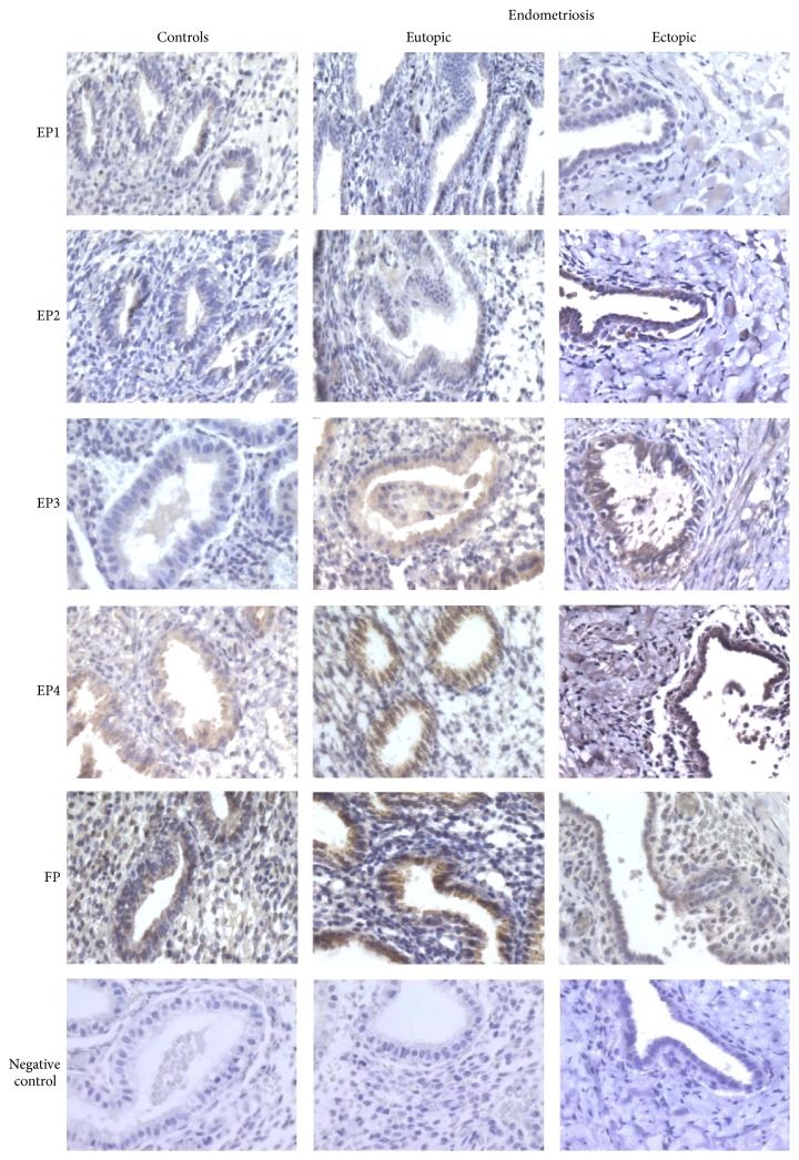
Cell membranes of ectopic endometriotic tissue and eutopic endometrium from endometriosis patients and from healthy subjects were examined using an immunohistochemical technique. Representative immunological staining of the receptors and transporters is shown in Figures 3 and 4. Staining of EP1 and EP2 in endometrial glands was weak. Clear staining of EP3, EP4, and FP was visible mainly in endometrial glands, but the adjacent stroma also appeared positive for these receptors. However, staining of FP was more intense in eutopic and ectopic endometrial tissues of endometriosis patients, whether in glandular cells or in surrounding stromal cells.
Figure 3.
Representative immunohistochemical staining of EP1, EP2, EP3, EP4, and FP in endometrium of healthy women and in eutopic and ectopic endometrium of endometriosis patients. No staining was observed in control sections incubated without the primary antibody or with an equivalent concentration of goat IgG. The original magnification was 400x.
Figure 4.
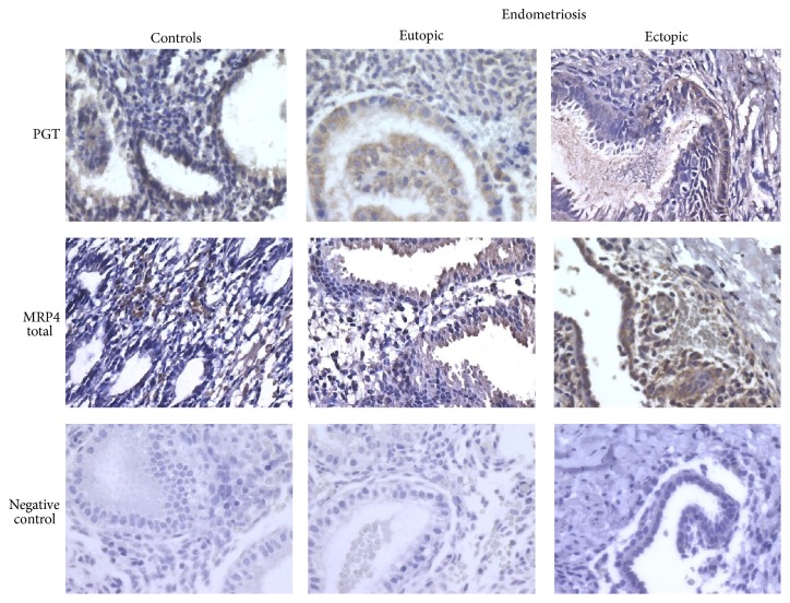
Representative immunohistochemical staining of PGT and total MRP4 in endometrium of healthy women and in eutopic and ectopic endometrium of endometriosis patients. No staining was observed in control sections incubated without the primary antibody or with an equivalent concentration of goat or rat IgG. The original magnification was 400x.
The pattern of PGT staining was similar in endometrial tissues from endometriosis patients and healthy subjects. Positive staining for MRP4 (total) was located mainly in stromal cells from control patients, but only in epithelial cells of eutopic endometrium from endometriosis patients. However, this staining was more intense in ectopic tissue, whether in glandular epithelial cells or in surrounding stromal cells.
4. Discussion
Prostaglandins E2 and F2α play major roles in the regulation of the cyclic changes of the endometrium and are also involved in diseases afflicting this tissue, in particular endometriosis. In this study, we showed that expression of mRNA encoding prostaglandin receptors EP3, EP4, and FP and of transporters PGT and MRP4 was increased in ectopic endometrium of women suffering from endometriosis. The expression of FP receptor was increased in both ectopic and eutopic endometrium of endometriosis patients, while expression of EP3 and EP4 was increased in ectopic endometrium only. No effect on expression of EP1 or EP2 receptors was observed. The menstrual cycle also had a significant effect, increasing EP3 receptor expression in the secretory phase both in the control group and in women with endometriosis. Although the cycle did not modulate overexpression of EP3, EP4, and FP in ectopic endometrium, it did appear to affect the eutopic endometrium of these patients, notably by causing decreased expression of FP in the secretory phase. This was not observed for any other receptor. While the increase in PGF2α during the secretory phase in healthy women is well documented, the regulation of PGE2 secretion during the menstrual cycle is less certain. Studies show that PGE2 level may increase during the secretory phase or the proliferative phase or may remain the same throughout the cycle [33–35]. Our data corroborate a previous observation of EP4 and FP expression increasing towards the end of the menstrual cycle and concomitant with the withdrawal of progesterone and sloughing of the functional layer of the endometrium, peaking during the mid-late proliferative phase and not in the secretory phase, coincident with an elevation in the expression of PGT [36]. In contrast, changes in EP3 expression across the menstrual cycle have not been noted previously [37–39].
The specific roles played by PGE2 and PGF2α in modulating reproductive physiology have been demonstrated using mice deficient in the corresponding receptors [40]. The most striking observations have been made using FP receptor and EP3 receptor knockout mice. It has thus been shown that the FP receptor is indispensable in female reproduction and that its ablation results in loss of parturition [41]. Studies of mice lacking individual prostaglandin receptors EP1–4 suggested strongly that EP3 was the principle receptor mediating pain [42, 43]. In addition, several in vitro studies demonstrated that expression of aromatase (an enzyme involved in the synthesis of estrogens) may be regulated through EP3 [44]. On the other hand, binding of PGE2 to the EP3 receptor regulates vascular function-dysfunction in ocular tissues and promotes vitreal neovascular diseases such as ischemic retinopathy [45] and also transcriptional upregulation of fibroblast growth factor 9 [46]. Although little is known about the angiogenic potential of other prostaglandin receptors, increased levels of EP4 and FP have been reported in perivascular cells in endometrial adenocarcinomas [27, 47]. More recent studies have demonstrated that selective blockade of EP4 signaling inhibits proliferation and adhesion of human endometriotic epithelial and stromal cells through suppression of integrin-mediated mechanisms [48–50]. It has also been shown that anomalies in cell adhesion, morphology, and proliferation can occur after binding of ligands to EP4 and FP or activation of downstream signaling pathways such as MAPK and PI3K [47, 51–53]. Our results suggest that the specific increase in expression of these receptors is not insignificant and that it may contribute to the principal symptoms associated with endometriosis. Very few authors have studied the role of MRP4 in endometriosis [54]. Increased MRP4 expression has been shown in malignant prostate tissue [55], in acute myeloid leukemia [56], and in colorectal neoplasia [57], while increased PGT expression has been associated with epithelial malignancy [58]. Our results show, for the first time, increased expression of both prostaglandin transporters in endometriotic tissues. We observed significant increases of PGT expression in eutopic endometrium during the proliferative phase of the menstrual cycle both in healthy women and in patients with endometriosis as well as in ectopic endometrium. As reported previously [54, 59], we also observed significantly increased expression of total MRP4 in ectopic tissue while MRP4 variant 1 showed no noticeable change. Recently described as a prostaglandin efflux transporter, MRP4 was expressed at much the same level throughout the menstrual cycle. Based on these results, we suggest that the observed overexpression of MRP4 is most likely due to variant 2 and not to the combination of the two variants, as is often reported. The increases in both transporter molecules appear concomitant, specifically in ectopic lesions. Although their actions seem to be different, these transporters might work in concert to improve prostaglandin dispatching.
Immunohistochemistry experiments largely support the mRNA expression results. In eutopic endometrium, PGE2 and PGF2α receptors and transporters were found at greater abundance in glandular epithelial cells than in stromal cells, consistent with increased epithelial MRP4 expression demonstrated in endometriosis [54] and cancer [55]. Furthermore, staining of PGT, FP, EP3, and EP4 in ectopic endometrial tissue was intense, as reported previously [60, 61]. Low levels of EP1 and EP2 were also found in the glandular epithelium and in ectopic tissue. Our results corroborate those of Arosh et al. [62], who reported that EP2 staining in the endometrium of cattle was expressed mainly in glandular epithelial cells.
Elevated MRP4 and PGT expression, particularly in epithelial glandular cells, may result in increased availability of PGE2 and PGF2α, which through engagement of their EP3, EP4, or FP receptors (also elevated in endometriosis) may activate intracellular signals such as the diacylglycerol or cyclic AMP pathways. Aberrant transport and signaling by prostaglandin receptors in the endometrium [47, 63] as observed in the present study therefore might promote uterine pathologies such as endometriosis. Although the modifications observed in the eutopic endometrium of endometriosis patients appear slight, they nevertheless affect sensitivity to PGE2 and PGF2α and thus disrupt normal function. The proinflammatory environment of the peritoneal cavity of women with endometriosis induces significant overexpression of the majority transporters and receptors required to regulate PGE2 and PGF2α. In addition to having a role in the pathogenesis of the disease, this could also disrupt the entire female reproductive tract. Since the female genitalia bathe in peritoneal liquid [64], an increased level of PGE2 and PGF2α could act on the whole system and thereby influence the reproductive process.
Although qualitative, immunohistochemistry has limitations that should not be overlooked. Immunohistochemical confirmation of mRNA expression reinforces the importance of our observations, especially in view of the inevitable variability associated with categorizing of clinical symptoms and/or staging of patients. In conclusion, this study reveals for the first time that endometriosis can affect the regulation of PGE2 and PGF2α activity at the points of reception on the cell surface and transport into the target cells.
Capsule. Eutopic and ectopic endometria of endometriosis patients display distinct anomalies in levels of expression of prostaglandins receptivity and uptake factors.
Acknowledgments
A special acknowledgement is due to the contribution of Dr. Ali Akoum, who passed away in July 2014. Dr. Akoum was a principal investigator in the endometriosis screening study, without which the present study would not have been possible. This paper was supported by Grants MOP-120769 and MOP-123259 to Ali Akoum from the Canadian Institutes for Health Research. Ali Akoum is also FRQ-S (Fonds de la Recherche du Québec-Santé) “National Researcher.” Halima Rakhila is the recipient of a doctoral studentship from the “Réseau Québécois en Reproduction.”
Conflict of Interests
The authors declare that they have no conflict of interests.
References
- 1.Giudice L. C. Clinical practice. Endometriosis. The New England Journal of Medicine. 2010;362(25):2389–2398. doi: 10.1056/nejmcp1000274. [DOI] [PMC free article] [PubMed] [Google Scholar]
- 2.Akoum A., Lemay A., Brunet C., Hebert J. Secretion of monocyte chemotactic protein-1 by cytokine-stimulated endometrial cells of women with endometriosis. Fertility and Sterility. 1995;63(2):322–328. doi: 10.1016/s0015-0282(16)57363-4. [DOI] [PubMed] [Google Scholar]
- 3.Yang Y., Degranpré P., Kharfi A., Akoum A. Identification of macrophage migration inhibitory factor as a potent endothelial cell growth-promoting agent released by ectopic human endometrial cells. The Journal of Clinical Endocrinology & Metabolism. 2000;85(12):4721–4727. doi: 10.1210/jc.85.12.4721. [DOI] [PubMed] [Google Scholar]
- 4.Khoufache K., Bondza P. K., Harir N., et al. Soluble human IL-1 receptor type 2 inhibits ectopic endometrial tissue implantation and growth: identification of a novel potential target for endometriosis treatment. The American Journal of Pathology. 2012;181(4):1197–1205. doi: 10.1016/j.ajpath.2012.06.022. [DOI] [PubMed] [Google Scholar]
- 5.Dawood M. Y., Khan-Dawood F. S., Wilson L., Jr. Peritoneal fluid prostaglandins and prostanoids in women with endometriosis, chronic pelvic inflammatory disease, and pelvic pain. American Journal of Obstetrics & Gynecology. 1984;148(4):391–395. doi: 10.1016/0002-9378(84)90713-0. [DOI] [PubMed] [Google Scholar]
- 6.Lee T.-C., Ho H.-C. Effects of prostaglandin E2 and vascular endothelial growth factor on sperm might lead to endometriosis-associated infertility. Fertility and Sterility. 2011;95(1):360–362. doi: 10.1016/j.fertnstert.2010.08.040. [DOI] [PubMed] [Google Scholar]
- 7.Koskimies A. I., Tenhunen A., Ylikorkala O. Peritoneal fluid 6-ketoprostaglandin F1α, thromboxane B2 in endometriosis and unexplained infertility. Acta Obstetricia et Gynecologica Scandinavica. 1984;63(123):19–21. doi: 10.3109/00016348409156975. [DOI] [PubMed] [Google Scholar]
- 8.Ferreira S. H. Prostaglandins, pain, and inflammation. Agents and Actions Supplements. 1986;19:91–98. [PubMed] [Google Scholar]
- 9.Arayne M. S., Ul Hasan S. S. Prostaglandins: in pain and inflammatiin. Journal of the Pakistan Medical Association. 1977;27(5):326–330. [PubMed] [Google Scholar]
- 10.Ylikorkala O., Koskimies A., Laatkainen T., Tenhunen A., Viinikka L. Peritoneal fluid prostaglandins in endometriosis, tubal disorders, and unexplained infertility. Obstetrics and Gynecology. 1984;63(5):616–620. [PubMed] [Google Scholar]
- 11.Pobedinskiǐ N. M., Khachikian M. A., Zykova V. P., Fanchenko N. D. Blood levels of prostaglandins in fertile women and in women with different variants of endocrine infertility. Akusherstvo i Ginekologiia. 1982;(8):14–18. [PubMed] [Google Scholar]
- 12.Schenken R. S., Asch R. H., Williams R. F., Hodgen G. D. Etiology of infertility in monkeys with endometriosis: measurement of peritoneal fluid prostaglandins. American Journal of Obstetrics and Gynecology. 1984;150(4):349–353. doi: 10.1016/s0002-9378(84)80136-2. [DOI] [PubMed] [Google Scholar]
- 13.Shirasuna K., Sasahara K., Matsui M., Shimizu T., Miyamoto A. Prostaglandin F2α differentially affects mRNA expression relating to angiogenesis, vasoactivation and prostaglandins in the early and mid corpus luteum in the cow. Journal of Reproduction and Development. 2010;56(4):428–436. doi: 10.1262/jrd.10-004O. [DOI] [PubMed] [Google Scholar]
- 14.Majima M., Amano H., Hayashi I. Endogenous prostaglandins and angiogenesis. Nihon Yakurigaku Zasshi. 2001;117(4):283–292. doi: 10.1254/fpj.117.283. [DOI] [PubMed] [Google Scholar]
- 15.Ulug U., Goldman S., Ben-Shlomo I., Shalev E. Matrix metalloproteinase (MMP)-2 and MMP-9 and their inhibitor, TIMP-1, in human term decidua and fetal membranes: the effect of prostaglandin F2α and indomethacin. Molecular Human Reproduction. 2001;7(12):1187–1193. doi: 10.1093/molehr/7.12.1187. [DOI] [PubMed] [Google Scholar]
- 16.Hsu H.-H., Hu W.-S., Lin Y.-M., et al. JNK suppression is essential for 17β-Estradiol inhibits prostaglandin E2-Induced uPA and MMP-9 expressions and cell migration in human LoVo colon cancer cells. Journal of Biomedical Science. 2011;18(1, article 61) doi: 10.1186/1423-0127-18-61. [DOI] [PMC free article] [PubMed] [Google Scholar]
- 17.Ota H., Igarashi S., Sasaki M., Tanaka T. Distribution of cyclooxygenase-2 in eutopic and ectopic endometrium in endometriosis and adenomyosis. Human Reproduction. 2001;16(3):561–566. doi: 10.1093/humrep/16.3.561. [DOI] [PubMed] [Google Scholar]
- 18.Carli C., Metz C. N., Al-Abed Y., Naccache P. H., Akoum A. Up-regulation of cyclooxygenase-2 expression and prostaglandin E2 production in human endometriotic cells by macrophage migration inhibitory factor: involvement of novel kinase signaling pathways. Endocrinology. 2009;150(7):3128–3137. doi: 10.1210/en.2008-1088. [DOI] [PMC free article] [PubMed] [Google Scholar]
- 19.Fan H., Fang X. L. Expression of cylooxygenase-2 in endometriosis. Zhong Nan Da Xue Xue Bao Yi Xue Ban. 2005;30(1):92–95. [PubMed] [Google Scholar]
- 20.Banu S. K., Lee J., Speights V. O., Jr., Starzinski-Powitz A., Arosh J. A. Cyclooxygenase-2 regulates survival, migration, and invasion of human endometriotic cells through multiple mechanisms. Endocrinology. 2008;149(3):1180–1189. doi: 10.1210/en.2007-1168. [DOI] [PubMed] [Google Scholar]
- 21.Willman E. A., Collins W. P., Clayton S. G. Studies in the involvement of prostaglandins in uterine symptomatology and pathology. BJOG. 1976;83(5):337–341. doi: 10.1111/j.1471-0528.1976.tb00839.x. [DOI] [PubMed] [Google Scholar]
- 22.Lundstrom V., Green K., Svanborg K. Endogenous prostaglandins in dysmenorrhea and the effect of prostaglandin synthetase inhibitors (PGSI) on uterine contractility. Acta Obstetricia et Gynecologica Scandinavica. 1979;58(87):51–56. doi: 10.3109/00016347909157790. [DOI] [PubMed] [Google Scholar]
- 23.Stromberg P., Akerlund M., Forsling M. L., Kindahl H. Involvement of prostaglandins in vasopressin stimulation of the human uterus. British Journal of Obstetrics & Gynaecology. 1983;90(4):332–337. doi: 10.1111/j.1471-0528.1983.tb08919.x. [DOI] [PubMed] [Google Scholar]
- 24.Badawy S. Z. A., Cuenca V., Marshall L. Peritoneal fluid prostaglandins in patients with endometriosis. Contributions to Gynecology and Obstetrics. 1987;16:60–65. [PubMed] [Google Scholar]
- 25.Badawy S. Z. A., Marshall L., Cuenca V. Peritoneal fluid prostaglandins in various stages of the menstrual cycle: role in infertile patients with endometriosis. International Journal of Fertility. 1985;30(2):48–52. [PubMed] [Google Scholar]
- 26.Rakhila H., Carli C., Daris M., Lemyre M., Leboeuf M., Akoum A. Identification of multiple and distinct defects in prostaglandin biosynthetic pathways in eutopic and ectopic endometrium of women with endometriosis. Fertility and Sterility. 2013;100(6):1650.e2–1659.e2. doi: 10.1016/j.fertnstert.2013.08.016. [DOI] [PubMed] [Google Scholar]
- 27.Jabbour H. N., Sales K. J. Prostaglandin receptor signalling and function in human endometrial pathology. Trends in Endocrinology and Metabolism. 2004;15(8):398–404. doi: 10.1016/j.tem.2004.08.006. [DOI] [PubMed] [Google Scholar]
- 28.Jabbour H. N., Sales K. J., Smith O. P. M., Battersby S., Boddy S. C. Prostaglandin receptors are mediators of vascular function in endometrial pathologies. Molecular and Cellular Endocrinology. 2006;252(1-2):191–200. doi: 10.1016/j.mce.2006.03.025. [DOI] [PubMed] [Google Scholar]
- 29.Lee J., McCracken J. A., Banu S. K., Arosh J. A. Intrauterine inhibition of prostaglandin transporter protein blocks release of luteolytic PGF2alpha pulses without suppressing endometrial expression of estradiol or oxytocin receptor in ruminants. Biology of Reproduction. 2013;89(2, article 27) doi: 10.1095/biolreprod.112.106427. [DOI] [PubMed] [Google Scholar]
- 30.Chi Y., Schuster V. L. The prostaglandin transporter PGT transports PGH2 . Biochemical and Biophysical Research Communications. 2010;395(2):168–172. doi: 10.1016/j.bbrc.2010.03.108. [DOI] [PMC free article] [PubMed] [Google Scholar]
- 31.Lacroix-Pépin N., Danyod G., Krishnaswamy N., et al. The multidrug resistance-associated protein 4 (MRP4) appears as a functional carrier of prostaglandins regulated by oxytocin in the bovine endometrium. Endocrinology. 2011;152(12):4993–5004. doi: 10.1210/en.2011-1406. [DOI] [PubMed] [Google Scholar]
- 32.Wu M.-H., Shoji Y., Chuang P.-C., Tsai S.-J. Endometriosis: disease pathophysiology and the role of prostaglandins. Expert Reviews in Molecular Medicine. 2007;9(2):1–20. doi: 10.1017/s146239940700021x. [DOI] [PubMed] [Google Scholar]
- 33.Lumsden M. A., Kelly R. W., Abel M. H., Baird D. T. The concentrations of prostaglandins in endometrium during the menstrual cycle in women with measured menstrual blood loss. Prostaglandins Leukotrienes and Medicine. 1986;23(2-3):217–227. doi: 10.1016/0262-1746(86)90189-7. [DOI] [PubMed] [Google Scholar]
- 34.Milne S. A., Perchick G. B., Boddy S. C., Jabbour H. N. Expression, localization, and signaling of PGE2 and EP2/EP4 receptors in human nonpregnant endometrium across the menstrual cycle. Journal of Clinical Endocrinology and Metabolism. 2001;86(9):4453–4459. doi: 10.1210/jc.86.9.4453. [DOI] [PubMed] [Google Scholar]
- 35.Rees M. C. P., Anderson A. B. M., Demers L. M., Turnbull A. C. Endometrial and myometrial prostaglandin release during the menstrual cycle in relation to menstrual blood loss. The Journal of Clinical Endocrinology & Metabolism. 1984;58(5):813–818. doi: 10.1210/jcem-58-5-813. [DOI] [PubMed] [Google Scholar]
- 36.Kang J., Chapdelaine P., Parent J., Madore E., Laberge P. Y., Fortier M. A. Expression of human prostaglandin transporter in the human endometrium across the menstrual cycle. Journal of Clinical Endocrinology and Metabolism. 2005;90(4):2308–2313. doi: 10.1210/jc.2004-1482. [DOI] [PubMed] [Google Scholar]
- 37.Namba T., Sugimoto Y., Negishi M., et al. Alternative splicing of C-terminal tail of prostaglandin E receptor subtype EP3 determines G-protein specificity. Nature. 1993;365(6442):166–170. doi: 10.1038/365166a0. [DOI] [PubMed] [Google Scholar]
- 38.Sakamoto K., Ezashi T., Miwa K., et al. Molecular cloning and expression of a cDNA of the bovine prostaglandin F2 α receptor. The Journal of Biological Chemistry. 1994;269(5):3881–3886. [PubMed] [Google Scholar]
- 39.Arosh J. A., Banu S. K., Chapdelaine P., et al. Molecular cloning and characterization of bovine prostaglandin E2 receptors EP2 and EP4: expression and regulation in endometrium and myometrium during the estrous cycle and early pregnancy. Endocrinology. 2003;144(7):3076–3091. doi: 10.1210/en.2002-0088. [DOI] [PubMed] [Google Scholar]
- 40.Narumiya S., Sugimoto Y., Ushikubi F. Prostanoid receptors: structures, properties, and functions. Physiological Reviews. 1999;79(4):1193–1226. doi: 10.1152/physrev.1999.79.4.1193. [DOI] [PubMed] [Google Scholar]
- 41.Narumiya S., FitzGerald G. A. Genetic and pharmacological analysis of prostanoid receptor function. The Journal of Clinical Investigation. 2001;108(1):25–30. doi: 10.1172/jci200113455. [DOI] [PMC free article] [PubMed] [Google Scholar]
- 42.Ueno A., Matsumoto H., Naraba H., et al. Major roles of prostanoid receptors IP and EP3 in endotoxin-induced enhancement of pain perception. Biochemical Pharmacology. 2001;62(2):157–160. doi: 10.1016/s0006-2952(01)00654-2. [DOI] [PubMed] [Google Scholar]
- 43.Minami T., Nakano H., Kobayashi T., et al. Characterization of EP receptor subtypes responsible for prostaglandin E2-induced pain responses by use of EP1 and EP3 receptor knockout mice. British Journal of Pharmacology. 2001;133(3):438–444. doi: 10.1038/sj.bjp.0704092. [DOI] [PMC free article] [PubMed] [Google Scholar]
- 44.Richards J. A., Brueggemeier R. W. Prostaglandin E2 regulates aromatase activity and expression in human adipose stromal cells via two distinct receptor subtypes. The Journal of Clinical Endocrinology & Metabolism. 2003;88(6):2810–2816. doi: 10.1210/jc.2002-021475. [DOI] [PubMed] [Google Scholar]
- 45.Sennlaub F., Valamanesh F., Vazquez-Tello A., et al. Cyclooxygenase-2 in human and experimental ischemic proliferative retinopathy. Circulation. 2003;108(2):198–204. doi: 10.1161/01.CIR.0000080735.93327.00. [DOI] [PubMed] [Google Scholar]
- 46.Chuang P.-C., Sun H. S., Chen T.-M., Tsai S.-J. Prostaglandin E2 induces fibroblast growth factor 9 via EPS-dependent protein kinase Cδ and Elk-1 signaling. Molecular and Cellular Biology. 2006;26(22):8281–8292. doi: 10.1128/mcb.00941-06. [DOI] [PMC free article] [PubMed] [Google Scholar]
- 47.Sales K. J., Milne S. A., Williams A. R. W., Anderson R. A., Jabbour H. N. Expression, localization, and signaling of prostaglandin F2α receptor in human endometrial adenocarcinoma: regulation of proliferation by activation of the epidermal growth factor receptor and mitogen-activated protein kinase signaling pathways. Journal of Clinical Endocrinology and Metabolism. 2004;89(2):986–993. doi: 10.1210/jc.2003-031434. [DOI] [PubMed] [Google Scholar]
- 48.Lee J., Banu S. K., Burghardt R. C., Starzinski-Powitz A., Arosh J. A. Selective inhibition of prostaglandin E2 receptors EP2 and EP4 inhibits adhesion of human endometriotic epithelial and stromal cells through suppression of integrin-mediated mechanisms. Biology of Reproduction. 2013;88(3):p. 77. doi: 10.1095/biolreprod.112.100883. [DOI] [PMC free article] [PubMed] [Google Scholar]
- 49.Lee J., Banu S. K., Rodriguez R., Starzinski-Powitz A., Arosh J. A. Selective blockade of prostaglandin E2 receptors EP2 and EP4 signaling inhibits proliferation of human endometriotic epithelial cells and stromal cells through distinct cell cycle arrest. Fertility and Sterility. 2010;93(8):2498–2506. doi: 10.1016/j.fertnstert.2010.01.038. [DOI] [PubMed] [Google Scholar]
- 50.Lee J., Banu S. K., Subbarao T., Starzinski-Powitz A., Arosh J. A. Selective inhibition of prostaglandin E2 receptors EP2 and EP4 inhibits invasion of human immortalized endometriotic epithelial and stromal cells through suppression of metalloproteinases. Molecular and Cellular Endocrinology. 2011;332(1-2):306–313. doi: 10.1016/j.mce.2010.11.022. [DOI] [PubMed] [Google Scholar]
- 51.Pierce K. L., Fujino H., Srinivasan D., Regan J. W. Activation of FP prostanoid receptor isoforms leads to rho-mediated changes in cell morphology and in the cell cytoskeleton. Journal of Biological Chemistry. 1999;274(50):35944–35949. doi: 10.1074/jbc.274.50.35944. [DOI] [PubMed] [Google Scholar]
- 52.Sheng H., Shao J., Washington M. K., DuBois R. N. Prostaglandin E2 increases growth and motility of colorectal carcinoma cells. Journal of Biological Chemistry. 2001;276(21):18075–18081. doi: 10.1074/jbc.m009689200. [DOI] [PubMed] [Google Scholar]
- 53.Capra V., Habib A., Accomazzo M. R., et al. Thromboxane prostanoid receptor in human airway smooth muscle cells: a relevant role in proliferation. European Journal of Pharmacology. 2003;474(2-3):149–159. doi: 10.1016/s0014-2999(03)02014-4. [DOI] [PubMed] [Google Scholar]
- 54.Gori I., Rodriguez Y., Pellegrini C., et al. Augmented epithelial multidrug resistance-associated protein 4 expression in peritoneal endometriosis: regulation by lipoxin A4. Fertility and Sterility. 2013;99(7):1965.e2–1973.e2. doi: 10.1016/j.fertnstert.2013.01.146. [DOI] [PubMed] [Google Scholar]
- 55.Lye L. H., Kench J. G., Handelsman D. J., et al. Androgen regulation of multidrug resistance-associated protein 4 (MRP4/ABCC4) in prostate cancer. The Prostate. 2008;68(13):1421–1429. doi: 10.1002/pros.20809. [DOI] [PubMed] [Google Scholar]
- 56.Copsel S., Garcia C., Diez F., et al. Multidrug resistance protein 4 (MRP4/ABCC4) regulates cAMP cellular levels and controls human leukemia cell proliferation and differentiation. The Journal of Biological Chemistry. 2011;286(9):6979–6988. doi: 10.1074/jbc.m110.166868. [DOI] [PMC free article] [PubMed] [Google Scholar]
- 57.Holla V. R., Backlund M. G., Yang P., Newman R. A., DuBois R. N. Regulation of prostaglandin transporters in colorectal neoplasia. Cancer Prevention Research. 2008;1(2):93–99. doi: 10.1158/1940-6207.CAPR-07-0009. [DOI] [PubMed] [Google Scholar]
- 58.Guda K., Fink S. P., Milne G. L., et al. Inactivating mutation in the prostaglandin transporter gene, SLCO2A1, associated with familial digital clubbing, colon neoplasia, and NSAID resistance. Cancer Prevention Research. 2014;7(8):805–812. doi: 10.1158/1940-6207.capr-14-0108. [DOI] [PMC free article] [PubMed] [Google Scholar]
- 59.Kumar R., Clerc A.-C., Gori I., et al. Lipoxin A4 prevents the progression of de novo and established endometriosis in a mouse model by attenuating prostaglandin E2 production and estrogen signaling. PLoS ONE. 2014;9(2) doi: 10.1371/journal.pone.0089742.e89742 [DOI] [PMC free article] [PubMed] [Google Scholar]
- 60.Keightley M. C., Sales K. J., Jabbour H. N. PGF2α-F-prostanoid receptor signalling via ADAMTS1 modulates epithelial cell invasion and endothelial cell function in endometrial cancer. BMC Cancer. 2010;10, article 488 doi: 10.1186/1471-2407-10-488. [DOI] [PMC free article] [PubMed] [Google Scholar]
- 61.Banu S. K., Arosh J. A., Chapdelaine P., Fortier M. A. Expression of prostaglandin transporter in the bovine uterus and fetal membranes during pregnancy. Biology of Reproduction. 2005;73(2):230–236. doi: 10.1095/biolreprod.105.039925. [DOI] [PubMed] [Google Scholar]
- 62.Arosh J. A., Banu S. K., Kimmins S., Chapdelaine P., MacLaren L. A., Fortier M. A. Effect of interferon-τ on prostaglandin biosynthesis, transport, and signaling at the time of maternal recognition of pregnancy in cattle: evidence of polycrine actions of prostaglandin E2 . Endocrinology. 2004;145(11):5280–5293. doi: 10.1210/en.2004-0587. [DOI] [PubMed] [Google Scholar]
- 63.Jabbour H. N., Milne S. A., Williams A. R. W., Anderson R. A., Boddy S. C. Expression of COX-2 and PGE synthase and synthesis of PGE2 in endometrial adenocarcinoma: a possible autocrine/paracrine regulation of neoplastic cell function via EP2/EP4 receptors. British Journal of Cancer. 2001;85(7):1023–1031. doi: 10.1038/sj.bjc.6692033. [DOI] [PubMed] [Google Scholar]
- 64.Oral E., Olive D. L., Arici A. The peritoneal environment in endometriosis. Human Reproduction Update. 1996;2(5):385–398. doi: 10.1093/humupd/2.5.385. [DOI] [PubMed] [Google Scholar]


