Abstract
The economic and clinical significance of apicomplexan parasites drives interest in their many evolutionary novelties. Distinctive intracellular organelles play key roles in parasite motility, invasion, metabolism, and replication, and understanding their relationship with the organelles of better-studied eukaryotic systems suggests potential targets for therapeutic intervention. Recent work has demonstrated divergent aspects of canonical eukaryotic components in the apicomplexa, including Golgi bodies and mitochondria. The apicoplast is a relict plastid of secondary endosymbiotic origin, harboring metabolic pathways distinct from those of host species. The inner membrane complex is derived from the cortical alveoli defining the superphylum Alveolata, but in apicomplexans functions in parasite motility and replication. Micronemes and rhoptries are associated with establishment of the intracellular niche, and define the apical complex for which the phylum is named. Morphological, cell biological and molecular evidence strongly suggest that these organelles are derived from the endocytic pathway.
Introduction
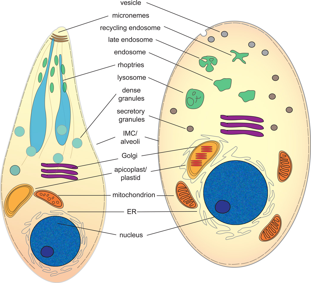
The apicomplexan lineage includes some of the world’s most abundant – and most devastating – protozoan parasites. Toxoplasma infects ~30% of the global human population [1], and while usually asymptomatic in otherwise healthy adults, acute disease can produce severe neurological disease or death during fetal infection and in immunocompromised patients [2]. Cryptosporidium is a prominent source of severe diarrhea in both cattle and human infants [3], and Eimeria, Neospora, Babesia and Theileria cause agricultural diseases of poultry and/or livestock (Cryptosporidium and Babesia are also opportunistic pathogens in humans). Plasmodium is responsible for at least 200 million cases of malaria, resulting in >660,000 deaths each year (http://www.who.int/malaria/publications/world_malaria_report_2012/en/) [2]. Most therapeutic development has focused on parasite-specific biochemical targets, but cell biological features of these microbial eukaryotes are perhaps their most distinctive attributes (Figure 1). Comparison with other eukaryotic systems provides insight into the origin and diversity of eukaryotic organelles, including distinctive cell biological aspects of the Apicomplexa that suggest novel targets for therapeutic intervention.
Figure 1.
Organelle homology, highlighting potential relationships between a schematized apicomplexan and a hypothetical comparative alveolate cell. Endosymbiotic organelles are indicated in warm tones, while endomembrane organelles are shown in cool tones.
Some organellar homologs are obvious, including the nucleus, ER and plasma membrane. Others display clear homology to ubiquitous eukaryotic organelles, including the Golgi and mitochondria, but have accrued unique biological traits. For others, homology was initially unclear, but recent studies have unmasked the apicoplast as a secondary endosymbiotic plastid, and the Inner Membrane Complex (IMC) as a homolog of ciliate alveoli, enhancing our understanding of apicomplexan cell biology, evolution and pathogenicity. The origin of other structures, including invasion organelles of the apical complex (micronemes, rhoptries) remains unresolved. This report briefly summarizes the divergent paths taken by the mitochondria and Golgi, and highlights recent work on the cell biology and evolutionary history of the apicoplast and IMC. It concludes with the idea that we now have sufficient evidence to say with some certainty that the apical complex organelles have an endolysosomal origin, although the precise nature of this homology remains an interesting point of enquiry.
Outgroups, ingroups and unambiguous organellar homologs
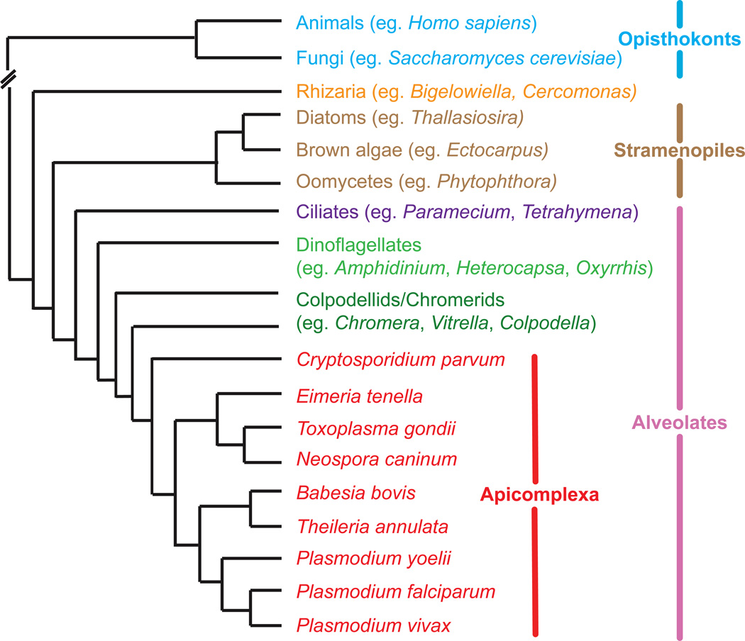
Studies on the supergroup Opisthokonta (animals and fungi; Fig 2) provide a wealth of cell biological knowledge, but porting this knowledge to the apicomplexans requires a map, with organismal sign-posts for reference. Fortunately, advances in eukaryotic molecular taxonomy now makes such mapping possible [4]. The ‘SAR’ supergroup includes Stramenopiles (diatoms, brown algae, oomycetes), Rhizaria, and Alveolates; the latter is comprised of three major lineages: the Ciliates, Dinoflagellates, and Apicomplexa. Recent environmental sampling [5] reveals a wealth of uncharacterized apicomplexans, as well as other groups such as the colpodellids, whose diversity is just beginning to be explored. Among those, the newly discovered basal apicomplexans Chromera velia and Vitrella brassicaformis [6,7] are free-living/symbiotic and photosynthetic organisms, and hold great promise for comparative studies on exclusively parasitic Apicomplexa.
Figure 2.
Phylogenetic position of the apicomplexan relative to selected outgroups. Broken line indicates the large evolutionary distance between SAR and opisthokont taxa. Note that this cladogram displays relative position only, with no indication of evolutionary distance or rates.
Toxoplasma gondii displays the least divergent set of organelles among well-studied and experimentally-tractable apicomplexans, providing a model for apicomplexan cell biology [8]. The stacked cisternae of the single T. gondii Golgi are closely associated with the endoplasmic reticulum at the apical end of the nucleus, providing a textbook example of this organelle, including trafficking via COP-I, COP-II and clathrin-coated vesicles [9]. The Golgi of Plasmodium (and many other apicomplexans) is more highly reduced (often just a single cisterna), and harbors divergent, lineage-specific trafficking factors [10,11]. As the central nexus of vesicular transport, the Golgi mediates targeting to both intracellular locations and the exterior. A better understanding of the apicomplexan Golgi is likely to provide useful insights into the biology and pathogenesis mediated by these parasites’ distinctive endomembrane organelles.
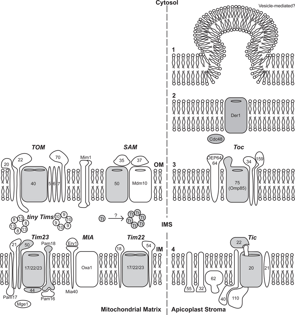
The Apicomplexa also harbor a mitochondrion (Figure 1) displaying several unusual features. The majority of apicomplexan mitochondrial genomes (including that of Vitrella) are very small (6–11 kb), and encode just three protein-coding genes (cox1, cox3, cob), along with extensively fragmented rRNA genes [12]. The genome organization varies according to species and is either monomeric linear (e.g. Theileria) or concatemeric (e.g. Plasmodium) [12]. Although the precise arrangement of genes may differ, no genome is greater than 11kb [12]. The exceptions are Cryptosporidium, which has entirely lost its mitochondrial genome and Chromera, which possesses an even more reduced mitochondrial genome than many apicomplexans, resembling the fragmented dinoflagellate mitochondrial genome [13]. Most mitochondrial proteins are imported via a greatly reduced import apparatus (Figure 3); the Cryptosporidium import system contains just seven proteins, representing one of the most reduced systems yet defined [14]. Apicomplexan mitochondria also display reduced metabolic capacity [15], including an unusual partitioning of heme biosynthesis also observed in chromerids [16]. Apicomplexan mitochondria also lack pyruvate dehydrogenase, generally supposed to be the entry point for energy metabolism, and conserved in other aerobic mitochondria. This absence is shared with dinoflagellates [17], suggesting loss prior to their divergence, and some members of both lineages contain pyruvate:ferridoxin oxidoreductase instead [15,18]. These genomic and metabolic differences highlight extreme divergence between parasite and host biology, providing exciting areas for further investigation.
Figure 3.
Import machinery for apicomplexan endosymbiotic organelles, highlighting conserved components in gray; white components are absent from at least some apicomplexa (based on bioinformatics searches; few of these proteins have been confirmed experimentally [14,28]). Left, mitochondrion (OM, outer membrane; IM, inner membrane; IMS, inter-membrane space). Complex names are indicated in italics; numbers indicate the protein identifier, i.e. 22 = Tom22. Cryptosporidium displays the most highly reduced mitochondrial import. Plasmodium retains additional proteins, but has dispensed with Tom20 & Tom70. Right, apicoplast (membranes numbered sequentially from exterior to interior; Plasmodium shown). Proteins likely cross the outer membrane by vesicle fusion [25], the second membrane using a translocon derived from the endoplasmic reticulum ERAD-system, and a reduced chloroplast import apparatus to cross the third and fourth membranes [27,28].
A relict chloroplast
Comparison with other eukaryotes has also been instrumental in characterizing the apicomplexan plastid (apicoplast; Figure 1) [19–21], a secondary endosymbiotic organelle surrounded by four membranes acquired when an ancestral alveolate engulfed a eukaryotic alga, and retained the algal plastid. Chromera [6] contains a descendent of this organelle retaining photosynthetic function, providing considerable insight into apicoplast origins [22–24]; careful phylogenetic analysis now strongly supports a red algal ancestry [22].
The majority of apicoplast genes are encoded in the nucleus [21]. Some proteins target the apicoplast using a tyrosine-based motif [25], but most exploit a classical secretory signal sequence mediating cotranslational translocation across the endoplasmic reticulum; vesicular fusion then mediates traversal of the first apicoplast membrane [21,26]. Phylogenetic and cell biological analyses have shown that the apicoplast has repurposed proteins from the ERAD system (normally used to remove misfolded proteins from the ER) as an apicoplast translocon [27,28]. Finally, a greatly reduced conventional chloroplast import apparatus (Fig 3) is exploited to cross the inner two (original plastid) membranes (see Deponte 2012 [28]for a more detailed review.)
Little is known about transcription and translation in the apicoplast, which encodes its own RNA polymerase, ribosomal RNAs, and many ribosomal proteins (others are encoded in the nucleus and imported, as above). Although plants and algal plastids also exploit a nuclear-encoded phage-type RNA polymerase, the only phage-type RNA polymerase reported in apicomplexa to date is presumed to be targeted to the mitochondrion [29]. Transcription in the apicoplast appears to be polycistronic [30] as observed in photosynthetic chloroplasts. Preliminary results (Dorrell, personal communication) suggest that both Chromera and Vitrella are able to distinguish between mRNA molecules encoding photosynthetic or nonphotosynthetic genes, by the addition of a polyU tail transcripts encoding proteins involved in photosynthesis. Such a mechanism provides an intriguing mechanism for the adaptation to parasitic lifestyle - the loss of the polyU polymerase would be sufficient to prevent photosynthesis.
Although no longer photosynthetic, the apicoplast carries out several biochemical processes including the synthesis of isoprenoids (via the xylulose pathway, rather than HMG CoA reductase used by humans and other opisthokonts), fatty acids (using a type II fatty acid synthase, rather than the type I FAS typical of opisthokonts), heme (partitioned unusually between the mitochondrion and apicoplast), and Fe-S cluster maturation (reviewed in [31]). The functional importance of these pathways has long been a mystery, however, particularly as Crypotosporidium has lost the apicoplast entirely, acquiring all relevant nutrients from its environment. A breakthrough article [32] recently demonstrated that the apicoplast can be eliminated from blood-stage Plasmodium, if the growth medium is supplemented with isopentenyl pyrophosphate. This implicates the isoprenoid synthesis as the sole essential apicoplast function in these parasites, although not necessarily implying the lack of other roles in other apicomplexans. Nonetheless, this strategy provides researchers with a powerful research tool for assessing drugs thought to target the apicoplast – an organelle with no counterpart in human or animal host species.
IMC: Homology with ciliate and dinoflagellate alveolae
Perhaps the most distinctive aspect of apicomplexan cell biology is their peculiar mechanism of replication, in which daughter parasites are assembled de novo, within the maternal cytoplasm, rather than dividing by binary fission [33,34]. This process, termed schizogony, involves an unusual membrane-cytoskeletal complex known as the Inner Membrane Complex. The IMC is derived from cortical alveolae [35,36] -- a morphological character defining the superphylum Alveolata, including apicomplexans, chromerids and colpodellids, ciliates, and dinoflagellates [7] (Fig 2).
Ciliate alveolae are specialized for storage and regulatory activities [35], while dinoflagellate alveolae have evolved into the armored plates characteristic of this phylum [36]. In the apicomplexa, the IMC forms a patchwork of Golgi-derived flattened membrane vesicles, closely apposed to the plasma membrane to yield a triple membrane [37,38]. The cytoplasmic face of the apicomplexan IMC associates with subpellicular cytoskeletal elements (microtubules and intermediate filament-like alveolins) [34,37,39]. The complex organization of this structure appears to be essential for the maintenance of cell shape and pellicle integrity [38,40,41]. In motile apicomplexan zoites, the IMC also serves to anchor the glideosome motility machinery [42].
It is unclear how the alveoli were coopted for the purpose of division in the Apicomplexa, but the IMC provides a practical solution to several fundamental problems facing by many apicomplexan parasites, including the strict requirement to maintain polarity during replication, the need to rapidly assemble multiple daughter parasites prior to bursting out of the infected host cell, and the challenges posed by the lack of classical lysosomes: all maternal organelles packaged into daughter parasites are the result of a positive ‘decision’; waste material (including the indigestible hemozoin polymer produced by degradation of hemoglobin in malaria parasites) is simply left behind [33,38].
Rhoptries and micronemes: divergent endolysosomal homologues?
Elicidating organelle homology in Apicomplexa has clearly helped to uncover their unique aspects. And yet, despite their tremendous global impact, and the scientific effort applied, there remain apicomplexan organelles for which the homology remains incompletely understood. The Apicomplexa are named for the Apical complex, a characteristic set of apical invasion organelles (Figure 1). This includes the microtubular conoid and the single-membrane bound rhoptries and micronemes. In mature cells, spherical or ellipsoidal micronemes localize to the apical end of the cell in close association with the conoid. The micronemes are first to discharge upon binding to the host cell. The club-shaped rhoptries, which occupy a large cellular area with the thinner neck portions (Figure 1) oriented toward the apical end of the cell [43], then discharge and mediate entry of the parasite to the host cell. The evolutionary origins of these organelles has been murky, but the most strongly supported hypothesis [36,44–46] is of a highly divergent endolysosomal origin.
This general idea is not new, with the first proposition almost a decade ago [47] that rhoptries are directly homologous to secretory lysosomes. A significant body of evidence, however, has now accumulated from diverse studies from morphology to trafficking to proteomics. pH-sensitive immunolocalization microscopy suggests that mature rhoptries are acidic, (pH 5 – 7), while pre-rhoptries are even more acidic (pH 3.5–5.5) [48]. Both rhoptries and micronemes are granular, and have dense staining areas under electron microscopy [49], similar to endosomes. Furthermore, early in their biogenesis, micronemes closely resemble multi-vesicular bodies or late endosomes. The key endosomal proteins AP-1 and Rab11A co-localize with rhoptries [50,51][35,36] and proteomic studies have identified various hydrolases in the rhoptry lumen, potentially similar to lysosomal hydrolases [52]. Likewise, the microneme appears to contain various endosomal membrane-trafficking proteins including protein homologues of VAMP, syntaxin 13, clathrin, sortillin and Eps15R [53].
Trafficking to micronemes and rhoptries has been an area of intense research, but is still not fully understood. Microneme proteins traffic through the endosomal system via signals in N-terminal prodomains which are cleaved en route via a Cathepsin L protease [54,55]. Rhoptry trafficking, though less-clearly characterized, also appears to rely on N-terminal prodomains, [56], or in the case of membrane proteins, specific residues within the N-terminal region [57]. It is clear though that trafficking to both organelles relies on apicomplexan homologues of sortilin and dynamin [58,59], and may rely on transmembrane cargo adaptors [60]. Additionally, Rab5A and 5C are important for both microneme protein trafficking and rhoptry biogenesis [45]. TgROP2 was initially reported to contain a YXXϕ motif in its cytoplasmic tail and traffic via AP-1 [51,61], although these results are contentious. Alternatively, the TgROP2 result may be explained by the notion that sortilin, in model eukaryotes, interacts with numerous trafficking factors including AP1 and AP2, clathrin, and components of the retromer complex [59].
Despite these similarities to endolysosomal organelles in model systems, it is clear that these apical organelles also possess unique aspects. Although proteomic studies identified endosomal membrane-trafficking machinery, they also showed a large proportion of organelle-specific proteins, eg. the MICs, ROPs, and more [53,62]. As well, Rab5 and Rab7, canonical markers for endosomes and lysosomes, localize to the late secretory pathway instead of the micronemes or rhoptries [45].
Though the evidence is strongly suggestive of homology between the apical complex invasion organelles (Rhoptries, micronemes) and the organelles of the endosomal system (early/recycling endosomes, secretory lysosomes), the nature of this homology is less clear. The presence of a dynamic vacuolar compartment in T. gondii similar to plant lytic vacuoles [63], and of the digestive vacuole in Plasmodium spp., further complicates any one-to-one assignment of homology. Furthermore, the variable presence of rhoptries and micronemes in early branching dinoflagellates such as perkinsids, colpedellids, and chromerids, and the presence of trichocysts in some ciliates and dinoflagellates, suggests not only that the invasion organelles of Apicomplexa pre-date the development of intracellular parasitism, but also that alveolates possess diverse modifications of their endocytic systems [7,36].
It is possible that these organelles are derived from an organellar expansion of either endosomes or lysosomes. It is also possible that there is a one to one correlation, possibly micronemes to endosomes, rhoptries to secretory lysosomes, and the apicomplexan system has diverged to such an extent that the homology has become difficult to assess. The apicomplexan endocytic membrane-trafficking machinery complement has certainly been modified via loss from a more complete canonical eukaryotic set. Key cargo adaptors (AP3), MVB machinery (ESCRTs I, II) and endocytic MTC complexes have been lost ([30,46], Klinger and Dacks unpublished) via a process that is best explained by lack of selection on the machinery associated with the endocytic functions of the endolysosomal organelles. This may also indicate a concurrent adaptive emphasis on their secretory functions.
Further work by molecular parasitology in apicomplexans is certainly warranted, but with the evidence now pointing to endolysosomal homology, the investigative path is, at least, somewhat clearer.
Conclusions
Whether from an organismal perspective (alveolates, colpodellids/chromerids, and between apicomplexans) or organelle by organelle, taking a comparative approach to apicomplexan cell biology has already produced important insight into pathogenesis on a cellular level. With new organelle homology hypotheses solidifying, and advances both in experimental tools and genome sequencing to explore alveolate diversity (notably in the colpodellids and chromerids), further work from this evolutionary cell biological approach will help lead to a better understanding of apicomplexan parasites and a way forward to combating these threats to our global health and well-being.
Highlights.
A apicomplexan mitochondrion is greatly reduced, lacking many significant enzymes.
The ‘apicoplast’ is a non-photosynthetic secondary endosymbiotic plastid, and potentially vulnerable to therapeutic intervention.
The ‘inner membrane complex’ is derived from cortical alveolae of the superphylum Alveolata, and central to these parasites’ distinctive replicative mechanism.
Microneme and rhoptry components of the apicomplexan ‘apical complex’ invasion machinery may be derived from secretory endosomes.
Understanding organellar origins provides context for functional studies.
Acknowledgements
CMK is funded by a studentship from Alberta Innovates Health Solutions and the Women and Children’s Health Research Institute. JBD is the CRC in Evolutionary Cell Biology. RERN is funded by the Wellcome Trust and DSR is funded by the National Institutes of Allergy and Infectious Diseases.
Footnotes
Publisher's Disclaimer: This is a PDF file of an unedited manuscript that has been accepted for publication. As a service to our customers we are providing this early version of the manuscript. The manuscript will undergo copyediting, typesetting, and review of the resulting proof before it is published in its final citable form. Please note that during the production process errors may be discovered which could affect the content, and all legal disclaimers that apply to the journal pertain.
Contributor Information
Christen M. Klinger, Email: cklinger@ualberta.ca.
Dinkorma T. Ouologuem, Email: eanaouologuem@yahoo.fr.
David S. Roos, Email: droos@sas.upenn.edu.
Joel B. Dacks, Email: dacks@ualberta.ca, joel.dacks@ualberta.ca.
References
- 1.Plattner F, Soldati-Favre D. Hijacking of host cellular functions by the Apicomplexa. Annu Rev Microbiol. 2008;62:471–487. doi: 10.1146/annurev.micro.62.081307.162802. [DOI] [PubMed] [Google Scholar]
- 2.Vidal JE, Hernandez AV, de Oliveira AC, Dauar RF, Barbosa SP, Jr, Focaccia R. Cerebral toxoplasmosis in HIV-positive patients in Brazil: clinical features and predictors of treatment response in the HAART era. AIDS Patient Care STDS. 2005;19:626–634. doi: 10.1089/apc.2005.19.626. [DOI] [PubMed] [Google Scholar]
- 3.Kotloff KL, Blackwelder WC, Nasrin D, Nataro JP, Farag TH, van Eijk A, Adegbola RA, Alonso PL, Breiman RF, Faruque AS, et al. The Global Enteric Multicenter Study (GEMS) of diarrheal disease in infants and young children in developing countries: epidemiologic and clinical methods of the case/control study. Clin Infect Dis. 2013;55(Suppl 4):S232–S245. doi: 10.1093/cid/cis753. [DOI] [PMC free article] [PubMed] [Google Scholar]
- 4.Adl SM, Simpson AG, Lane CE, Lukes J, Bass D, Bowser SS, Brown MW, Burki F, Dunthorn M, Hampl V, et al. The revised classification of eukaryotes. J Eukaryot Microbiol. 2012;59:429–493. doi: 10.1111/j.1550-7408.2012.00644.x. [DOI] [PMC free article] [PubMed] [Google Scholar]
- 5. Janouskovec J, Horak A, Barott KL, Rohwer FL, Keeling PJ. Global analysis of plastid diversity reveals apicomplexan-related lineages in coral reefs. Curr Biol. 2012;22:R518–R519. doi: 10.1016/j.cub.2012.04.047. * Environmental sampling reveals that the diversity of free-living alveolates has been greatly underestimated.
- 6.Moore RB, Obornik M, Janouskovec J, Chrudimsky T, Vancova M, Green DH, Wright SW, Davies NW, Bolch CJ, Heimann K, et al. A photosynthetic alveolate closely related to apicomplexan parasites. Nature. 2008;451:959–963. doi: 10.1038/nature06635. [DOI] [PubMed] [Google Scholar]
- 7. Obornik M, Modry D, Lukes M, Cernotikova-Stribrna E, Cihlar J, Tesarova M, Kotabova E, Vancova M, Prasil O, Lukes J. Morphology, ultrastructure and life cycle of Vitrella brassicaformis n. sp., n. gen., a novel chromerid from the Great Barrier Reef. Protist. 2011;163:306–323. doi: 10.1016/j.protis.2011.09.001. **This paper, along with Moore et al 2008, describes Vitrella and Chromera as free-living photosynthetic relatives of apicomplexans. These species will be invaluable for understanding the transition to parasitism in the Apicomplexa.
- 8.Joiner KA, Roos DS. Secretory traffic in the eukaryotic parasite Toxoplasma gondii: less is more. J Cell Biol. 2002;157:557–563. doi: 10.1083/jcb.200112144. [DOI] [PMC free article] [PubMed] [Google Scholar]
- 9.Pelletier L, Stern CA, Pypaert M, Sheff D, Ngo HM, Roper N, He CY, Hu K, Toomre D, Coppens I, et al. Golgi biogenesis in Toxoplasma gondii. Nature. 2002;418:548–552. doi: 10.1038/nature00946. [DOI] [PubMed] [Google Scholar]
- 10.Ayong L, DaSilva T, Mauser J, Allen CM, Chakrabarti D. Evidence for prenylation-dependent targeting of a Ykt6 SNARE in Plasmodium falciparum. Mol Biochem Parasitol. 2011;175:162–168. doi: 10.1016/j.molbiopara.2010.11.007. [DOI] [PMC free article] [PubMed] [Google Scholar]
- 11.Struck NS, Herrmann S, Langer C, Krueger A, Foth BJ, Engelberg K, Cabrera AL, Haase S, Treeck M, Marti M, et al. Plasmodium falciparum possesses two GRASP proteins that are differentially targeted to the Golgi complex via a higher- and lower-eukaryote-like mechanism. J Cell Sci. 2008;121:2123–2129. doi: 10.1242/jcs.021154. [DOI] [PubMed] [Google Scholar]
- 12.Hikosaka K, Kita K, Tanabe K. Diversity of mitochondrial genome structure in the phylum Apicomplexa. Mol Biochem Parasitol. 2013;188:26–33. doi: 10.1016/j.molbiopara.2013.02.006. [DOI] [PubMed] [Google Scholar]
- 13.Flegontov P, Lukes J. Mitochondrial Genome Evolution. L M-D: Elsevier Ltd: Academic Press; 2012. Mitochondrial genomes of photosynthetic euglenids and alveolates; pp. 127–153. [Google Scholar]
- 14.Heinz E, Lithgow T. Back to basics: a revealing secondary reduction of the mitochondrial protein import pathway in diverse intracellular parasites. Biochim Biophys Acta. 2013;1833:295–303. doi: 10.1016/j.bbamcr.2012.02.006. [DOI] [PubMed] [Google Scholar]
- 15.Mogi T, Kita K. Diversity in mitochondrial metabolic pathways in parasitic protists Plasmodium and Cryptosporidium. Parasitol Int. 2010;59:305–312. doi: 10.1016/j.parint.2010.04.005. [DOI] [PubMed] [Google Scholar]
- 16.Koreny L, Sobotka R, Janouskovec J, Keeling PJ, Obornik M. Tetrapyrrole synthesis of photosynthetic chromerids is likely homologous to the unusual pathway of apicomplexan parasites. Plant Cell. 2011;23:3454–3462. doi: 10.1105/tpc.111.089102. [DOI] [PMC free article] [PubMed] [Google Scholar]
- 17.Butterfield ER, Howe CJ, Nisbet RE. An analysis of dinoflagellate metabolism using EST data. Protist. 2013;164:218–236. doi: 10.1016/j.protis.2012.09.001. [DOI] [PubMed] [Google Scholar]
- 18.Hug LA, Stechmann A, Roger AJ. Phylogenetic distributions and histories of proteins involved in anaerobic pyruvate metabolism in eukaryotes. Mol Biol Evol. 2010;27:311–324. doi: 10.1093/molbev/msp237. [DOI] [PubMed] [Google Scholar]
- 19.Kohler S, Delwiche CF, Denny PW, Tilney LG, Webster P, Wilson RJ, Palmer JD, Roos DS. A plastid of probable green algal origin in Apicomplexan parasites. Science. 1997;275:1485–1489. doi: 10.1126/science.275.5305.1485. [DOI] [PubMed] [Google Scholar]
- 20.Lemgruber L, Kudryashev M, Dekiwadia C, Riglar DT, Baum J, Stahlberg H, Ralph SA, Frischknecht F. Cryo-electron tomography reveals four-membrane architecture of the Plasmodium apicoplast. Malar J. 2013;12:25. doi: 10.1186/1475-2875-12-25. [DOI] [PMC free article] [PubMed] [Google Scholar]
- 21.Roos DS, Crawford MJ, Donald RG, Kissinger JC, Klimczak LJ, Striepen B. Origin, targeting, and function of the apicomplexan plastid. Curr Opin Microbiol. 1999;2:426–432. doi: 10.1016/S1369-5274(99)80075-7. [DOI] [PubMed] [Google Scholar]
- 22. Burki F, Flegontov P, Obornik M, Cihlar J, Pain A, Lukes J, Keeling PJ. Re-evaluating the green versus red signal in eukaryotes with secondary plastid of red algal origin. Genome Biol Evol. 2012;4:626–635. doi: 10.1093/gbe/evs049. ** This paper helps to elucidate the complex evolutionary history of the apicoplast.
- 23.Janouskovec J, Horak A, Obornik M, Lukes J, Keeling PJ. A common red algal origin of the apicomplexan, dinoflagellate, and heterokont plastids. Proc Natl Acad Sci U S A. 2010;107:10949–10954. doi: 10.1073/pnas.1003335107. [DOI] [PMC free article] [PubMed] [Google Scholar]
- 24.Woehle C, Dagan T, Martin WF, Gould SB. Red and problematic green phylogenetic signals among thousands of nuclear genes from the photosynthetic and apicomplexa-related Chromera velia. Genome Biol Evol. 2011;3:1220–1230. doi: 10.1093/gbe/evr100. [DOI] [PMC free article] [PubMed] [Google Scholar]
- 25.DeRocher AE, Karnataki A, Vaney P, Parsons M. Apicoplast targeting of a Toxoplasma gondii transmembrane protein requires a cytosolic tyrosine-based motif. Traffic. 2012;13:694–704. doi: 10.1111/j.1600-0854.2012.01335.x. [DOI] [PMC free article] [PubMed] [Google Scholar]
- 26.Foth BJ, Ralph SA, Tonkin CJ, Struck NS, Fraunholz M, Roos DS, Cowman AF, McFadden GI. Dissecting apicoplast targeting in the malaria parasite Plasmodium falciparum. Science. 2003;299:705–708. doi: 10.1126/science.1078599. [DOI] [PubMed] [Google Scholar]
- 27.Agrawal S, Chung DW, Ponts N, van Dooren GG, Prudhomme J, Brooks CF, Rodrigues EM, Tan JC, Ferdig MT, Striepen B, et al. An apicoplast localized ubiquitylation system is required for the import of nuclear-encoded plastid proteins. PLoS Pathog. 2013;9:e1003426. doi: 10.1371/journal.ppat.1003426. [DOI] [PMC free article] [PubMed] [Google Scholar]
- 28.Deponte M, Hoppe HC, Lee MC, Maier AG, Richard D, Rug M, Spielmann T, Przyborski JM. Wherever I may roam: protein and membrane trafficking in P. falciparum-infected red blood cells. Mol Biochem Parasitol. 2012;186:95–116. doi: 10.1016/j.molbiopara.2012.09.007. [DOI] [PubMed] [Google Scholar]
- 29.Ke H, Morrisey JM, Ganesan SM, Mather MW, Vaidya AB. Mitochondrial RNA polymerase is an essential enzyme in erythrocytic stages of Plasmodium falciparum. Mol Biochem Parasitol. 2012;185:48–51. doi: 10.1016/j.molbiopara.2012.05.001. [DOI] [PMC free article] [PubMed] [Google Scholar]
- 30.Wilson RJ, Denny PW, Preiser PR, Rangachari K, Roberts K, Roy A, Whyte A, Strath M, Moore DJ, Moore PW, et al. Complete gene map of the plastid-like DNA of the malaria parasite Plasmodium falciparum. J Mol Biol. 1996;261:155–172. doi: 10.1006/jmbi.1996.0449. [DOI] [PubMed] [Google Scholar]
- 31.Seeber F, Soldati-Favre D. Metabolic pathways in the apicoplast of apicomplexa. Int Rev Cell Mol Biol. 2010;281:161–228. doi: 10.1016/S1937-6448(10)81005-6. [DOI] [PubMed] [Google Scholar]
- 32. Yeh E, DeRisi JL. Chemical rescue of malaria parasites lacking an apicoplast defines organelle function in blood-stage Plasmodium falciparum. PLoS Biol. 2011;9:e1001138. doi: 10.1371/journal.pbio.1001138. **Demonstrates that IPP is necessary and sufficient for retention of the apicoplast, identifying its synthesis as the essential function of this organelle.
- 33.Nishi M, Hu K, Murray JM, Roos DS. Organellar dynamics during the cell cycle of Toxoplasma gondii. J Cell Sci. 2008;121:1559–1568. doi: 10.1242/jcs.021089. [DOI] [PMC free article] [PubMed] [Google Scholar]
- 34.Hu K, Mann T, Striepen B, Beckers CJ, Roos DS, Murray JM. Daughter cell assembly in the protozoan parasite Toxoplasma gondii. Mol Biol Cell. 2002;13:593–606. doi: 10.1091/mbc.01-06-0309. [DOI] [PMC free article] [PubMed] [Google Scholar]
- 35.Stelly N, Mauger JP, Claret M, Adoutte A. Cortical alveoli of Paramecium: a vast submembranous calcium storage compartment. J Cell Biol. 1991;113:103–112. doi: 10.1083/jcb.113.1.103. [DOI] [PMC free article] [PubMed] [Google Scholar]
- 36.Gubbels MJ, Duraisingh MT. Evolution of apicomplexan secretory organelles. Int J Parasitol. 2012;42:1071–1081. doi: 10.1016/j.ijpara.2012.09.009. [DOI] [PMC free article] [PubMed] [Google Scholar]
- 37.Morrissette NS, Murray JM, Roos DS. Subpellicular microtubules associate with an intramembranous particle lattice in the protozoan parasite Toxoplasma gondii. J Cell Sci. 1997;110(Pt 1):35–42. doi: 10.1242/jcs.110.1.35. [DOI] [PubMed] [Google Scholar]
- 38.Kono M, Herrmann S, Loughran NB, Cabrera A, Engelberg K, Lehmann C, Sinha D, Prinz B, Ruch U, Heussler V, et al. Evolution and architecture of the inner membrane complex in asexual and sexual stages of the malaria parasite. Mol Biol Evol. 2012;29:2113–2132. doi: 10.1093/molbev/mss081. [DOI] [PubMed] [Google Scholar]
- 39.Gould SB, Tham WH, Cowman AF, McFadden GI, Waller RF. Alveolins, a new family of cortical proteins that define the protist infrakingdom Alveolata. Mol Biol Evol. 2008;25:1219–1230. doi: 10.1093/molbev/msn070. [DOI] [PubMed] [Google Scholar]
- 40.Khater EI, Sinden RE, Dessens JT. A malaria membrane skeletal protein is essential for normal morphogenesis, motility, and infectivity of sporozoites. J Cell Biol. 2004;167:425–432. doi: 10.1083/jcb.200406068. [DOI] [PMC free article] [PubMed] [Google Scholar]
- 41.Tremp AZ, Khater EI, Dessens JT. IMC1b is a putative membrane skeleton protein involved in cell shape, mechanical strength, motility, and infectivity of malaria ookinetes. J Biol Chem. 2008;283:27604–27611. doi: 10.1074/jbc.M801302200. [DOI] [PMC free article] [PubMed] [Google Scholar]
- 42.Frenal K, Polonais V, Marq JB, Stratmann R, Limenitakis J, Soldati-Favre D. Functional dissection of the apicomplexan glideosome molecular architecture. Cell Host Microbe. 2010;8:343–357. doi: 10.1016/j.chom.2010.09.002. [DOI] [PubMed] [Google Scholar]
- 43.Paredes-Santos TC, de Souza W, Attias M. Dynamics and 3D organization of secretory organelles of Toxoplasma gondii. J Struct Biol. 2012;177:420–430. doi: 10.1016/j.jsb.2011.11.028. [DOI] [PubMed] [Google Scholar]
- 44.Diekmann Y, Pereira-Leal JB. Evolution of intracellular compartmentalization. Biochem J. 2013;449:319–331. doi: 10.1042/BJ20120957. [DOI] [PubMed] [Google Scholar]
- 45. Kremer K, Kamin D, Rittweger E, Wilkes J, Flammer H, Mahler S, Heng J, Tonkin CJ, Langsley G, Hell SW, et al. An overexpression screen of Toxoplasma gondii Rab-GTPases reveals distinct transport routes to the micronemes. PLoS Pathog. 2013;9:e1003213. doi: 10.1371/journal.ppat.1003213. *Provides evidence for the involvement of endocytic Rabs in protein trafficking to the micronemes and rhopties, suggesting endolysosomal homology.
- 46.Nevin WD, Dacks JB. Repeated secondary loss of adaptin complex genes in the Apicomplexa. Parasitol Int. 2009;58:86–94. doi: 10.1016/j.parint.2008.12.002. [DOI] [PubMed] [Google Scholar]
- 47.Ngo HM, Yang M, Joiner KA. Are rhoptries in Apicomplexan parasites secretory granules or secretory lysosomal granules? Mol Microbiol. 2004;52:1531–1541. doi: 10.1111/j.1365-2958.2004.04056.x. [DOI] [PubMed] [Google Scholar]
- 48.Shaw MK, Roos DS, Tilney LG. Acidic compartments and rhoptry formation in Toxoplasma gondii. Parasitology. 1998;117(Pt 5):435–443. doi: 10.1017/s0031182098003278. [DOI] [PubMed] [Google Scholar]
- 49.Blackman MJ, Bannister LH. Apical organelles of Apicomplexa: biology and isolation by subcellular fractionation. Mol Biochem Parasitol. 2001;117:11–25. doi: 10.1016/s0166-6851(01)00328-0. [DOI] [PubMed] [Google Scholar]
- 50.Agop-Nersesian C, Naissant B, Ben Rached F, Rauch M, Kretzschmar A, Thiberge S, Menard R, Ferguson DJ, Meissner M, Langsley G. Rab11A-controlled assembly of the inner membrane complex is required for completion of apicomplexan cytokinesis. PLoS Pathog. 2009;5:e1000270. doi: 10.1371/journal.ppat.1000270. [DOI] [PMC free article] [PubMed] [Google Scholar]
- 51.Hoppe HC, Ngo HM, Yang M, Joiner KA. Targeting to rhoptry organelles of Toxoplasma gondii involves evolutionarily conserved mechanisms. Nat Cell Biol. 2000;2:449–456. doi: 10.1038/35017090. [DOI] [PubMed] [Google Scholar]
- 52.Bradley PJ, Ward C, Cheng SJ, Alexander DL, Coller S, Coombs GH, Dunn JD, Ferguson DJ, Sanderson SJ, Wastling JM, et al. Proteomic analysis of rhoptry organelles reveals many novel constituents for host-parasite interactions in Toxoplasma gondii. J Biol Chem. 2005;280:34245–34258. doi: 10.1074/jbc.M504158200. [DOI] [PubMed] [Google Scholar]
- 53.Lal K, Prieto JH, Bromley E, Sanderson SJ, Yates JR, 3rd, Wastling JM, Tomley FM, Sinden RE. Characterisation of Plasmodium invasive organelles; an ookinete microneme proteome. Proteomics. 2009;9:1142–1151. doi: 10.1002/pmic.200800404. [DOI] [PMC free article] [PubMed] [Google Scholar]
- 54.Gaji RY, Flammer HP, Carruthers VB. Forward targeting of Toxoplasma gondii proproteins to the micronemes involves conserved aliphatic amino acids. Traffic. 2011;12:840–853. doi: 10.1111/j.1600-0854.2011.01192.x. [DOI] [PMC free article] [PubMed] [Google Scholar]
- 55.Parussini F, Coppens I, Shah PP, Diamond SL, Carruthers VB. Cathepsin L occupies a vacuolar compartment and is a protein maturase within the endo/exocytic system of Toxoplasma gondii. Mol Microbiol. 2010;76:1340–1357. doi: 10.1111/j.1365-2958.2010.07181.x. [DOI] [PMC free article] [PubMed] [Google Scholar]
- 56.Hajagos BE, Turetzky JM, Peng ED, Cheng SJ, Ryan CM, Souda P, Whitelegge JP, Lebrun M, Dubremetz JF, Bradley PJ. Molecular dissection of novel trafficking and processing of the Toxoplasma gondii rhoptry metalloprotease toxolysin-1. Traffic. 2012;13:292–304. doi: 10.1111/j.1600-0854.2011.01308.x. [DOI] [PMC free article] [PubMed] [Google Scholar]
- 57.Cabrera A, Herrmann S, Warszta D, Santos JM, John Peter AT, Kono M, Debrouver S, Jacobs T, Spielmann T, Ungermann C, et al. Dissection of minimal sequence requirements for rhoptry membrane targeting in the malaria parasite. Traffic. 2012;13:1335–1350. doi: 10.1111/j.1600-0854.2012.01394.x. [DOI] [PubMed] [Google Scholar]
- 58.Breinich MS, Ferguson DJ, Foth BJ, van Dooren GG, Lebrun M, Quon DV, Striepen B, Bradley PJ, Frischknecht F, Carruthers VB, et al. A dynamin is required for the biogenesis of secretory organelles in Toxoplasma gondii. Curr Biol. 2009;19:277–286. doi: 10.1016/j.cub.2009.01.039. [DOI] [PMC free article] [PubMed] [Google Scholar]
- 59.Sloves PJ, Delhaye S, Mouveaux T, Werkmeister E, Slomianny C, Hovasse A, Dilezitoko Alayi T, Callebaut I, Gaji RY, Schaeffer-Reiss C, et al. Toxoplasma sortilin-like receptor regulates protein transport and is essential for apical secretory organelle biogenesis and host infection. Cell Host Microbe. 2012;11:515–527. doi: 10.1016/j.chom.2012.03.006. [DOI] [PubMed] [Google Scholar]
- 60.Richard D, Kats LM, Langer C, Black CG, Mitri K, Boddey JA, Cowman AF, Coppel RL. Identification of rhoptry trafficking determinants and evidence for a novel sorting mechanism in the malaria parasite Plasmodium falciparum. PLoS Pathog. 2009;5:e1000328. doi: 10.1371/journal.ppat.1000328. [DOI] [PMC free article] [PubMed] [Google Scholar]
- 61.Ngo HM, Yang M, Paprotka K, Pypaert M, Hoppe H, Joiner KA. AP-1 in Toxoplasma gondii mediates biogenesis of the rhoptry secretory organelle from a post-Golgi compartment. J Biol Chem. 2003;278:5343–5352. doi: 10.1074/jbc.M208291200. [DOI] [PubMed] [Google Scholar]
- 62.Oakes RD, Kurian D, Bromley E, Ward C, Lal K, Blake DP, Reid AJ, Pain A, Sinden RE, Wastling JM, et al. The rhoptry proteome of Eimeria tenella sporozoites. Int J Parasitol. 2013;43:181–188. doi: 10.1016/j.ijpara.2012.10.024. [DOI] [PubMed] [Google Scholar]
- 63.Miranda K, Pace DA, Cintron R, Rodrigues JC, Fang J, Smith A, Rohloff P, Coelho E, de Haas F, de Souza W, et al. Characterization of a novel organelle in Toxoplasma gondii with similar composition and function to the plant vacuole. Mol Microbiol. 2010;76:1358–1375. doi: 10.1111/j.1365-2958.2010.07165.x. [DOI] [PMC free article] [PubMed] [Google Scholar]


