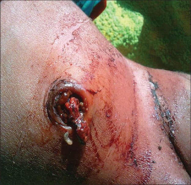
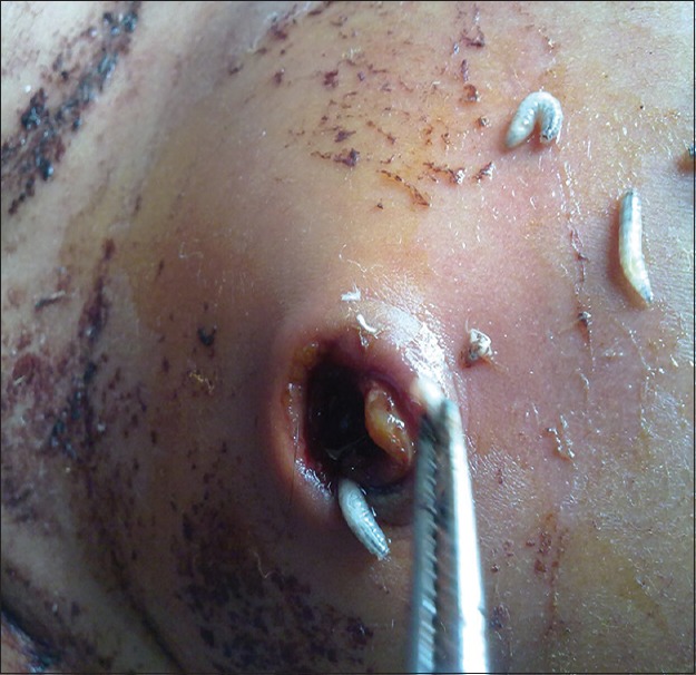
An 8-day-old 2.900 kg term male baby presented to us with umbilical discharge, mild periumbilical erythema and poor sucking. The baby was wrapped in a foul smelling dirty piece of cloth. On examination, reflexes were subnormal, although vital parameters were stable. Movement of some organisms was noted in the umbilical region of the baby [Figure 1]. The gangrenous cord was excised, and several maggots were removed with a forceps after local application of ether [Figure 2]. He was started on intravenous fluid and intravenous cloxacillin and amikacin empirically after obtaining blood samples. The sepsis screen was found to be positive although blood culture yielded no growth. Umbilical discharge revealed growth of Staphylococcus aureus. The patient improved over the next three days and the umbilicus became dry. He was discharged after 10 days of antimicrobial therapy.
Figure 1.

Maggot crawling out of a gangrenous umbilical area
Figure 2.

Maggots being removed after local instillation of ether
Myiasis is a rare form of infestation by larvae of the dipterous fly, found mainly in tropical and subtropical regions where basic sanitation is lacking. It is defined as an animal or human disease caused by parasitic dipterous fly larvae feeding on the host's necrotic or living tissue. Although infestation by fly larvae is much more prevalent in animals, it is a relatively frequent occurrence among humans in rural, tropical and subtropical regions. Poor hygienic condition and low socioeconomic status are important predisposing factors for the development of myiasis. Warm and moist nature of umbilical stump may attract gravid female flies to lay eggs on it if a neonate is kept in an unhygienic and filthy environment. The accompanying omphalitis provides the required nutritive support for hatching of eggs and further development into the larval form.
Myiasis of the neonatal umbilicus is a rare disease with only a few reported cases in the literature.[1,2,3,4,5] Other sites reported among neonates are ear, nasopharynx, periorbital region, vagina, skin and intestine.[6,7] Treatment of myiasis consists of removal of the larvae and control of local and systemic infection, if any. The larvae can be killed by suffocating them with ether. Subsequently they can be removed from the affected site of the host by irrigation, manipulation or surgery as any dead or decaying deep seated larvae can cause secondary infection or sepsis.
Footnotes
Source of Support: Nil
Conflict of Interest: None declared.
REFERENCES
- 1.Duro EA, Mariluis JC, Mulieri PR. Umbilical myiasis in a human newborn. J Perinatol. 2007;27:250–1. doi: 10.1038/sj.jp.7211654. [DOI] [PubMed] [Google Scholar]
- 2.Ghosh T, Nayek K, Ghosh N, Ghosh MK. Umbilical myiasis in newborn. Indian Pediatr. 2011;48:321–3. [PubMed] [Google Scholar]
- 3.Patra S, Purkait R, Basu R, Konar MC, Sarkar D. Umbilical myiasis associated with Staphylococcus aureus sepsis in a neonate. J Clin Neonatol. 2012;1:42–3. doi: 10.4103/2249-4847.92229. [DOI] [PMC free article] [PubMed] [Google Scholar]
- 4.Ambey R, Singh A. Umbilical myiasis in a healthy newborn. Paediatr Int Child Health. 2012;32:56–7. doi: 10.1179/1465328111Y.0000000043. [DOI] [PubMed] [Google Scholar]
- 5.Kumar V, Gupta SM. Umbilical myiasis in a neonate. Paediatr Int Child Health. 2012;32:58–9. doi: 10.1179/1465328111Y.0000000022. [DOI] [PubMed] [Google Scholar]
- 6.Koh TH. Neonatal myiasis: A case report and a role of the Internet. J Perinatol. 1999;19:528–9. doi: 10.1038/sj.jp.7200211. [DOI] [PubMed] [Google Scholar]
- 7.Shekhawat PS, Joshi KR, Shekhawat R. Contaminated milk powder and intestinal myiasis. Indian Pediatr. 1993;30:1138–9. [PubMed] [Google Scholar]


