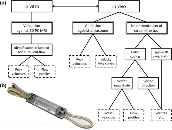
Fig. 1.

a Workflow diagram of the present study: first, validation of 4D PC MRI in vitro against slice-selective 2D PC MRI in a flow phantom, which enables the identification of laminar and turbulent flows inside the phantom from peak velocities and reconstructed flow profiles; second, validation of the method in vivo in mice against the gold-standard ultrasound in terms of peak flow velocity quantification and velocity–time curve reconstruction; third, implementation of a streamline analysis tool for high-quality visualization and improved analysis of flow behaviors. Color encoding for either vector magnitude or vector direction representation provides information regarding vessel velocities and flow profiles as well as structures in the blood flow such as vortices or helical streams. b Photograph of the flow phantom, comprising a water-filled acryl glass body with four parallel tubes (internal diameter: 3 mm), which are connected to each other in series and attached to an adjustable peristaltic pump
