Abstract
Laminins are important basement membrane (BM) components with crucial roles in development. The numbers of laminin isoforms in various organisms are determined by the composition of the different α, β, and γ chains, and their coding genes, which are variable across spieces. In insects, only two α, one β, and one γ chains have been identified thus far. Here, we isolated a novel laminin β gene, BmLanB1-w, by positional cloning of the mutant (crayfish, cf) with blistered wings in silkworm. Gene structure analysis showed that a 2 bp deletion of the BmLanB1-w gene in the cf mutant caused a frame-shift in the open reading frame (ORF) and generated a premature stop codon. Knockdown of the BmLanB1-w gene produced individuals exhibiting blistered wings, indicating that this laminin gene was required for cell adhesion during wing development. We also identified laminin homologs in different species and showed that two copies of β laminin likely originated in Lepidoptera during evolution. Furthermore, phylogenetic and gene expression analyses of silkworm laminin genes revealed that the BmLanB1-w gene is newly evolved, and is required for wing-specific cell adhesion. This is the first report showing the tissue specific distribution and functional differentiation of β laminin in insects.
Laminins, a family of heterotrimeric glycoproteins, are the major biologically active components in basement membranes (BMs) and effectors of tissue architecture1,2. They are made of three different subunits, α, β, and γ chains, which assemble into a cruciform structure with three short arms and one long arm2. The short arms are associated with the polymerization of the molecule and the long arm is capable of binding to cellular receptors3. Thus, the laminins interact with other extracellular proteins and adhere to cells via receptors, such as integrins, dystroglycan, heparan sulfates, and sulfated glycolipids, thereby promoting adhesion, motility, proliferation, survival and differentiation of various cell types4,5,6.
Laminins are highly conserved across evolution, although the numbers of laminin isoforms and their coding genes are variable across spieces. In lower organisms, such as Hydra, there is only one laminin isoform, which is composed of one α, one β, and one γ gene7. Four laminin genes have been identified in protosomia (nematodes and insects) thus far. For example, it was discovered that the Drosophila genome contains two α genes, wing blister (wb, α1,2) and laminin A (LanA, α3,5), one β gene, laminin B1 (LanB1), and one γ gene, laminin B2 (LanB2).The four laminin genes are capable of assembling into two different heterotrimers. In complex organisms such as mammals, there are five α, four β, and three γ chains that give rise to at least 16 different isoforms8,9. It is presumed that the increase in the number of laminin genes through evolution could have occurred through a series of gene duplications and modifications.
Laminin isoforms differ in their composition of α, β, γ chains and may have cell- and tissue-specific distribution that reflects diverse biological functions. For example, in mammals, laminin 1, containing α1, β1, and γ1, has an essential function during early embryogenesis10, while laminin 2, made of α2, β1, and γ1 chains, is mainly expressed in muscle cells11. However, the laminin isoform composed of α3, β2, and γ3 chains, is essential for the CNS synaptic organization12.
Studies have suggested that the differential temporal and spatial distribution of laminins is mainly determined by variations in the expression of the α chain9. However, other studies have disproved this theory. In higher and lower species, absence or mutation in any laminin chain has resulted in embryonic lethality or severe disease condition that affects organ morphogenesis7,13,14. For instance, lack or partial loss of laminin α2 led to variation in skeletal muscle fibers and muscle fiber necrosis in mice and humans15,16. Mice with a mutation in laminin β1 or γ1 chain lack embryonic BMs and cannot survive past the late embryonic stage (day E5.5). In Drosophila melanogaster, subtle change in the wb gene causes a mild phenotype well known for wing blister, in which the dorsal and ventral wing surfaces separate. However, total lack of the gene results in early embryonic lethality17. LanA deficient mutants exhibit embryonic lethality with defects in the morphogenesis of heart, trachea and somatic muscle18. Besides, the elimination of LanB1 (β) prevents the normal morphogenesis of most organs and tissues, including the gut, trachea, muscles and nervous system, thereby resulting in mortality at the end of embryogenesis19. Thus, these evidences strongly support the notion that the spatio-temporal differentiation of laminins is due to all three chains.
In this study, we identified a new laminin β subunit gene (named BmLanB1-w) in silkworm by cloning crayfish (cf), a spontaneous recessive mutation with blister wing in homozygous mutants and no visible defect in any other tissues or stages. Previous studies have shown that in insecta, only one laminin β subunit is present, while we identified two β subunit genes in the silkworm genome. BmLanB1-w RNAi showed that the gene was essential for the interaction between the two chitinous wing layers in silk moths. Furthermore, the gene expression profile of silkworm laminin genes and phylogenetic analysis revealed that BmLanB1-w gene is a new member of the insect laminin β gene family and that it is abundantly expressed in the wing tissue.
Results
Phenotype analysis
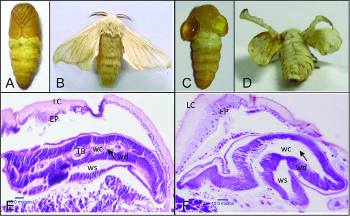
The phenotypes of cf mutant (Fig. 1C,D) and wild-type (WT) (Fig. 1A,B) individuals were recorded using a digital camera (Canon EOS 5D Mark III). Wings of the cf mutant pupae are similar to the two large chelipeds in crayfish. Homozygous mutations of the cf locus led to one or two pairs of blister wings in the pupae and adult moths (Fig. 1C,D). The blistered pupal wings are filled with hemolymph between the dorsal and ventral layers. They are extremely fragile and can be easily wounded by slightly wobbling or touching, and result in bleeding to death.
Figure 1. Phenotype of wild-type (Dazao) and cf mutant strain.
Pupal and moth stages of wild-type (A,B) and cf (C,D) are shown. Hematoxylin–eosin stained paraffin sections of silkworm wing discs in late larval stage (day 6 of 5th instar) of wild-type (E) and cf (F) are also shown. LC: larval cuticle, EP: epidermis, wc: wing cavity, ws: wing sac, wd: wing disc, TB: trachea tube.
To analyze how the wing discs of cf are different from the WT, the wing discs of both strains were dissected every day after the last larval molt. Visible differences were not observed in size and shape of the wing discs between the two strains. Further analysis of the inner structure of wing discs using paraffin sections revealed that in the WT the wing bilayer was attached all the time, and the trachea was located on the site of attachment (Fig. 1E). However, in the cf mutants the two wing disc layers failed to adhere to each other from late larval stage (day 5–6 in 5th instar) (Fig. 1F). These results indicate that the cf defect occurred before larval-pupal metamorphosis, and that the phenotype was due to the separation of dorsal and ventral wing surfaces.
Positional cloning of cf locus in Bombyx mori
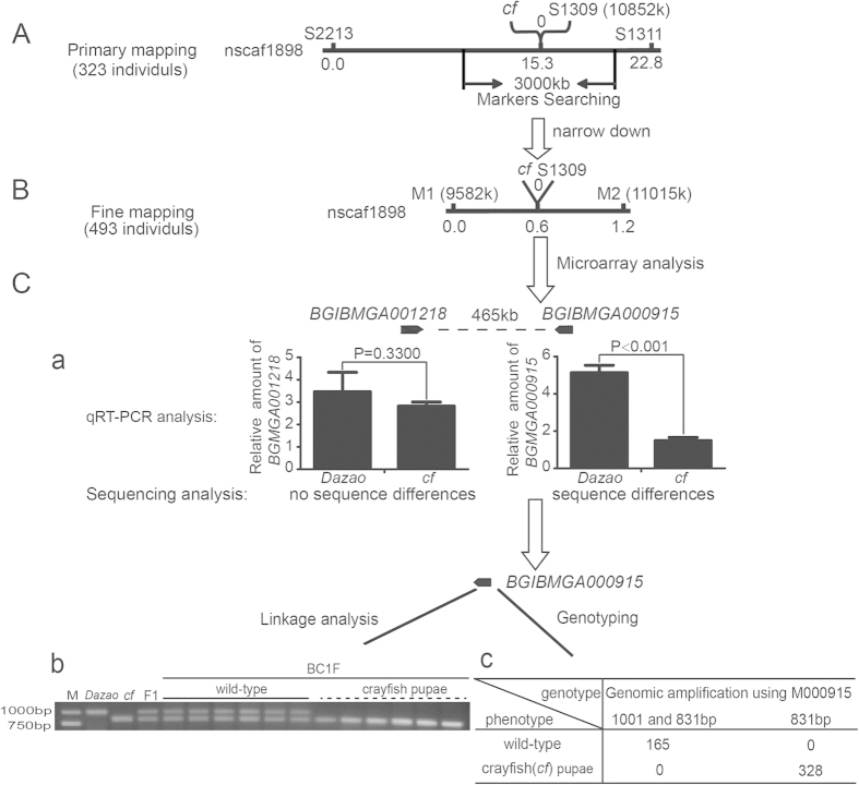
To identify the candidate gene responsible for the cf phenotype, we performed primary mapping of the 323 BC1M progeny (a cross between cf ♀ × (Dazao × cf) ♂) using the SSR markers20. First, we roughly mapped the cf locus between the markers, S2213 and S1311, within scaffold nscaf1898 on the 13th chromosome. The distances from cf locus to these markers were 15.3 cM and 7.5 cM, respectively, and we found that marker S1309 was closely linked to the cf locus (Fig. 2A). We then designed new primer sets at 1500 kb upstream and downstream of S1309 flanking sequences based on the silkworm genome database. Genotyping using 493 BC1M individuals, further delimited the cf locus between markers M1 and M2, and the marker S1309 was still tightly linked to the cf locus (Fig. 2B). As a result, 63 genes were predicted to be present within this region (1433 kb).
Figure 2. Mapping, bioinformatics and expression profile analysis of the cf locus.
(A) Preliminary mapping of the cf locus. The cf locus was mapped between markers S2213 and S1311. The marker S1309 was tightly linked with the cf locus. Marker search in the genome was carried out within a 1500 kb sequence length both upstream and downstream of S1309. (B) Fine mapping of the cf locus. The cf locus was mapped between M1 and M2 using 493 BC1M individuals. S1309 was still tightly linked with the cf locus. (C) Screening the candidate gene and genotyping using polymorphism markers. Microarray analysis showed that there were only two genes (BGIBMGA001218 and BGIBMGA000915) separated by a distance of 465 kb in the mapped region with different expression levels in the wild-type and the cf mutant. (a) qRT-PCR analysis of BGIBMGA001218 revealed no significant difference in gene expression between the cf mutant and wild-type (The P value equals 0.3300, Student’s t-test. N = 3). Gene BGIBMGA000915 within the mapped region was expressed much lower in cf than in wild-type based on both microarray and qRT-PCR analysis. Sequencing analysis of the two genes revealed that BGIBMGA001218 was not different between the cf mutant and wild-type, but BGIBMGA000915 differed. (b) Linkage analysis of the polymorphism marker M000915, which is 450 bp downstream of BGIBMGA000915. F1: first filial generation. BC1F: first backcross generation for linkage analysis. (c) Genotyping of the M000915 marker and cf locus using 493 BC1M (first backcross generation for recombination analysis) individuals. There is no recombination between M000915 and the cf locus. The results indicate that M000915 was also tightly linked with the cf locus.
To help identify the candidate gene for the cf phenotype, microarray analysis was performed using the wing discs of Dazao (WT) and cf at W0 stage (<5 hours after the start of larval wandering). The result showed that the expression levels of the two genes, BGIBMGA001218 and BGIBMGA000915, in the narrowed region exhibited different patterns in the WT and the cf mutant (supplementary dataset). Using BLASTX search, we further found that BGIBMGA001218 (nscaf1898:10187163…10194437) encoded a heat shock cognate protein, while, BGIBMGA000915 (nscaf1898:10659797…10665195) encoded a laminin protein. To verify the microarray data, we performed qRT-PCR analysis on the genes within the mapped region. The results showed that only BGIBMGA000915 had a significant change in expression level in the mutant wings (supplementary dataset). The expression levels of BGIBMGA001218 varied considerably among individuals, and was not significantly different between the cf mutant and WT (The P value equals 0.33002) (Fig. 2C–a), Further sequences analysis of BGIBMGA001218 revealed no sequence divergence between the cf mutant and WT silkworm. In Drosophila, the laminin homolog was reported to be essential for wing development, and a defect in laminin resulted in blistered wings17,21,22. Consistent with this finding in Drosophila, BGIBMGA000915 was expressed at extremely lower level in the wing discs of the cf mutant when compared to the WT (Fig. 2C–a). Furthermore, genotyping revealed that another polymorphism marker, M000915, located at 450 bp downstream of the BGIBMGA000915 termination codon (Fig. 3A) was tightly linked to the cf locus (Fig. 2C–b,c). Thus, we found that the BGIBMGA000915 gene was located between these two closely linked markers, corresponding to 10852 k (S1309) and 10658 k (M000915), respectively in the nasca1898 scaffold. These results suggest that BGIBMGA000915 was more likely the candidate gene for the cf phenotype. Based on the homologs in other organisms, we named it BmLanB1-w.
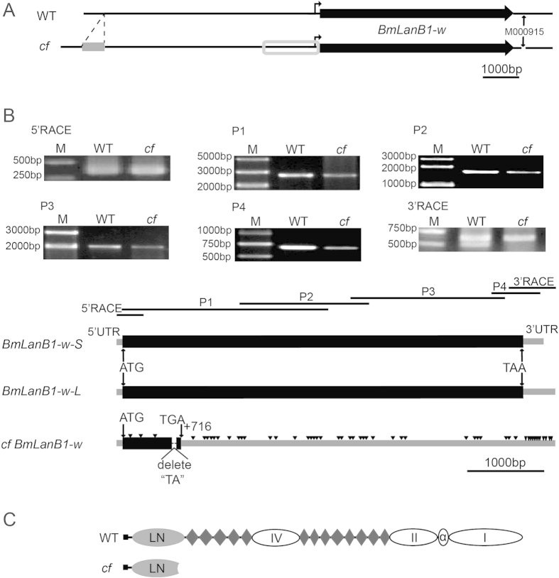
Figure 3. Schematic diagram of BmLanB1-w in wild-type (Dazao) and cf mutant.
(A) Differences in the upstream and downstream sequences of BmLanB1-w between Dazao and cf. Gray rectangle indicates a non-LTR retrotransposon located 5.9 kb upstream of BmLanB1-w in the cf mutant. Gray box indicates the region upstream of BmLanB1-w in cf with sequences differing from Dazao. M000915 is a polymorphism marker 450 bp downstream of the BGIBMGA000915 gene. Bold arrows indicate the location and transcriptional orientation of BmLanB1-w. (B) Cloning and characterization of the BmLanB1-w transcripts in Dazao and cf strains. We cloned the full-length cDNA sequences of the BmLanB1-w in Dazao and cf by RT-PCR and RACE techniques, using six primers (5′RACE, P1, P2, P3, P4, 3′RACE). The black horizontal lines indicate the location of the cloned fragments in BmLanB1-w, respectively. There are two types of transcripts (BmLanB1-w-L and BmLanB1-w-S) in Dazao, but only one type in cf. Black rectangles and gray bars indicate coding and untranslated regions of the BmLanB1-w exon, respectively. A 2 bp (“TA”) deletion was found in BmLanB1-w in the cf strain, and this deletion introduced a premature stop codon (TGA). Downward-pointing triangles represent nucleotide substitution in the BmLanB1-w in the cf strain. The scale bar corresponds to 1000 bp. (C) Predicted protein structure of BmLanB1-w. Gray oval represents LanB1 N-terminal domain, diamond represents laminin-type EGF-like domains, black rectangle represents signal peptide, and white oval represents other conserved domains.
Gene structure and expression analysis of the candidate gene
To clarify the structural features of the candidate gene, we cloned the full-length cDNA sequences of the BmLanB1-w in WT and cf by RT-PCR and RACE (rapid-amplification of cDNA ends) (Fig. 3A,B). Two types of transcripts, BmLanB1-w-L (5709 bp, Accession number: KP057212) and BmLanB1-w-S (5587 bp, Accession number: KP057213), were amplified from the wing discs of WT (Fig. 3B). Both had only one exon, which yielded the same BmLanB1-w protein (1779 amino acids) with a typical LanB1 N-terminal domain and 13 laminin-type EGF-like domains (Fig. 3C). However, only one BmLanB1-w (5707 bp) was detected in the cf mutant (Accession number: KP057211). We also examined the sequences upstream (~6 kb) of the transcription start site and found a non-LTR retrotransposon (560 bp) located 5.9 kb upstream of BmLanB1-w in the cf mutant (Fig. 3A, gray rectangle). In addition, there were a number of single nucleotide substitutions and short fragment insertions or deletions in the vicinity of the transcription start site (Fig. 3A, gray box) in the mutant. Compared to the BmLanB1-w-L coding sequence in the WT, BmLanB1-w in the cf mutant had 52 single nucleotide substitutions, and a 2 bp deletion, causing a frame-shift in the ORF and generating a premature stop codon (TGA) (Fig. 3B). The cf-type protein was predicted to contain a defective LanB1 N-terminal domain and lack all EGF-like domains (Fig. 3C). Multiple sequence alignments also suggested that the mutated protein most likely lacked the function of the WT protein.
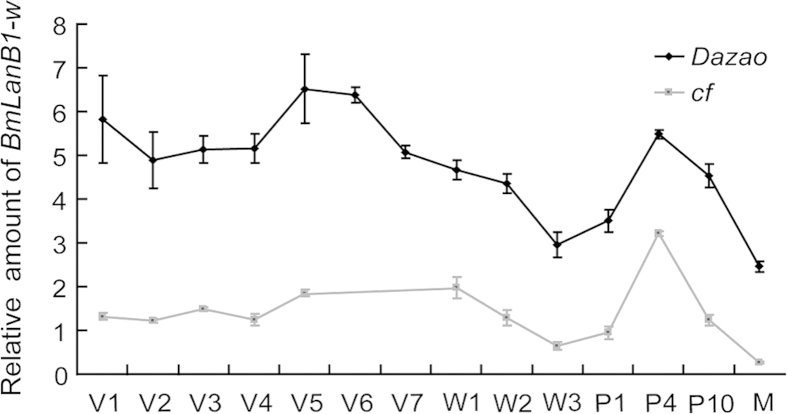
Next, we compared the expression profiles of BmLanB1-w in the WT and cf mutant during wing morphogenesis by quantitative RT-PCR (qRT-PCR). In WT, the BmLanB1-w gene was expressed in the wing discs during all stages analyzed, from the 1st day of the 5th instar larval stage to the newly emerged moth. We observed two peaks; one at the end of the larval stage and the second at the mid-pupal stage, respectively. In contrast, expression of the BmLanB1-w gene had a fluctuating pattern that was similar between the cf mutant and the WT but the expression level was significantly lower in the cf mutant when compared to the WT (Fig. 4). Taken together, the predicted structural abnormality and the decreased expression level of BmLanB1-w in cf mutant strongly suggest that the BmLanB1-w gene was responsible for the cf phenotype.
Figure 4. Temporal expression patterns of BmLanB1-w in Dazao and cf mutant.
Quantitative RT-PCR analysis of BmLanB1-w was performed in wing discs and wings of wild-type (Dazao) and cf at different developmental stages. The BmLanB1-w gene expression had similar fluctuating patterns in both strains but was expressed at a significantly lower level in the cf mutant compared to the wild-type. All data are mean ± S.D (n = 3). V1-V7 represent 1st day to 7th of 5th instar, respectively; W1-W3 represent day 1-day 3 of the wandering and spinning stages, respectively; P1, P4, P10 represent day 1, day 4, and day 10 of the pupae, respectively; M represents 1st day after eclosion. In wild-type (Dazao) the 5th instar lasts 7 days, while in cf it is only 5 days.
RNAi of BmLanB1-w
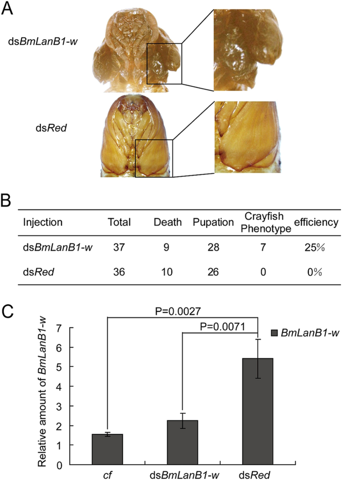
To verify whether the deficiency of BmLanB1-w could cause the blistered wing phenotype, we performed RNAi experiments to knockdown the expression of the gene in WT. We injected BmLanB1-w dsRNA (dsBmLanB1-w) twice into each 5th instar larvae. The first injection was carried out at the beginning of wandering stage, and the second injection was performed 1 day later. The dsRNA of red fluorescent protein (dsRed) was used as a negative control. After injection, many larvae did not complete the larval-to-pupal metamorphosis but died because the bouble injections induced intractable wounds in the wandering larvae. The results obtained from the surviving larvae showed that similar to the cf mutant, 25% (7 of 28) of the WT individuals injected with dsBmLanB1-w developed wings with blisters after pupation (Fig. 5A,B). In contrast, there was no effect on the wing shape after injecting dsRed (n = 26). We then performed real-time PCR to investigate the expression level of BmLanB1-w after dsRNA injections. Compared to the negative control (dsRed injection), the level of BmLanB1-w transcription in the dsBmLanB1-w treated group was significantly lower and was as low as in the cf mutant (Fig. 5C). These results suggest that the blistered wing of cf is a result of BmLanB1-w loss of function.
Figure 5. RNAi of BmLanB1-w.
(A) Phenotypes of BmLanB1-w RNAi: wings with blisters were similar to wings in the cf mutant. DsRed control did not show cf phenotype. (B) RNAi statistics. (C) Analysis of BmLanB1-w transcripts. All data are mean ± S.D (n = 3). Statistical analyses were performed using Student’s t-test (n = 3).
Laminin gene homologs in silkworm and phylogenetic analysis
As an essential regulator of morphogenesis of most organs, laminins require the simultaneous expression of all three chains and their assembly into α-β-γ heterotrimers to perform their roles in cell adhesion, migration, and rearrangement. In Drosophila, the elimination of LanB1 prevents the normal morphogenesis of most organs and tissues, including the gut, trachea, muscles, and nervous system19. However, in the silkworm cf mutant, the BmLanB1-w defect did not affect any other tissue morphogenesis except the wings.
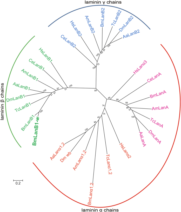
In order to investigate whether another BmLanB1 gene in silkworm genome could compensate for the absence of the BmLanB1-w transcript, we identified all the laminin genes in silkworm by searching the conserved motifs in entire silkworm genome. We performed BLASTP and TBLASTN searches against the predicted silkworm gene in the Silkworm Genome Database, SilkDB (http://www.silkdb.org/silkdb/)23. We identified five laminin genes in the silkworm genome. To deduce the function and evolution of these laminin genes, we performed phylogenetic analysis of laminin proteins from different species by the Maximum likelihood method using MEGA624. These laminin genes were also identified in the genomes of C. elegans, H. sapiens and four other insects including D. melanogaster, A. aegypti, A. mellifera, and T. castaneum by performing BLASTP and TBLASTN searches. The results were consistent with the laminin proteins in GenBank (http://www.ncbi.nih.gov/GenBank/). Therefore, we downloaded the amino acid sequences of the laminin subunits of the five insect species from GenBank for phylogenetic analysis. The resulting phylogenetic tree showed the division of laminins into three distinct clusters: laminin α chains, laminin β chains, laminin γ chains (Fig. 6). Based on sequence similarity, the five silkworm laminin genes were defined as two laminin α genes (BmLanA, BmLanα1, 2), two laminin β genes (BmLanB1-w, BmLanB1), and one laminin γ gene (BmLanB2). It is noteworthy to mention that BmLanB1-w and BmLanB1 shared 82.7% amino acid sequence identity and were grouped into one sub-clade, paralleling the other β chain genes in the subgroup. This indicates that the two laminin β genes were indeed present in the silkworm. To date, several laminin β paralogs have been reported only in the genome of Deuterostomia, and no additional β gene has been identified in lower Protostomia, which includes nematodes and insects. To investigate whether the duplication of laminin β also occurred in other lepidopteran species, we searched for the laminin genes in three other insect genomes, P. xylostella, H. melpomene, and D. plexippus, and compared the numbers of laminin genes in lepidopterans and other species (Fig. S1). As shown in the Fig. S1, the numbers of laminins are different among the four lepidoptera insects. Plutella xylostella has one additional α chain while silkworm has one extra β chain, suggesting that novel laminins originated independently in lepidopterans during evolution.
Figure 6. Phylogenetic analysis of laminins in various species.
MEGA 6.024 was used to construct the phylogenetic tree using the Maximum likelihood method. Numbers in the cladogram indicate bootstrap values. Abbreviations, Bm: Bombyx mori, Dm: Drosophila melanogaster, Am: Apis mellifera, Aa: Aedes aegypti, Tc: Tribolium castaneum, Hs: Homo sapiens, Ce: Caenorhabditis elegans. Accession numbers are listed in Table S2.
The high sequence similarity of two silkworm β subunits implied that they were generated through gene duplication. In order to examine the gene duplication events that generated new insect laminin genes, we used molecular phylogenetic analysis and compared gene contents between different species (Fig. S2). The phylogenetic tree was divided into two distinct orthologous groups; insects and vertebrates, respectively. In the insect clade, the two silkworm β genes formed a monophyletic subgroup and then clustered with each homolog in other insects. This suggested that the two silkworm laminin β genes were likely formed by the duplication within insects and could share a conserved function due to the high sequence similarity.
Together, these results support our hypothesis that in the cf mutant, another BmLanB1 chain probably compensated for the absence of BmLanB1-w chain in other organs and tissues except the wings.
Spatial expression profile of silkworm laminin genes
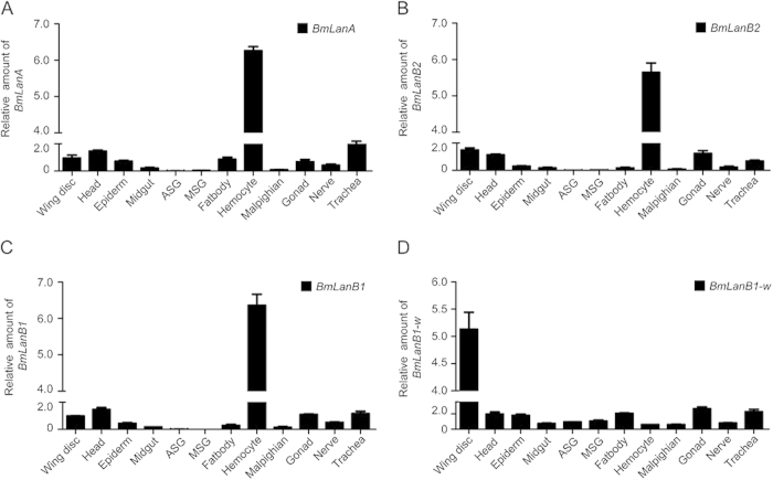
Since two laminin β genes exist in the silkworm genome, we investigated their expression profiles in various tissues by qRT-PCR. Total RNA were extracted from 12 different tissues from day 3 of 5th instar larvae. These included the wing disc, head, epidermis, midgut, fat body, hemocyte, malpighian tubule, gonad, trachea, anterior silk gland (ASG), middle silk gland (MSG), and nerve. The BmLanB1 gene was expressed robustly in the hemocyte and at much lower levels in the wing disc, head, trachea, and other organs, which was consistent with the expression pattern of two other laminin subunits, laminin α gene (BmLanA) and laminin γ gene (BmLanB2). In contrast, the BmLanB1-w gene was transcribed much strongly in the wing discs and weakly in other tissues (Fig. 7). This comparison revealed the wing-specific abundance of BmLanB1-w and its importance in wing morphogenesis. Moreover, the co-expression of BmLanB1, BmLanA, and BmLanB2 were likely sufficient to form α-β-γ heterotrimers in other tissues.
Figure 7. Spatial expression patterns of laminin genes in wild-type (Dazao).
Quantitative RT-PCR analysis of laminin genes in 12 tissues in the 3rd day of 5th instar. The BmLanB1-w gene had robust expression in the wing discs and weak expression in other tissues. In contrast, the BmLanB1 gene was expressed strongly in the hemocyte and weakly in wing discs and other organs, which was consistent with the expression patterns of laminin α (BmLanA) and laminin γ (BmLanB2) genes. Abbreviations, ASG: anterior silk glands, MSG: middle silk glands, Malpighian: malpighian tubule. The data indicate the mean ± S.D (n = 3).
Discussion
BmLanB1-w is responsible for cf mutant
To identify the gene responsible for the cf mutant of silkworm, genetic mapping and microarray analysis were performed. The results showed that two genes, one heat shock cognate protein (Hsc70) gene (BGIBMGA001218) and one laminin beta gene (BGIBMGA000915), of the mapped region changed their expression levels in the cf mutant wings. In Drosophila, several heat shock proteins are involved in wing sheet adhesion. For example, Hsp83 RNAi was implicated in wing blistering, causing “swarovski” or “stump” wings25. The BGIBMGA001218 gene is a homologue of a Hsc70 gene (FlyBase ID: FBgn0026418), and down-regulation of Hsc70-4 didn’t cause wing blisters in D. melanogaster25. The qRT-PCR analysis verified that only the BGIBMGA000915 gene, but not the Hsc70 gene, significantly change its expression level in the mutant wings. The inaccurate description of the Hsc70 gene expression in the microarray analysis is because only one set of comparable samples were used, which didn’t detect the expression variation of this gene among individuals. Considering no significant differences in expression level and sequence of the heat shock cognate protein gene between cf mutant and WT, and no similar phenotype in Drosophila when its homologue was knocked down, we presumed that the “heat shock cognate protein” gene (BGIBMGA001218) might be not responsible for the cf mutant.
Then, we focused on the laminin beta gene, BmLanB1-w, of which homolog in Drosophila was essential for wing sheet adhesion. By cloning and comparing the sequences of the BmLanB1-w in WT and cf, a whole lot of aberrations in this gene were identified in the cf mutant, including nucleotide substitutions, frame shift, and retrotransposon insertion. Numerous aberrations in the related mutant gene is comprehensible, because the crayfish (cf) is a spontaneous mutant strain. Furthermore, it was expressed at extremely lower level in the wing discs of the cf mutant compared to WT, which might be caused by the non-LTR retrotransposon located in upstream of BmLanB1-w in cf, or by nonsense-mediated decay (NMD) of mRNA containing a premature stop codon as previous reports26,27,28,29, or by these two effects or some other reasons. RNAi experiments demonstrated that the deficiency of BmLanB1-w induced the blistered wing phenotype, suggesting that the BmLanB1-w is responsible for the cf mutant. This is the first report of a novel laminin β gene involved in wing-specific cell adhesion in silkworm, Bombyx mori.
New laminin subunit in Insecta
Laminins are a large family of conserved proteins with crucial roles in development, adhesion and cell migration30. A number of studies have reported that the numbers of the laminin subunit genes increased from lower organisms (1 α, 1 β, and 1 γ chains) to higher organisms (5 α, 4 β, and 3 γ chains)7,8,9. However, only four laminin genes have been identified in insects thus far, including two α, one β, and one γ chains. In recent years, whole genome sequences from a variety of animal species have become available to identify laminins homologs in these species to better understand the evolution of laminins. Interestingly, we found that new laminin genes had emerged in lepidoptera insects. In the present study, a novel α chain gene and a novel β chain gene were identified in P. xylostella and B. mori, respectively. Phylogenetic analysis demonstrated that the laminin components in all lineages consistently had three subuints, suggesting that α, β and γ chains were the ancestral forms of laminin. The different subunits also have distinct functions, which are conserved during evolution. Lastly, the three ancestral laminin chains appear to have undergone multiple bifurcations during evolution to generate several paralogs by gene duplication. Taking the β chains as an example, the phylogenetic tree was divided into a vertebrate branch and an insect branch. In vertebrates, the β gene number increased to 3–6 copies. Lanβ1 and lanβ2 were clustered more closely in the phylogenetic tree, while, lanβ3 and lanβ4 belonged to another sub-group. Furthermore, evolutionary analysis suggested that lanβ1 and lanβ2 were generated by gene duplication before species divergence after which lanβ1 had duplicated again to yield lanβ1a and lanβ1b. Lanβ1a was absent in humans likely because of the loss of some chains as reported previously31. In contrast, lanβ3 and lanβ4 emerged by duplication after species formation. However, the two silkworm laminin β genes may have evolved from the same ancestral sequence by gene duplication during evolution of Lepidotera, probably within the silk moth lineage. Future sequencing of intermediate species may confirm such evolution. These results collectively indicate that laminin genes had undergone multiple independent duplications in evolutionary history. The duplication of laminin β during evolution of insects was independent of its duplication in vertebrates, such as D. rerio and H. sapiens. In summary, our data indicates that new laminin subunits evolved in insecta, likely due to an increased rate of evolution in this taxon, and the laminin β gene duplications occurred recently during evolution.
Duplicated laminin β genes preserve original ancestral function
After gene duplications, the possible functions of the duplicated genes include conservation, neofunctionalization, subfunctionalization, and specialization32,33. In the present study, sequence analysis suggested that the two laminin β genes, BmLanB1 and BmLanB1-w, were generated by gene duplication. This raised the question whether there is any functional diversity between the two genes.
BmLanB1-w gene was wing-specifically abundant during wing development. Its expression profile in the wing discs presented two peaks; one at the end of larval stage and another in the mid-pupal stage, respectively. The former expression peak may contribute to the elongation of wing discs and the association between the wing discs and the tracheal system. The later peak occurred simultaneously during vein formation, which suggests that it could improve vein development. This hypothesis was supported by observations in Drosophila that LanB1 was expressed at the basal surface of veins from 32 to 44 h after pupation19. Furthermore, loss-of-function of the BmLanB1-w induced blistered wings, which demonstrated that BmLanB1-w was essential for cell adhesion between the dorsal and ventral wing surfaces. In Drosophila, flies carrying wing cell mutation for LanB1 showed a strong wing blister phenotype consistent with the mutations disrupting the laminin α1,2 (wb) or laminin α3,5 chains (LanA)17,21,34. These similarities suggest that the BmLanB1-w gene had a conserved role in cell adhesion throughout evolution.
The next question was why are two laminin β genes required for silkworms? Deletion of Drosophila LanB1 caused late embryonic lethality and defects in most organs and tissue morphogenesis19. In contrast, we found that BmLanB1-w was not required for the silkworm embryo and other tissues during development because no obvious defects were observed in the cf mutant or in individuals after BmLanB1-w knockdown. The spatial expression profile of different laminin genes in silkworm revealed that the expression pattern of BmLanB1 was consistent with α and γ subunits in other tissues. Although BmLanB1-w was also detected in other organs, its expression level was not comparable with that in BmLanB1 (data not shown). Therefore, it appears that BmLanB1 is capable of compensating for the absence of BmLanB1-w transcript in other tissues but not in wings. In other words, the BmLanB1 is required to fulfill the original function in border cells and tissues. Further, the co-expression of BmLanB1 with other chains suggested that BmLanB1 might be the ancestral copy, while the wing-specific abundance and the effect of BmLanB1-w gene suggested that it could be the newly generated copy by gene duplication. From these observations, we presume that the ancestral silkworm laminin β functions had undergone tissue differentiation. The newly evolved BmLanB1-w was necessary for the wing-specific cell adhesion, and both BmLanB1 and BmLanB1-w were required to preserve all ancestral functions.
Tissue-specific cell adhesion of laminins
The expression patterns of laminin isoforms are often tissue- and temporal-specific during development35. However, the underlying molecular mechanisms regulating their expression still remain unknown. Many previous studies suggested that the differential expression is mainly (but not exclusively) determined by variations in the expression of α chains9. For example, in mammals, the α1 chain is highly expressed in the epithelia of early embryos, and in adult reproductive organs including kidney and liver36. The α2 chain is mostly localized in the neuromuscular system37. The α3 chain is mainly found in the skin and other epithelia38,39. The reason for the tissue specific expression of α is presumed to be due to the large size of this chain, which contains the major cell-adhesive sites. For example, its long arm on the C-terminal end consists the LG1–5 domains, which are involved in the interactions with cellular receptors such as integrins and dystroglycans. The N-terminal end of the short arm is also capable of binding to integrin receptors40. Here, we demonstrated that the tissue-specificity of laminins could also be determined by the β chain, which contains two C-terminal globular domains that may also link the laminin molecules to the cell surface through direct or indirect interactions with cellular receptors, such as integrin, collagen, and sulfated glycolipids. This hypothesis is supported by reports that the β3 chain of laminin 5 binds to collagen VII41, and that the β2 chain could modulate integrin binding affinities of laminins42. An alternative explanation is that some cis-regulatory elements of laminin gene could regulate the spatiotemporal expression.
Mechanism leading to cf mutation
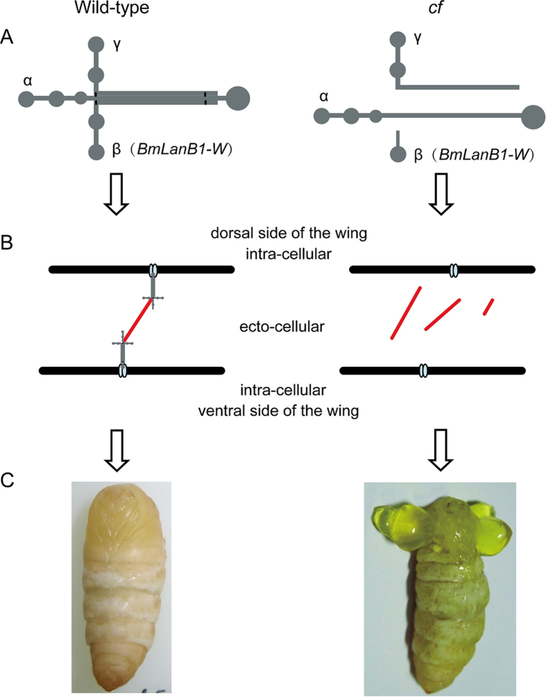
Based on our findings we predict the following mechanism that leads to cf mutation. The wing is composed of two epithelial sheets that adhere tightly to each other. Laminin is the secreted protein localized in the extracellular basal membrane of the epidermis43. In the wild type imaginal wing discs, the β chain encoded by BmLanB1-w gene is assembled with α and γ chains to form the heterotrimeric laminin. Then, heterotimers are self-assembled into a cell-associated network through the ternary nodes, which is formed by the common domain structure in the short arms of each chain. Finally, polymerized laminins bind to the cell surface through interactions with cellular receptors, such as collagen and integrin, making dorsal and ventral sides of wings attached together thus enabling normal wings to withstand the pressure of the hemolymph (Fig. 8). However, in the cf mutant, ablation of the BmLanB1-w chain in wings prevents laminin heterotrimer formation and basement membrane assembly, causing a failure in adhesion between the two layers of the wing. The wings are filled with hemolymph between the two sheets, eventually forming the cf blistered wings phenotype (Fig. 8).
Figure 8. Schematic representation of the mechanism leading to cf phenotype.
(A) Laminin structure in wild-type and cf mutant. In the cf mutant, laminin trimer cannot form due to the defect in BmLanB1-w. (B) Model diagram depicting the adhesion of dorsal and ventral sides of the wings in wild-type and cf. In the cf mutant, dorsal and ventral sides of the wings are not linked because normal basement membrane cannot form due to dysfunctional laminin trimer, which does not allow wings to withstand the hemolymph pressure thus resulting in wing blisters. Light blue oval represents integrins, gray pattern represents laminin trimer, red line indicates proteins such as collagen IV that are linked to the laminin trimer to enable basement membrane assembly. (C) Phenotypes of wild-type and cf.
In conclusion, our results demonstrated that loss-of-function of a novel laminin family gene, BmLanB1-w, is responsible for the cf phenotype with blistered wings in B. mori. We also showed the existence of two laminin β genes in silkworm. This is the first report of two laminin β subunits in insecta. The spatial expression profile analysis suggested that the two genes might perform sub-functions of the ancestral gene and may have diverged in a spatial pattern. BmLanB1 maintained the function in a broad range of tissues, while the new laminin gene was evolved to perform tissue specific functions in cellular adhesion during wing organogenesis. Further studies are needed to address how laminins can perform such different functions during development. The information derived from these studies should advance our understanding in the evolutionary history and functional differentiation of laminin genes.
Materials and Methods
Silkworm Strains
The crayfish mutant (cf) and the Dazao wild-type (WT) silkworm were provided by the Silkworm Gene Bank in Southwest University, China. The silkworms were reared on fresh mulberry leaves under a 12 h/12 h light/dark photoperiod at 25 °C during the experiments.
Paraffin section of silkworm wing discs
To detect the phenotype differences in the wing discs of WT (Dazao) and cf, the anterior and posterior wing discs were dissected from the two strains every day after the last larval molt. First, we cut the integument near the wing discs with a razor blade. Then, wing discs were extracted carefully, fixed in Bouin’s fixative at room temperature for 24 h, dehydrated first in a graded series of ethanol followed by a series of xylene, and eventually embedded in paraffin wax. A series of continuous sections (5 μm) were obtained using a LEICA RM2235 microtome (Germany) and stained with hematoxylin-eosin using standard protocols44. Subsequently, sections were observed through a Nikon C-DSS230 stereomicroscope (Japan).
Mapping of cf Locus
Two silkworm strains, Dazao (+cf/+cf) and cf (cf/cf), were selected as parents for genetic mapping. F1 offspring were obtained from the cross between female WT (Dazao) and male cf mutant. For linkage analysis, 20 BC1F progeny (10 WT and 10 mutant) from the cross (Dazao × cf) ♀ × cf♂ were used. For recombination analysis, the BC1M offspring of cf♀ × (Dazao × cf) ♂ were used. SSR markers from the published SSR molecular linkage map were used for preliminary mapping. Based on the preliminary mapping results, we analyzed sequences both upstream and downstream of the SSR marker that were tightly linked with the cf locus. New primers were designed based on the sequences adjacent to the predicted genes, and markers that exhibited polymorphism between parents were used to genotype the 493 BC1M progeny. For this analysis, we used the silkworm 9 × assembly genome database (http://www.silkdb.org/silkdb/), BLAST45, and primer 5.0. The primers used for mapping are listed in Table S1.
Microarray Analysis
Total RNA from the wing discs of Dazao and cf at W0 (<5 h after the start of larval wandering) were extracted using EZ.N.ATM Microelute Total RNA Kit (Omega). Microarray was performed by CapitalBio Corporation (Beijing, China). Microarray data analysis was performed as described previously46.
cDNA sequence cloning
Total RNA were extracted from the wing discs of Dazao and cf at W0. Primers were designed based on the sequences of the predicted genes (BGIBMGA000915 and BGIBMGA001218) from silkDB (http://www.silkdb.org/silkdb/). Full-length cDNA of the genes was obtained using GeneRacerTM Kit (Invitrogen) according to the manufacturer’s protocol. PCR products were cloned into the pMD19-T vector (Takara) for sequencing. Primers used for cloning are listed in Table S1.
Quantitative RT-PCR
ABI Prism7000 sequence detecting system (Applied Biosystems, Foster City, CA, USA) and SYBR Premix EX Taq-kit (Takara) were used for qRT-PCR to investigate the expression levels of the candidate gene in the wing discs at different developmental stages (from 1st day of 5th instar to 1st day of eclosion). Total RNA from the wing discs and wings were extracted using the EZ.N.ATM Microelute Total RNA Kit (Omega), and reverse transcribed into cDNA using oligo (dT) primer and M-MLV reverse transcriptase (Promega) according to the manufacturer’s protocol. The analysis of spatial gene expression profile consisted of various tissues from the 3rd day of 5th instar Dazao strain including wing disc, anterior silk gland, middle silk gland, midgut, malpighian tubule, fat body, gonad, head, nerve, integument, hemocyte, and trachea. The Bombyx mori eucaryotic translation initiation factor 4 A (microarray ID: sw22934) was used as an internal control. All experiments were performed in three independent biological sets and results were presented as mean ± S.D. Statistical analyses were performed using Student’s t-test (n = 3), and P values less than 0.05 were considered statistically significant. Primers used in qRT-PCR are listed in Table S1.
RNAi
The dsBmLanB1-w and dsRed (red fluorescent protein, used as control) were synthesized using RiboMAXTM Large Scale RNA Production System-T7 kit (Promega). Based on the developing periods of wings and the BmLanB1-w temporal expression pattern, we performed RNAi injections two times to each individual: first injection was in the 1st hour after wandering initiated and the second was 24 h after the start of wandering. Each injection dose was 60 μg/individual. Phenotypes were observed 6 h after pupation and expression level of BmLanB1-w was analyzed by qRT-PCR.
Homology search and Phylogenetic Analysis
Homologs of α, β, and γ subunits in other silkworm species were identified in SilkDB using TBLASTN and BLASTP searches. We also performed TBLASTN and BLASTP searches in the genomes of C. elegans and five other insects (P. xylostella, H. melpomene, D. plexippus, T. castaneum, and A. mellifera) to identify laminin genes. The amino acid sequences of predicted laminin genes in the related genome were consistent with the laminin proteins in GenBank (http://www.ncbi.nih.gov/GenBank/). Therefore, we downloaded the amino acid sequences of the laminin α subunit (LanA, Lanα1,2), laminin β subunit (LanB1), and laminin γ subunit (LanB2) of these species from GenBank. Phylogenetic trees were constructed by the Maximum likelihood method using MEGA624 with the default settings. To evaluate branch strength in the phylogenetic tree, a bootstrap analysis of 1000 replicates was performed.
Additional Information
How to cite this article: Tong, X. et al. A novel laminin β gene BmLanB1-w regulates wing-specific cell adhesion in silkworm, Bombyx mori. Sci. Rep. 5, 12562; doi: 10.1038/srep12562 (2015).
Supplementary Material
Acknowledgments
We thank M.S. Maoke Su for helping with paraffin section, Dr. Liang Qiao and M.S. Kunpeng Lu for their assistant of the genetic mapping, RNAi, and gene expression analysis. This work was funded by Hi-Tech Research and Development 863 Program of China Grant (No. 2013AA102507), National Natural Science Foundation of China (No. 31472153, No. 30901053 and No. 31372379) and Fundamental Research Funds for the Central Universities in China (No. XDJK2013A001). All authors read and commented on the paper. The authors declare no conflict of interest.
Footnotes
Author Contributions All listed authors contributed to the idea generation, design, and completion of this work. X.T. and S.H. contributed equally to the idea generation, experimental work and manuscript preparation. J.C. contributed in positional clone of cf locus. F.D. and H.H. contributed in preparation of the cf samples. Z.X., C.L. and F.D. guided the idea generation, experimental work and manuscript preparation. All authors read and commented on the paper.
References
- Burgeson R. E. et al. A new nomenclature for the laminins. Matrix Biol 14, 209–211 (1994). [DOI] [PubMed] [Google Scholar]
- Timpl R. et al. Laminin—a glycoprotein from basement membranes. J Biol Chem 254, 9933–9937 (1979). [PubMed] [Google Scholar]
- Patarroyo M., Tryggvason K. & Virtanen I. Laminin isoforms in tumor invasion, angiogenesis and metastasis. Semin Cancer Biol 12, 197–207 (2002). [DOI] [PubMed] [Google Scholar]
- Adams J. C. & Watt F. M. Regulation of development and differentiation by the extracellular matrix. Development 117, 1183–1198 (1993). [DOI] [PubMed] [Google Scholar]
- Colognato H. & Yurchenco P. D. Form and function: the laminin family of heterotrimers. Dev Dyn 218, 213–234 (2000). [DOI] [PubMed] [Google Scholar]
- Hohenester E. & Yurchenco P. D. Laminins in basement membrane assembly. Cell Adh Migr 7, 56–63 (2013). [DOI] [PMC free article] [PubMed] [Google Scholar]
- Zhang X. et al. Structure and function of an early divergent form of laminin in hydra: a structurally conserved ECM component that is essential for epithelial morphogenesis. Dev Genes Evol 212, 159–172 (2002). [DOI] [PubMed] [Google Scholar]
- Miner J. H. Laminins and their roles in mammals. Microsc Res Tech 71, 349–356 (2008). [DOI] [PubMed] [Google Scholar]
- Durbeej M. Laminins. Cell Tissue Res 339, 259–268 (2010). [DOI] [PubMed] [Google Scholar]
- Yurchenco P. D. & Wadsworth W. G. Assembly and tissue functions of early embryonic laminins and netrins. Curr Opin Cell Biol 16, 572–579 (2004). [DOI] [PubMed] [Google Scholar]
- Miyagoe Y. et al. Laminin alpha2 chain-null mutant mice by targeted disruption of the Lama2 gene: a new model of merosin (laminin 2)-deficient congenital muscular dystrophy. FEBS Lett 415, 33–39 (1997). [DOI] [PubMed] [Google Scholar]
- Egles C. et al. Laminins containing the beta2 chain modulate the precise organization of CNS synapses. Mol Cell Neurosci 34, 288–298 (2007). [DOI] [PubMed] [Google Scholar]
- Sarras M. P. Jr. et al. Cloning and biological function of laminin in Hydra vulgaris. Dev Biol 164, 312–324 (1994). [DOI] [PubMed] [Google Scholar]
- Shimizu H. et al. Epithelial morphogenesis in hydra requires de novo expression of extracellular matrix components and matrix metalloproteinases. Development 129, 1521–1532 (2002). [DOI] [PubMed] [Google Scholar]
- Helbling-Leclerc A. et al. Mutations in the laminin alpha 2-chain gene (LAMA2) cause merosin-deficient congenital muscular dystrophy. Nat Genet 11, 216–218 (1995). [DOI] [PubMed] [Google Scholar]
- Pulkkinen L. & Uitto J. Mutation analysis and molecular genetics of epidermolysis bullosa. Matrix Biol 18, 29–42 (1999). [DOI] [PubMed] [Google Scholar]
- Martin D. et al. wing blister, a new Drosophila laminin alpha chain required for cell adhesion and migration during embryonic and imaginal development. J Cell Biol 145, 191–201 (1999). [DOI] [PMC free article] [PubMed] [Google Scholar]
- Yarnitzky T. & Volk T. Laminin is required for heart, somatic muscles, and gut development in the Drosophila embryo. Dev Biol 169, 609–618 (1995). [DOI] [PubMed] [Google Scholar]
- Urbano J. M. et al. Drosophila laminins act as key regulators of basement membrane assembly and morphogenesis. Development 136, 4165–4176 (2009). [DOI] [PMC free article] [PubMed] [Google Scholar]
- Miao X. X. et al. Simple sequence repeat-based consensus linkage map of Bombyx mori. Proc Natl Acad Sci USA 102, 16303–16308 (2005). [DOI] [PMC free article] [PubMed] [Google Scholar]
- Henchcliffe C., Garcia-Alonso L., Tang J. & Goodman C. S. Genetic analysis of laminin A reveals diverse functions during morphogenesis in Drosophila. Development 118, 325–337 (1993). [DOI] [PubMed] [Google Scholar]
- Kuttner B. J. & Woodruff J. J. Selective adherence of lymphocytes to myelinated areas of rat brain. J Immunol 122, 1666–1671 (1979). [PubMed] [Google Scholar]
- Duan J. et al. SilkDB v2.0: a platform for silkworm (Bombyx mori ) genome biology. Nucleic Acids Res 38, D453–456 (2010). [DOI] [PMC free article] [PubMed] [Google Scholar]
- Tamura K., Stecher G., Peterson D., Filipski A. & Kumar S. MEGA6: Molecular Evolutionary Genetics Analysis version 6.0. Mol Biol Evol 30, 2725–2729 (2013). [DOI] [PMC free article] [PubMed] [Google Scholar]
- Bilousov O., Koval A., Keshelava A. & Katanaev V. L. Identification of novel elements of the Drosophila blisterome sheds light on potential pathological mechanisms of several human diseases. PLoS One 9, e101133 (2014). [DOI] [PMC free article] [PubMed] [Google Scholar]
- Lu C., Dai F. & Xiang Z. Studies on the Mutation Strains of the Bombyx mori Gene Bank. Sci Agric Sin 36, 968–975 (2003). [Google Scholar]
- Lisch D. How important are transposons for plant evolution? Nat Rev Genet 14, 49–61 (2013). [DOI] [PubMed] [Google Scholar]
- Feschotte C. Transposable elements and the evolution of regulatory networks. Nat Rev Genet 9, 397–405 (2008). [DOI] [PMC free article] [PubMed] [Google Scholar]
- Kervestin S. & Jacobson A. NMD: a multifaceted response to premature translational termination. Nat Rev Mol Cell Biol 13, 700–712 (2012). [DOI] [PMC free article] [PubMed] [Google Scholar]
- Aumailley M. & Krieg T. Laminins: a family of diverse multifunctional molecules of basement membranes. J Invest Dermatol 106, 209–214 (1996). [DOI] [PubMed] [Google Scholar]
- Domogatskaya A., Rodin S. & Tryggvason K. Functional diversity of laminins. Annu Rev Cell Dev Biol 28, 523–553 (2012). [DOI] [PubMed] [Google Scholar]
- Innan H. & Kondrashov F. The evolution of gene duplications: classifying and distinguishing between models. Nat Rev Genet 11, 97–108 (2010). [DOI] [PubMed] [Google Scholar]
- He X. & Zhang J. Rapid subfunctionalization accompanied by prolonged and substantial neofunctionalization in duplicate gene evolution. Genetics 169, 1157–1164 (2005). [DOI] [PMC free article] [PubMed] [Google Scholar]
- Urbano J. M., Dominguez-Gimenez P., Estrada B. & Martin-Bermudo M. D. PS integrins and laminins: key regulators of cell migration during Drosophila embryogenesis. PLoS One 6, e23893 (2011). [DOI] [PMC free article] [PubMed] [Google Scholar]
- Vaananen A., Risteli J. & Liesi P. Spatial and temporal distribution of laminins in permanent focal ischemic brain damage of the adult rat. J Neurosci Res, 10.1002/jnr.21723 (2008). [DOI] [PubMed] [Google Scholar]
- Ekblom P., Lonai P. & Talts J. F. Expression and biological role of laminin-1. Matrix Biol 22, 35–47 (2003). [DOI] [PubMed] [Google Scholar]
- Brandenberger R., Kammerer R. A., Engel J. & Chiquet M. Native chick laminin-4 containing the beta 2 chain (s-laminin) promotes motor axon growth. J Cell Biol 135, 1583–1592 (1996). [DOI] [PMC free article] [PubMed] [Google Scholar]
- Rousselle P., Lunstrum G. P., Keene D. R. & Burgeson R. E. Kalinin: an epithelium-specific basement membrane adhesion molecule that is a component of anchoring filaments. J Cell Biol 114, 567–576 (1991). [DOI] [PMC free article] [PubMed] [Google Scholar]
- Doliana R. et al. The human alpha3b is a ‘full-sized’ laminin chain variant with a more widespread tissue expression than the truncated alpha3a. FEBS Lett 417, 65–70 (1997). [DOI] [PubMed] [Google Scholar]
- Tzu J. & Marinkovich M. P. Bridging structure with function: structural, regulatory, and developmental role of laminins. Int J Biochem Cell Biol 40, 199–214 (2008). [DOI] [PMC free article] [PubMed] [Google Scholar]
- Chen M. et al. NC1 domain of type VII collagen binds to the beta3 chain of laminin 5 via a unique subdomain within the fibronectin-like repeats. J Invest Dermatol 112, 177–183 (1999). [DOI] [PubMed] [Google Scholar]
- Taniguchi Y. et al. The C-terminal region of laminin beta chains modulates the integrin binding affinities of laminins. J Biol Chem 284, 7820–7831 (2009). [DOI] [PMC free article] [PubMed] [Google Scholar]
- Fristrom D., Wilcox M. & Fristrom J. The distribution of PS integrins, laminin A and F-actin during key stages in Drosophila wing development. Development 117, 509–523 (1993). [DOI] [PubMed] [Google Scholar]
- Fischer A. H., Jacobson K. A., Rose J. & Zeller R. Hematoxylin and eosin staining of tissue and cell sections. CSH Protoc 2008, 10.1101/pdb.prot4986 (2008). [DOI] [PubMed] [Google Scholar]
- Altschul S. F. et al. Gapped BLAST and PSI-BLAST: a new generation of protein database search programs. Nucleic Acids Res 25, 3389–3402 (1997). [DOI] [PMC free article] [PubMed] [Google Scholar]
- Wu P. Y., Phan J. H. & Wang M. D. Exploring the feasibility of next-generation sequencing and microarray data meta-analysis. Conf Proc IEEE Eng Med Biol Soc 2011, 7618–7621 (2011). [DOI] [PMC free article] [PubMed] [Google Scholar]
Associated Data
This section collects any data citations, data availability statements, or supplementary materials included in this article.


