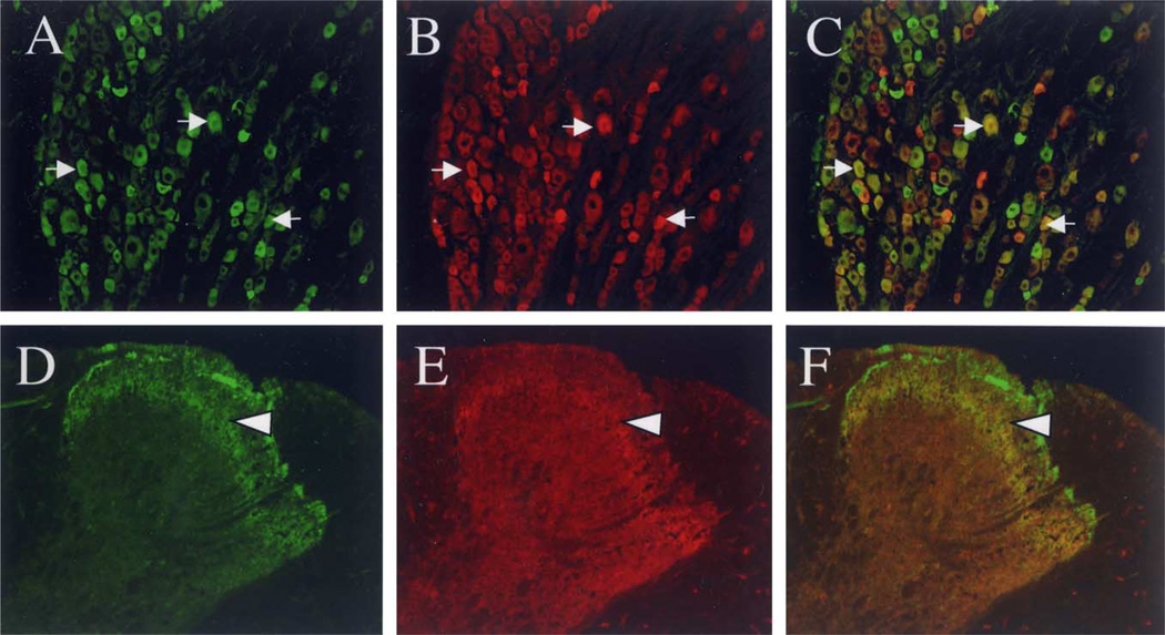
Fig. 5.
Co-localization of VR1 immunoreactivity with Y1 immunoreactivity in DRG neurons and in the superficial dorsal horn. (A–C) Double labeling of VR1 (A; green) with Y1 (B; red) and overlay (C) in DRG (magnification = 20×). Co-localization is shown in yellow and by arrows. (D–F) Double labeling of VR1 (D; green) with Y1 (E; red) and overlay (F; magnification = 20×). Immunoreactivity for Y1 and VR1 are both localized in inner lamina II, as indicated by yellow and arrowhead. These images were digitally sharpened using Adobe Photoshop 5.0.

