Abstract
Background:
Various root canal systems are available commercially with each manufacturer stating superior characteristics of their respective systems. The fifth generation root canal systems claim to have better flexibility and superior debris elimination due to their offset design. This study aims to compare the effects of fifth generation rotary systems on canal curvature, transportation and centering ratio of curved mesial root canals of mandibular molar via cone-beam computed tomographic (CBCT) imaging.
Materials and Methods:
With curvature ranging from 20° to 40°, 60 mandibular first molars with mesiobuccal root angle were divided into three groups with 20 canals each. Before instrumentation, the groups were balanced with respect to the angle of canal curvature based on CBCT images taken. All root canals were shaped to an apical size of 25: OneShape (OS) (Micro Mega, Besancon, France), ProTaper Next (PTN) (Dentsply Maillefer), Revo S (RS) (Micro Mega, Besancon, France). CBCT assessment was done post instrumentation. SPSS version 16 software was used for statistical analysis. The significance level was set at P = 0.05.
Results:
The RS system maintained better canal centricity and less transportation as compared to PTN and OS. There was no significant difference among the three groups in canal curvature after instrumentation.
Conclusions:
All file systems used straightened the root canal curvature similarly. RS instrumentation exhibited superior performance compared with the OS and PTN systems with respect to transportation and centering ratio.
Keywords: Cone-beam computed tomographic imaging, One Shape, ProTaper Next, Revo S, root canal transportation
Introduction
Objective in root canal preparation is to develop a shape that tapers from apical to coronal, maintaining the original canal shape.1 With advent of Instruments manufactured from nickel titanium alloys (NiTi),2 there was significant improvement of quality of root canal shaping, with predictable results and less iatrogenic damage, even in severely curved canals.3
Recently a new concept for NiTi files has recently been introduced with asymmetric cross sections that finish root canal shaping.
The fifth generation single file system, OneShape (OS) file (Micro Mega, Besancon, France) is few single file instruments used in continuous clockwise rotational motion for quick and safe root canal preparation. The OS file has an asymmetric cross-sectional geometry that generates traveling waves of motion along the active part of the file.4
The ProTaper Next (PTN) (Dentsply Maillefer, Ballaigues, Switzerland) is another NiTi file system; which has three significant design features, including progressive percentage tapers on a single file, M-wire technology, and the fifth generation of continuous improvement, the off set design which in turn decreases the dangerous taper lock and screw effect by minimizing the contact between a file and dentin.5
Revo S (RS) is a sequence of 3 NiTi with an asymmetric cross section which facilitates penetration by a snake like movement and offers root canal shaping which is adapted to biological and ergonomic imperatives. This sequence has debris elimination, cutting and cleaning cycle which improves root canal cleaning by facilitating upward removal of generated dentin debris.6,7
In order to investigate the efficacy of instruments and techniques developed for root canal preparation, numerous methods have been used to evaluate the canal shape before and after instrumentation. Cone-beam computed tomography (CBCT) imaging is a non-invasive technique for analysis of canal geometry and efficiency of shaping techniques. Using CBCT it becomes possible to compare the an anatomic structure of root canal before and after root canal preparation.1,8,9
The present study was set out to determine canal centering ability, transportation and curvature using fifth generation NiTi rotary systems as mentioned above.
Materials and Methods
Inclusion criteria
Curved mesial roots of mandibular molars extracted because of reasons irrelevant to this investigation such as periodontitis, grossly decayed teeth, etc. Radiographs of teeth in both the buccolingual and mesiodistal directions were taken for selecting the right samples. Teeth with two separated mesial canals and no significant calcifications were included.
Exclusion criteria
Teeth with open apex, root curvature below 20°, as well as above 40° and thin narrow calcified canals.
A 60 teeth were mounted in an acrylic block keeping their apices open in order to allow their scanning for morphometric evaluation of the pre instrumented root canals by using CBCT imaging, (i-CAT, Insight CBCT Centre, Mumbai). Canal curvature angles of the teeth were measured according to Schneider.10 The specimens were allocated to 1 of 3 groups (n = 20) based on the canal curvature angle. Teeth were accessed with a diamond bur, and the working length determination of mesiobuccal canals was determined by inserting a size 10 K-type file to the root canal terminus and reducing 1 mm from this measurement. A glide path was performed via a size 15 K-type file. RC Help (Prime Dental Products Pvt. Ltd.) was used in all canal preparations, and the root canal was irrigated with 2 ml 2.5% sodium hypochlorite solution after change of each instrument. Use of each instrument was restricted to four canals only. Apical preparation was completed with a size 25 instrument by using the instrument order specified by the manufacturer.
The preparation sequences were as follows:
Group 1: OS file having a taper of 0.06 and a size of 25 was used with in and out movements without pressure at a rotational speed of 400 rpm along with torque values of 400 gcm. When apical resistance was encountered, the instrument was removed and cleaned, and the root canal was irrigated
Group 2: PTN files were used with the sequence PU SX, PTN X1, and X2 at a rotational speed of 300 rpm along with torque values of 200 gcm. Each file was used with a brushing motion similar to the PU files
Group 3: RS was used with slow and unique downward movement in free progression and without pressure (up and down movement) with a rotation speed ranging between 250 and 400 rpm.
After instrumentation, specimens were scanned under the same conditions as initial scans and the post-operative images were captured in the same way as mentioned before. Canal curvatures of samples were measured with the same protocol. Crosssectional planes of images at 2, 5, and 8 mm from the apical end of the root were analyzed before and after instrumentation for transportation and centering ratio. Transportation at each level was calculated using the following formula:11
(x1−x2)−(y1−y2)
The canal centering ratio at each level was calculated using the following formula:11
(x1−x2)/(y1−y2) or (y1−y2)/(x1−x2)
x1 and x2 showed the shortest mesial distances from the outside of the curved root to the periphery of the un-instrumented and instrumented canal, respectively, and y1 and y2 represented the shortest distal distances from the outside of the curved root to the periphery of the un-instrumented and instrumented canal, respectively (Figure 1a and b). The smallest distance from the outer periphery of the curved root to the periphery of the instrumented canal was also recorded. The distribution of teeth among the three groups for pre-instrumentation canal curvature was assessed by using analysis of variance (ANOVA). Changes in canal transportation and centering ratio data were analyzed using the Kruskal–Wallis ANOVA which showed statistical significant difference and pairwise comparison was done by Mann–Whitney U-test. The significance level was set at P = 0.05. Statistical analysis was performed with SPSS statistics version.
Figure 1.
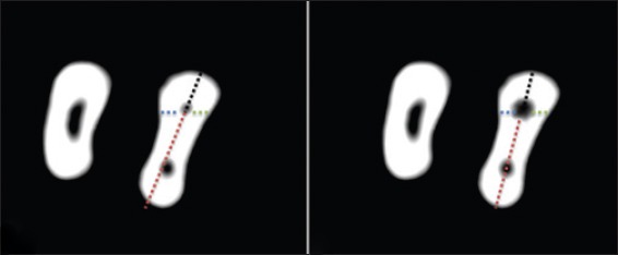
Measurements of root canal transportation, (a) Before instrumentation, (b) after instrumentation
Graph 1.
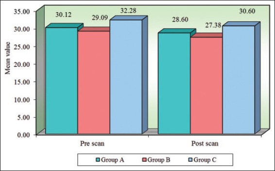
Comparison of three groups (A, B, C) with respect to curvature scores at pre- and post-scan.
Results
Table 1 shows the pre- and post-instrumentation curvature of curved canals.
Table 1.
Comparison of three groups (A, B, C) with respect to curvature scores at pre- and post-scan by one-way ANOVA.
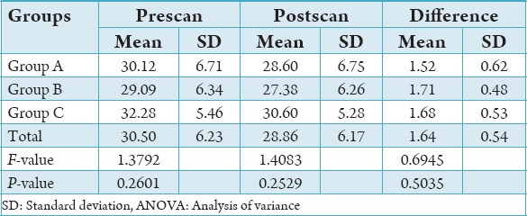
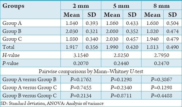
No files fractured during the study. There was no significant difference among the three groups in canal curvature (Table 1 and Graph 1). The RS showed least canal transportation and better centering ability at 2 mm as compared to PTN and OS (Tables 2 and 3, Graphs 2 and 3).
Table 2.
Comparison of three groups (A, B, C) with respect to transportation scores at 2 mm, 5 mm, and 8 mm by Kruskal–Wallis ANOVA.
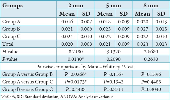
Table 3.
Comparison of three groups (A, B, C) with respect to canal centering ratio scores at 2 mm, 5 mm, and 8 mm by Kruskal–Wallis ANOVA.
Graph 2.
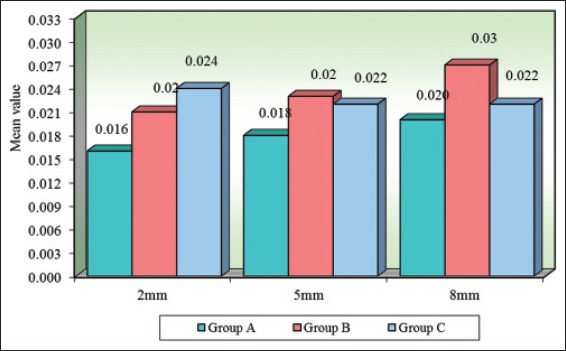
Comparison of three groups (A, B, C) with respect to transportation scores at 2 mm, 5 mm, and 8 mm.
Graph 3.
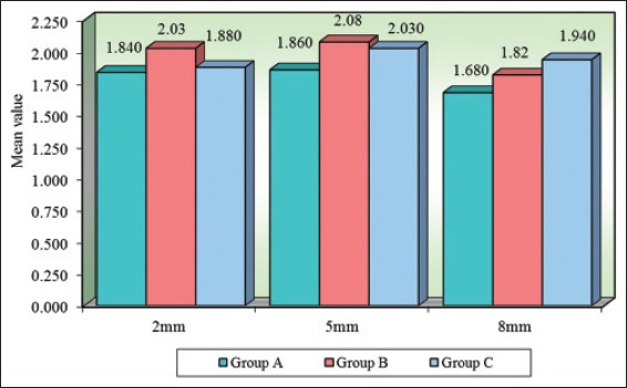
Comparison of three groups (A, B, C) with respect to canal centering ratio scores at 2 mm, 5 mm, and 8 mm.
Discussion
NiTi rotary instruments are important in endodontics because of their ability to shape root canals with minimum complications.12 As the rotary NiTi instruments maintained the original canal curvature, particularly in the apical region of the root canal better than stainless steel hand instruments, studies compared the shaping ability of different rotary NiTi systems with different designs.
In this study, an evaluation on the effects of three newly developed file systems belonging to the fifth generation that have different cutting blade designs, metallurgies, manufacturing process along with sequence and number of files on the parameters of canal transportation, centering ratio, and canal curvature by using CBCT imaging was performed.
In previous studies, two experimental models were usually preferred: Simulated canals vs. extracted teeth. Using extracted teeth has an advantage over resin blocks because they provide conditions closer to clinical situations.13 Even the hardness and abrasion behavior of acrylic resin and root dentin are not identical,14 and the heat generated may soften the resin material.15 Therefore, we used extracted teeth in this study to compare the shaping ability of rotary NiTi instruments which were used to provide clinical conditions.
The distribution of the three groups with respect to canal curvature showed no statistical significant difference (Table 1). The curvatures of root canals ranged between 20° and 40°. Changes in canal curvature after the use of the different NiTi file systems were not statistically significant. This is in agreement with the findings of previous studies.16-19 All the instruments have non-cutting tips that work with minimal apical pressure and function only as a guide to allow easy penetration.15 Single-file shaping technique may simplify instrumentation protocols and avoid the risk of cross-contamination. Moreover, the use of only one NiTi instrument is more cost-effective, and the learning curve is considerably reduced. Numerous studies comparing the shaping ability of single-file systems and conventional ones using a full range of instruments have shown, similar to our study, that both systems result in suitable preservation of the original canal shape.20-22 One of the study showed canals prepared with the F360 and OS systems were better centering ability viz., with the Reciproc and WaveOne systems.23 Our results showed no significant difference at 5 mm and 8 mm with all three systems. Single file system is as convenient as a multiple file system.
To check center of mass and/or the center of rotation, the fifth generation of shaping files has been designed. To generate mechanical wave of motion that travels along the active length of the file in rotation, the files that have an off set design are useful. This off set design serves to further minimize the engagement between the file and dentin.24 PTN files were used in sequence: PU SX followed by X1 (17/0.04), X2 (25/0.06). Revo-S NiTi instrument system includes three shaping instruments: the shaping and cleaning instrument (SC) number 1 (SC1) (#25/0.06), SC2 (#25/0.04), and the universal shaper (#25/0.06) which was used. The OS file from Micro-Mega (Besançon, France) is the only single-file NiTi instrument in continuous rotation available as (#25/0.06) for root canal preparations.
One of the study suggests, newly manufactured rotary systems with size 25 and a taper of 0.06 (PN, TFA, OS) or 0.08 (WO, R, PU) could be used in curved canals because of minimal transportation.25 We found that the RS system with a constant 0.06 taper removed less dentin in apical third than the PTN instrument (0.06 taper at the apical 2 mm) and OS (with 0.06 taper). Some authors emphasize that several factors may affect fl exibility and performance of rotary instruments. One study found increased tendency for canal transportation as diameter of file is increased,26 while other referred taper to be one of the factor responsible for canal transportation and suggested that Protaper Universal F3 and F4 to be used with caution owing to taper.27 Further it has been said that, NITi files with tapers more than 0.06 should not be used for the apical enlargement of curved canals, because of stiffness of the files.28,29
The comparable apical transportation was less and mean centering ability was superior of RS at 2 mm which may be due to asymmetrical cutting profile of RS which facilitates penetration by a snake like movement and offers a root canal shaping and apical finishing that is closely adapted to the anatomical and ecological criteria of the canal. Probably, other than above mentioned reason, RS has one cutting edge which makes it less aggressive as compared to others. Further, studies are required to understand the benefits of each file system.
Conclusion
This study showed that the three different file systems straighten root canal curvature similarly and RS was seen to maintain canal centricity along with less transportation at 2 mm followed by OS and PTN which may contributed to improved helical angle, adapted pitch and positive rake angle. CBCT is a good visualizing aid.9
Acknowledgments
The authors thank Micro Mega for providing the OS instruments.
References
- 1.Gergi R, Rjeily JA, Sader J, Naaman A. Comparison of canal transportation and centering ability of twisted files, Path file-ProTaper system, and stainless steel hand K-files by using computed tomography. J Endod. 2010;36(5):904–7. doi: 10.1016/j.joen.2009.12.038. [DOI] [PubMed] [Google Scholar]
- 2.Walia HM, Brantley WA, Gerstein H. An initial investigation of the bending and torsional properties of Nitinol root canal files. J Endod. 1988;14(7):346–51. doi: 10.1016/s0099-2399(88)80196-1. [DOI] [PubMed] [Google Scholar]
- 3.Peters OA. Current challenges and concepts in the preparation of root canal systems: A review. J Endod. 2004;30(8):559–67. doi: 10.1097/01.don.0000129039.59003.9d. [DOI] [PubMed] [Google Scholar]
- 4.Gernhardt CR. One shape - A single file system for root canal instrumentation used in continuous rotation. ENDO (Lond Engl) 2013;7:211–6. [Google Scholar]
- 5.Clifford JR, Pierre M, John DW. The shaping movement 5th generation technology. Adv Endod. 2013;32(4):94–9. [Google Scholar]
- 6.Arora A, Taneja S, Kumar M. Comparative evaluation of shaping ability of different rotary NiTi instruments in curved canals using CBCT. J Conserv Dent. 2014;17(1):35–9. doi: 10.4103/0972-0707.124127. [DOI] [PMC free article] [PubMed] [Google Scholar]
- 7.Mallet JP. An instrument innovation for primary endodontic treatment: The Revo-S sequence. Smile Dent J. 2009;4(4):3. [Google Scholar]
- 8.Bernardes RA, Rocha EA, Duarte MA, Vivan RR, de Moraes IG, Bramante AS, et al. Root canal area increase promoted by the endo sequence and pro taper systems: Comparison by computed tomography. J Endod. 2010;36(7):1179–82. doi: 10.1016/j.joen.2009.12.033. [DOI] [PubMed] [Google Scholar]
- 9.Venskutonis T, Plotino G, Juodzbalys G, Mickeviciene L. The importance of cone-beam computed tomography in the management of endodontic problems: A review of the literature. J Endod. 2014;40(12):1895–901. doi: 10.1016/j.joen.2014.05.009. [DOI] [PubMed] [Google Scholar]
- 10.Schneider SW. A comparison of canal preparations in straight and curved root canals. Oral Surg Oral Med Oral Pathol. 1971;32(2):271–5. doi: 10.1016/0030-4220(71)90230-1. [DOI] [PubMed] [Google Scholar]
- 11.Gambill JM, Alder M, del Rio CE. Comparison of nickel-titanium and stainless steel hand-file instrumentation using computed tomography. J Endod. 1996;22(7):369–75. doi: 10.1016/S0099-2399(96)80221-4. [DOI] [PubMed] [Google Scholar]
- 12.Young GR, Parashos P, Messer HH. The principles of techniques for cleaning root canals. Aust Dent J. 2007;52(1 Suppl):S52–63. doi: 10.1111/j.1834-7819.2007.tb00526.x. [DOI] [PubMed] [Google Scholar]
- 13.Schäfer E, Vlassis M. Comparative investigation of two rotary nickel-titanium instruments: ProTaper versus RaCe. Part 1. Shaping ability in simulated curved canals. Int Endod J. 2004;37(4):229–38. doi: 10.1111/j.0143-2885.2004.00786.x. [DOI] [PubMed] [Google Scholar]
- 14.Paqué F, Musch U, Hülsmann M. Comparison of root canal preparation using RaCe and pro taper rotary Ni-Ti instruments. Int Endod J. 2005;38(4):8–16. doi: 10.1111/j.1365-2591.2004.00889.x. [DOI] [PubMed] [Google Scholar]
- 15.Kum KY, Spängberg L, Cha BY, Il-Young J, Msd, Seung-Jong L, et al. Shaping ability of three pro file rotary instrumentation techniques in simulated resin root canals. J Endod. 2000;26(12):719–23. doi: 10.1097/00004770-200012000-00013. [DOI] [PubMed] [Google Scholar]
- 16.Celik D, Tasdemir T, Er K. Comparative study of 6 rotary nickel-titanium systems and hand instrumentation for root canal preparation in severely curved root canals of extracted teeth. J Endod. 2013;39(2):278–82. doi: 10.1016/j.joen.2012.11.015. [DOI] [PubMed] [Google Scholar]
- 17.Marzouk AM, Ghoneim AG. Computed tomographic evaluation of canal shape instrumented by different kinematics rotary nickel-titanium systems. J Endod. 2013;39(7):906–9. doi: 10.1016/j.joen.2013.04.023. [DOI] [PubMed] [Google Scholar]
- 18.You SY, Kim HC, Bae KS, Baek SH, Kum KY, Lee W. Shaping ability of reciprocating motion in curved root canals: A comparative study with micro-computed tomography. J Endod. 2011;37(9):1296–300. doi: 10.1016/j.joen.2011.05.021. [DOI] [PubMed] [Google Scholar]
- 19.Bürklein S, Benten S, Schäfer E. Shaping ability of different single-file systems in severely curved root canals of extracted teeth. Int Endod J. 2013;46(6):590–7. doi: 10.1111/iej.12037. [DOI] [PubMed] [Google Scholar]
- 20.Bürklein S, Hinschitza K, Dammaschke T, Schäfer E. Shaping ability and cleaning effectiveness of two single-file systems in severely curved root canals of extracted teeth: Reciproc and wave one versus Mtwo and pro taper. Int Endod J. 2012;45(5):449–61. doi: 10.1111/j.1365-2591.2011.01996.x. [DOI] [PubMed] [Google Scholar]
- 21.Berutti E, Chiandussi G, Paolino DS, Scotti N, Cantatore G, Castellucci A, et al. Canal shaping with wave one primary reciprocating files and pro taper system: A comparative study. J Endod. 2012;38(4):505–9. doi: 10.1016/j.joen.2011.12.040. [DOI] [PubMed] [Google Scholar]
- 22.Junaid A, Freire LG, da Silveira Bueno CE, Mello I, Cunha RS. Influence of single-file endodontics on apical transportation in curved root canals: An ex vivo micro-computed tomographic study. J Endod. 2014;40(5):717–20. doi: 10.1016/j.joen.2013.09.021. [DOI] [PubMed] [Google Scholar]
- 23.Saleh AM, Vakili Gilani P, Tavanafar S, Schäfer E. Shaping ability of 4 different single-file systems in simulated S-shaped canals. J Endod. 2015;41(4):548–52. doi: 10.1016/j.joen.2014.11.019. [DOI] [PubMed] [Google Scholar]
- 24.Goel A, Rastogi R, Rajkumar B. An overview of modern endodontic NiTi systems. Med Sci. 2015;4:2277–8179. [Google Scholar]
- 25.Capar ID, Ertas H, Ok E, Arslan H, Ertas ET. Comparative study of different novel nickel-titanium rotary systems for root canal preparation in severely curved root canals. J Endod. 2014;40(6):852–6. doi: 10.1016/j.joen.2013.10.010. [DOI] [PubMed] [Google Scholar]
- 26.López FU, Fachin EV, Camargo Fontanella VR, Barletta FB, Só MV, Grecca FS. Apical transportation: A comparative evaluation of three root canal instrumentation techniques with three different apical diameters. J Endod. 2008;34(12):1545–8. doi: 10.1016/j.joen.2008.07.027. [DOI] [PubMed] [Google Scholar]
- 27.Kunert GG, Camargo Fontanella VR, de Moura AA, Barletta FB. Analysis of apical root transportation associated with ProTaper Universal F3 and F4 instruments by using digital subtraction radiography. J Endod. 2010;36(6):1052–5. doi: 10.1016/j.joen.2010.02.004. [DOI] [PubMed] [Google Scholar]
- 28.Schäfer E, Dzepina A, Danesh G. Bending properties of rotary nickel-titanium instruments. Oral Surg Oral Med Oral Pathol Oral Radiol Endod. 2003;96(6):757–63. doi: 10.1016/s1079-2104(03)00358-5. [DOI] [PubMed] [Google Scholar]
- 29.Aydin C, Inan U, Murside G. Comparison of shaping ability of TF with PU and RS Ni-Ti instruments in simulated canals. J Dent Sci. 2012;7:283–8. [Google Scholar]


