Abstract
Traumatic neuromas are tumors resulting from hyperplasia of axons and nerve sheath cells after section or injury to the nervous tissue1. We present a case of this tumor, confirmed by anatomopathological examination, in a male patient with history of circumcision. Knowledge of this entity is very important in achieving the differential diagnosis with other lesions that affect the genital area such as condyloma acuminata, bowenoid papulosis, lichen nitidus, sebaceous gland hyperplasia, achrochordon and pearly penile papules.
Keywords: Circumcision, male; Neuroma; Penis
INTRODUCTION
Traumatic neuromas are tumors arising from hyperplasia of axons and nerve sheath cells after section or injury to the nerve.1 They may be located in any anatomical site and result from disorganized growth of the proximal portion of the lesioned axon. They are rare in the genital region. In cases of penile location the majority is associated to circumcision, even though any type of trauma may originate the tumor. Clinically, penile traumatic neuromas present themselves as skin-colored or erythematous papules, a diameter which varies from 1 to 7 mm, located in the glans, body of penis or in the transition area of both. The histopathological picture shows multiple hyperplastic nerve branches, forming entanglements, located beneath the epithelium. Differential diagnosis is important with other penile-located diseases such as condyloma acuminata, bowenoid papulosis, sebaceous gland hyperplasia, pearly penile papules, lichen nitidus and acrochorda.
CASE REPORT
Male patient, 22 years old, white, hospitalized at the dermatology ambulatory of Complexo Hospitalar Padre Bento de Guarulhos - SP. He reported history of asymptomatic, skin-colored papular lesions on the penis, for one and a half year. He also reported a circumcision when he was eight years old and denied comorbidities.
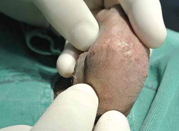
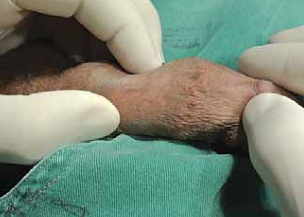
At the dermatological examination he presented multiple and small normochromic papules on the body of penis and the transition area to the glans, with approximately 2mm in diameter (Figures 1 and 2).
FIGURE 1.

Diminutive papules in the body of penis
FIGURE 2.

Papules on the body of penis. Notice location of area corresponding to insertion of foreskin
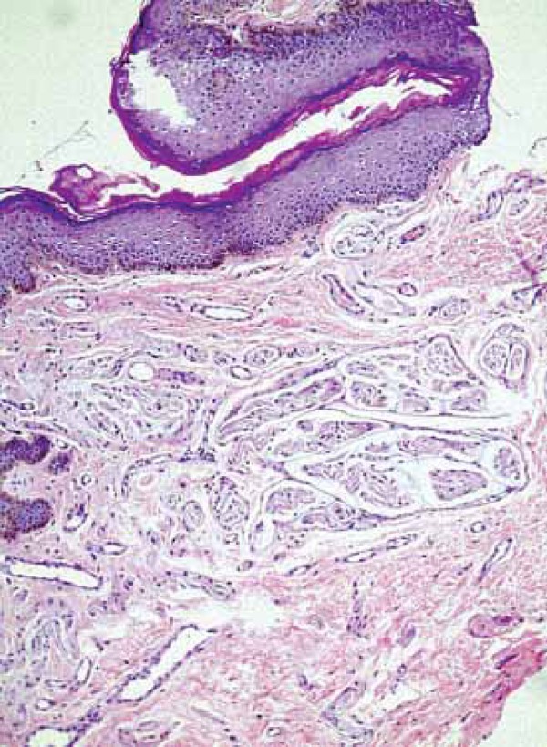
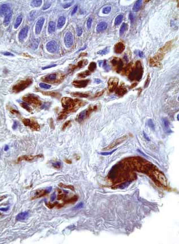
An excision of the lesion was performed with a 2mm punch and the histopathological examination revealed absence of acanthosis or papillomatosis; in the lamina propria, there was presence of multiple proliferated nerve branches form`ing "entanglements" (Figures 3 and 4). Immunoperoxidase technique for S-100 antigen confirmed they were neural branches (Figure 4).
FIGURE 3.

HE-100x. Proliferation of neural branches in the lamina propria. Observe the squamous epithelium without acanthosi
FIGURE 4.

Immunoperoxidase S-100 400x. Detail of proliferated neural branches
DISCUSSION
Neuromas are neural tissue proliferations on which Schwann cells and axonal components are found in almost equal amounts. They are subdivided in traumatic or amputation neuroma and palisade encapsulated neuroma. Traumatic neuromas are regenerative proliferations of nerve fibers secondary to lesions. The palisade encapsulated neuromas are hamartomatous proliferations of nerve fibers with no apparent previous tissue damage.2
Any extrinsic lesion may originate traumatic neuromas, and these occur in any age and in both sexes; however, they are uncommon. They present themselves clinically as solid papules, skin-colored or erythematous-purplish at the sites of the lesions, surgical scars or amputations3. The initial lesions are usually asymptomatic but can evolve with pain, paresthesia and pruritus in the course of months to years of its onset. The patient in question did not present any symptoms related to the lesions.
The penile traumatic neuroma was described only in 1990 by Montgomery et al, who described these lesions in a patient with penile trauma.4 Brehmer and Anderson in 1999 and Rossi and Franchella in 2005 have also reported cases, with three and one case described, respectively.5,6 Salcedo et al in 2009 described a series of 17 cases, all related to biopsies or circumcision7. In this report the authors listed the different types of cutaneous neuromas as: traumatic, rudimentary (observed in polydactyly) and solitary neuroma (palisade encapsulated neuroma). In 2009 Marinho and Bouscarat described a case of multiple papules over the circumcision scar of a 23-year-old man, and the anatomopathological examination revealed it to be a traumatic neuroma.8
Enzinger and Weiss also mention mucosal neuroma, which is related to multiple endocrine neoplasia syndrome type IIb (MEN Iib); in this patient, however, other lesions or comorbidities were not observed. 9
Even though the clinical aspects (surgeries, previous biopsies) are important for the diagnosis of this lesion, some histopathological aspects allow its characterization in light of other possible differential diagnoses, such as Morton's neuroma and pearly penile papule. Morton's neuroma is characterized by the presence of inflammatory and reparative phenomena (respectively lymphocitary inflammatory infiltrate and fibrosis, both perineural) and absence of proliferative phenomena, which characterize the traumatic neuroma. In pearly penile papule, fibrosis and vascular ectasias in the lamina propria are observed, without proliferation of neural branches.10
The differential clinical diagnoses of penile location lesions include condyloma acuminata, bowenoid papulosis, pearly penile papules, lichen nitidus, sebaceous gland hyperplasia (preputial glands), among others.
Even though it is considered a rare entity, the traumatic neuroma must be remembered and considered in the differential diagnosis of penile lesions, especially in circumcised patients and/or those with history of trauma or biopsy of penis. Surgical excision with later clinical follow-up is the treatment of choice for this entity, which portrays an excellent prognosis.
Footnotes
Financial Support: None.
Conflict of Interest: None.
How to cite this article: Cardoso TA, Santos KR, Franzotti AM, Avelar JCD, Tebcherani AJ, Pegas JRP. Traumatic neuroma of the penis after circumcision - Case report. An Bras Dermatol. 2015; 90(3):397-9.
Work performed at Complexo Hospitalar Padre Bento de Guarulhos (CHPBG) – Guarulhos (SP), Brazil.
Reference
- 1.Reed JR, Argenyi Z. Tumors of neural tissue. In: Elder D E, Elenitsas R, Johnson BL Jr, Murphy GF, editors. Lever's Histopathology of the skin. 9th ed. Philadelphia (PA):: Lippincott Williams & Wilkins; 2005. pp. 1134–1137. [Google Scholar]
- 2.Weedon D. Neural and neuroendocrine tumors. Skyn pathology. 2nd ed. Edinburgh: Churchill Livingstone; 2002. p. 977. [Google Scholar]
- 3.Argenyi ZB. Bolognia JL, Jorizzo JL, Rapini RP. Dermatologia. Rio De Janeiro: Elsevier; 2011. Neoplasias neurais e neuroendócrinas (exceto neurofibromatose) [Google Scholar]
- 4.Montgomery BS, Fletcher CD, Palfrey EL, Lloyd-Davies RW. Traumatic neuroma of the penis. Br J Urol. 1990;65:420–421. doi: 10.1111/j.1464-410x.1990.tb14770.x. [DOI] [PubMed] [Google Scholar]
- 5.Brehmer-Andersson EE. Penile neuromas with multiple Meissner's corpuscles. Histopathology. 1999;34:555–556. doi: 10.1111/j.1365-2559.1999.0696b.x. [DOI] [PubMed] [Google Scholar]
- 6.Rossi E, Franchella A. Amputation neuroma following a circumcision: a case report. Eur J Pediatr Surg. 2006;16:288–290. doi: 10.1055/s-2006-924342. [DOI] [PubMed] [Google Scholar]
- 7.Salcedo E, Soldano AC, Chen L, Rokhsar CK, Tam ST, Meehan SA, et al. Traumatic neuromas of the pênis : a clinical, histopathological and immunohistochemical study of 17 cases. J Cutan Pathol. 2009;36:229–233. doi: 10.1111/j.1600-0560.2008.01014.x. [DOI] [PubMed] [Google Scholar]
- 8.Marinho E, Bouscarat F. Multiple papules on a circumcision wound. Ann Dermatol Venereol. 2009;136:199–201. doi: 10.1016/j.annder.2008.12.011. [DOI] [PubMed] [Google Scholar]
- 9.Weiss SW, Goldblum JR. Enzinger & Weiss Soft Tissue Tumours. 5th ed. Philadelphia: Mosby Elesevier; 2008. pp. 829–832. [Google Scholar]
- 10.Sittart JAS. Contribuição ao estudo das pápulas perladas penianas em indivíduos das raças caucasoide e negróide e circuncidados e não circuncidados. São Paulo (SP): Hospital do Servidor Público Estadual de São Paulo; 1993. [tese] [Google Scholar]


