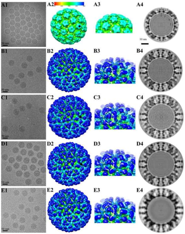
FIGURE 1. Cryo-EM reconstructions of HPV16 and HPV16-Fab complexes.
Representative regions of cryo-EM micrographs are shown for (A1) the HPV16 capsid (EM Database, EMD 5993) (16), (B1) HPV16 capsids complexed with H16.V5 (EM Database, EMD 5994) (16), (C1) H16.1A, (D1) H16.14J, and (E1) H16.263A2. (A2–E2) The 3-D complexes were radially colored according to the distance from the center of the capsid (color bar indicated) and surface rendered at 1σ. A small black pentagon indicates the position of a pentavalent capsomer. (A3–E3) In the zoomed in views of the complex maps the separate heavy chain and light chain Fab densities can be distinguished. (A4–E4) The central sections through the cryo-EM density maps were displayed in the same sequence as above. Capsids were cut vertically through the 2-, 3- and 5-fold icosahedral symmetry axes (black lines), with the central 2-fold axis appearing at the 12 o’clock position, and a scale bar equal to 10 nm.

