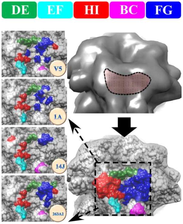
FIGURE 6. The location of the antibody binding groove on the surface of capsomer was made up of FG and DE loops with lesser contributions by the BC, EF, and HI loops.
All four epitopes map to a “groove” feature on the surface of the capsomer (upper right, dashed outline). The combined four footprints are color coded according to loop identity (lower right, dashed box). The left column shows the zoomed view of the individual epitopes of H16.V5, H16.1A, H16.14J and H263.A2 with specific loop contributions indicated by color (Top: color code bar for DE, EF, HI, BC, and FG).

