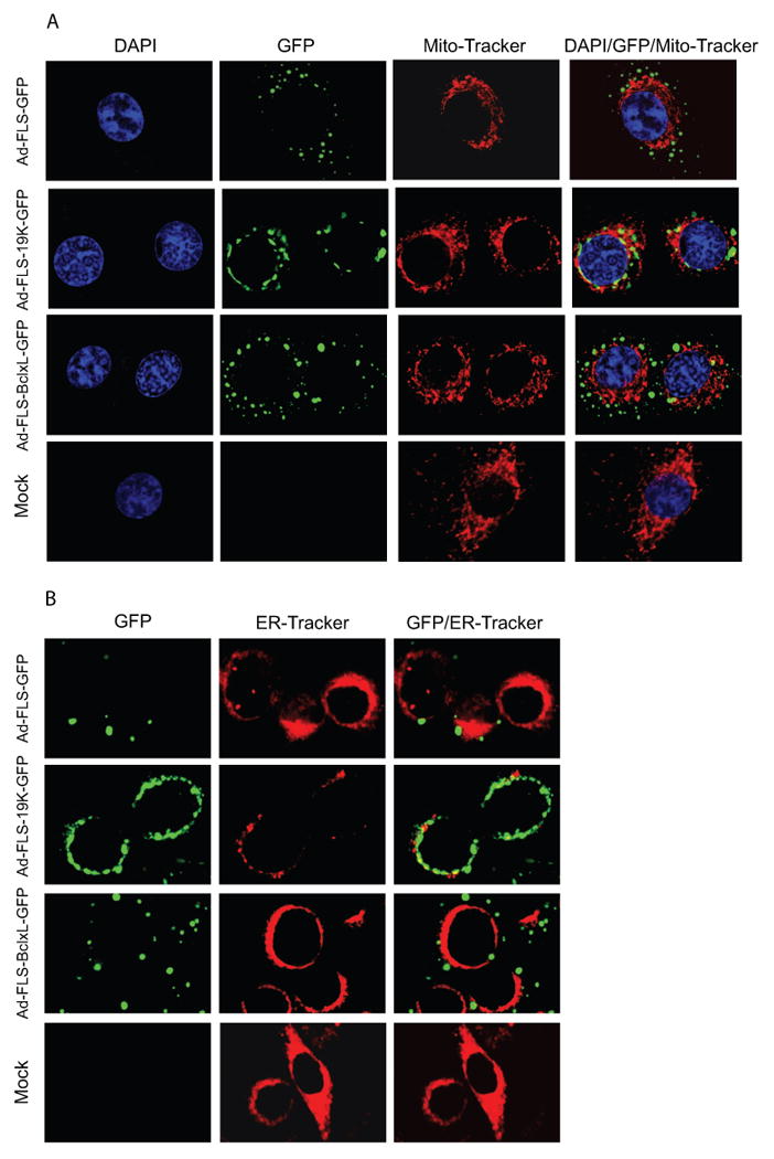
Fig. 2.

Mitochondrial and ER localization of chimeric proteins. Cells were infected with various viruses and after 24 hr of infection, mito-Tracker (A) or ER-Tracker (B) were added to the cells in medium and Hank's buffer respectively for 30 min, fixed and analyzed using a confocal microscope.
