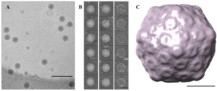
Figure 4.
Single particle reconstructions of φPsa17. (a) Representative cryo-EM field of view. Scale bar 200 nm; (b) A gallery of cryo-EM images representing full capsids (left), capsids with visible tails (middle), and empty capsids (right), scale bar 20 nm; (c) 3D reconstruction of the φPsa17 phage capsid, scale bar 20 nm.

