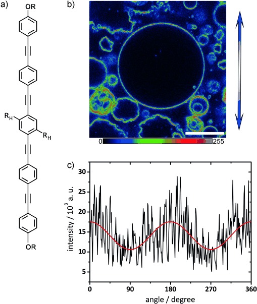
Figure 2.

a) Structure of the BP core (substituents OR as in Scheme 1 and RH=OC12H25). b) Confocal slice of GUVs prepared from a 1:10 ratio of E12/7 and DPPC, excited with polarized λ=405 nm laser light. The direction of polarization is indicated by the blue double arrow. A multicolor palette (as depicted below) is used to highlight fluorescence intensity variations. Scale bar=20 μm. Imaging was performed at room temperature (22 °C). c) The fluorescence intensity along the perimeter of the largest GUV in b) as a function of angle γ (black line: data; red line: cosine-squared fit function, I=a+b cos2 (γ)) shows maxima at the top and bottom of the image and minima to the right and the left; this indicates an approximately transmembrane incorporation of the BP into the bilayer.
