

Abstract
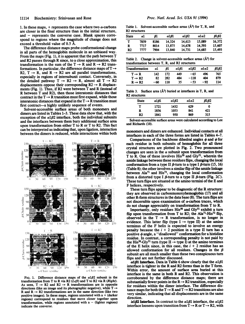
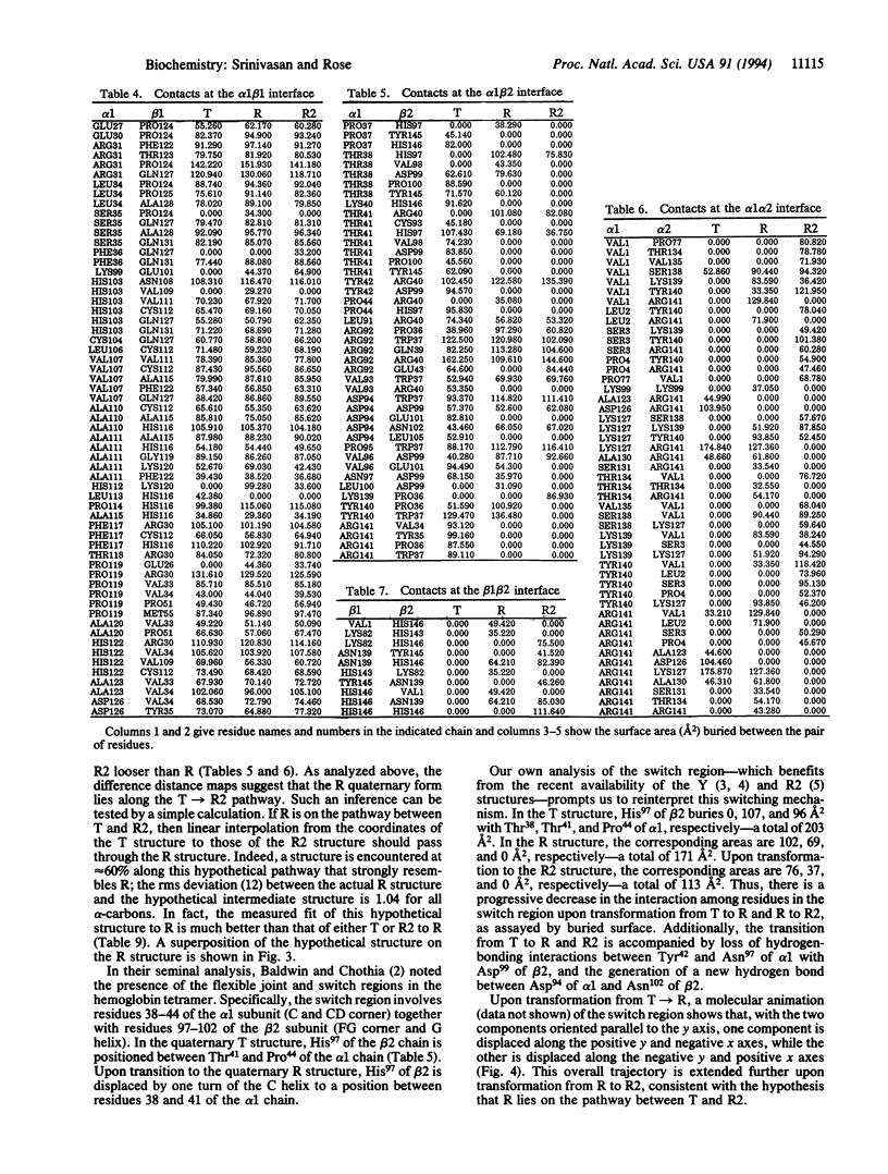
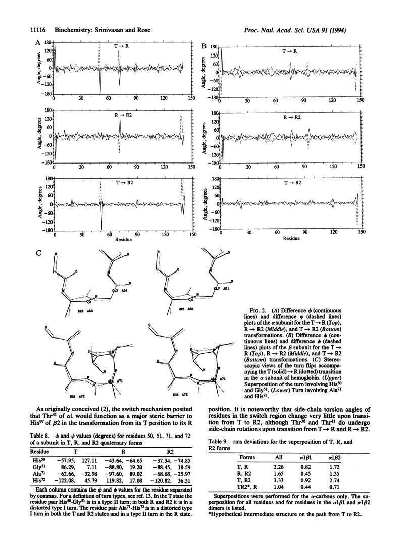
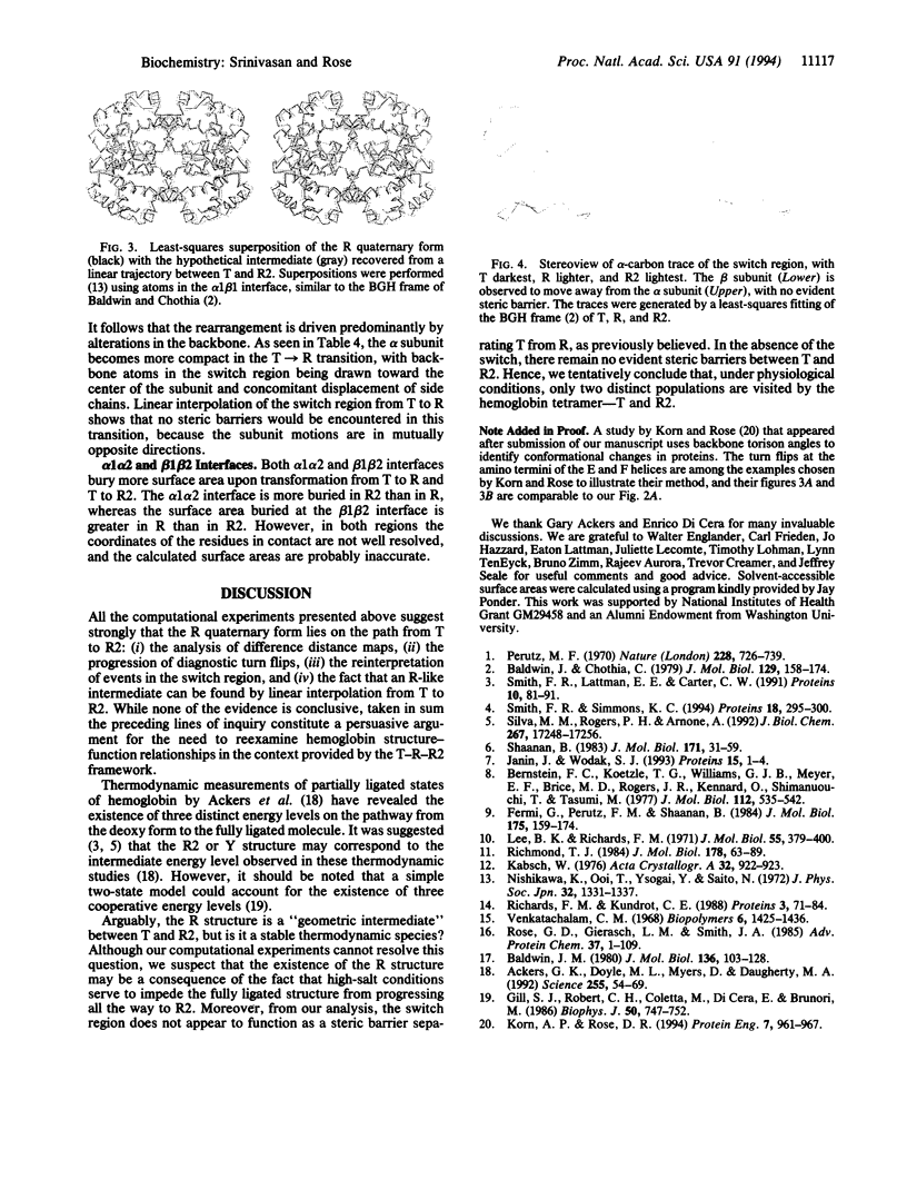
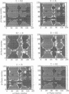
The relationship between the T, R, and R2 quaternary forms of hemoglobin is examined by computational experiments. Contrary to previous suggestions, we propose that the R quaternary form may lie on the pathway from T to R2. This proposal is consistent with four independent observations. (i) Difference distance maps are used to identify those parts of the molecule that undergo conformational change upon oxygenation. The simplest interpretation of these maps brackets R between T and R2. (ii) Linear interpolation from T to R2 passes through R. (iii) The well-known "switch" region (so called because, upon transition between the T and R quaternary forms, a residue from the beta 2 subunit toggles between two stable positions within the alpha 1 subunit) progresses from T through R to R2, successively. (iv) A hitherto-undocumented feature, diagnostic of the R structure, is noted within the alpha subunit: upon transformation from T to R, the beta-turns at the amino terminal of the E and F helices flip from one turn type to another. Upon transformation from R to R2, the latter turn--a strained conformation--flips back again.
Full text
PDF
Images in this article

Selected References
These references are in PubMed. This may not be the complete list of references from this article.
- Ackers G. K., Doyle M. L., Myers D., Daugherty M. A. Molecular code for cooperativity in hemoglobin. Science. 1992 Jan 3;255(5040):54–63. doi: 10.1126/science.1553532. [DOI] [PubMed] [Google Scholar]
- Baldwin J. M. The structure of human carbonmonoxy haemoglobin at 2.7 A resolution. J Mol Biol. 1980 Jan 15;136(2):103–128. doi: 10.1016/0022-2836(80)90308-3. [DOI] [PubMed] [Google Scholar]
- Bernstein F. C., Koetzle T. F., Williams G. J., Meyer E. F., Jr, Brice M. D., Rodgers J. R., Kennard O., Shimanouchi T., Tasumi M. The Protein Data Bank: a computer-based archival file for macromolecular structures. J Mol Biol. 1977 May 25;112(3):535–542. doi: 10.1016/s0022-2836(77)80200-3. [DOI] [PubMed] [Google Scholar]
- Fermi G., Perutz M. F., Shaanan B., Fourme R. The crystal structure of human deoxyhaemoglobin at 1.74 A resolution. J Mol Biol. 1984 May 15;175(2):159–174. doi: 10.1016/0022-2836(84)90472-8. [DOI] [PubMed] [Google Scholar]
- Gill S. J., Robert C. H., Coletta M., Di Cera E., Brunori M. Cooperative free energies for nested allosteric models as applied to human hemoglobin. Biophys J. 1986 Oct;50(4):747–752. doi: 10.1016/S0006-3495(86)83514-7. [DOI] [PMC free article] [PubMed] [Google Scholar]
- Janin J., Wodak S. J. The quaternary structure of carbonmonoxy hemoglobin ypsilanti. Proteins. 1993 Jan;15(1):1–4. doi: 10.1002/prot.340150102. [DOI] [PubMed] [Google Scholar]
- Korn A. P., Rose D. R. Torsion angle differences as a means of pinpointing local polypeptide chain trajectory changes for identical proteins in different conformational states. Protein Eng. 1994 Aug;7(8):961–967. doi: 10.1093/protein/7.8.961. [DOI] [PubMed] [Google Scholar]
- Lee B., Richards F. M. The interpretation of protein structures: estimation of static accessibility. J Mol Biol. 1971 Feb 14;55(3):379–400. doi: 10.1016/0022-2836(71)90324-x. [DOI] [PubMed] [Google Scholar]
- Perutz M. F. Stereochemistry of cooperative effects in haemoglobin. Nature. 1970 Nov 21;228(5273):726–739. doi: 10.1038/228726a0. [DOI] [PubMed] [Google Scholar]
- Richards F. M., Kundrot C. E. Identification of structural motifs from protein coordinate data: secondary structure and first-level supersecondary structure. Proteins. 1988;3(2):71–84. doi: 10.1002/prot.340030202. [DOI] [PubMed] [Google Scholar]
- Richmond T. J. Solvent accessible surface area and excluded volume in proteins. Analytical equations for overlapping spheres and implications for the hydrophobic effect. J Mol Biol. 1984 Sep 5;178(1):63–89. doi: 10.1016/0022-2836(84)90231-6. [DOI] [PubMed] [Google Scholar]
- Rose G. D., Gierasch L. M., Smith J. A. Turns in peptides and proteins. Adv Protein Chem. 1985;37:1–109. doi: 10.1016/s0065-3233(08)60063-7. [DOI] [PubMed] [Google Scholar]
- Shaanan B. Structure of human oxyhaemoglobin at 2.1 A resolution. J Mol Biol. 1983 Nov 25;171(1):31–59. doi: 10.1016/s0022-2836(83)80313-1. [DOI] [PubMed] [Google Scholar]
- Silva M. M., Rogers P. H., Arnone A. A third quaternary structure of human hemoglobin A at 1.7-A resolution. J Biol Chem. 1992 Aug 25;267(24):17248–17256. [PubMed] [Google Scholar]
- Smith F. R., Lattman E. E., Carter C. W., Jr The mutation beta 99 Asp-Tyr stabilizes Y--a new, composite quaternary state of human hemoglobin. Proteins. 1991;10(2):81–91. doi: 10.1002/prot.340100202. [DOI] [PubMed] [Google Scholar]
- Smith F. R., Simmons K. C. Cyanomet human hemoglobin crystallized under physiological conditions exhibits the Y quaternary structure. Proteins. 1994 Mar;18(3):295–300. doi: 10.1002/prot.340180310. [DOI] [PubMed] [Google Scholar]
- Venkatachalam C. M. Stereochemical criteria for polypeptides and proteins. V. Conformation of a system of three linked peptide units. Biopolymers. 1968 Oct;6(10):1425–1436. doi: 10.1002/bip.1968.360061006. [DOI] [PubMed] [Google Scholar]