Abstract
Fallopian tube is now generally considered the dominant site of origin for high-grade serous ovarian carcinoma. However, the molecular pathogenesis of fallopian tube-derived serous carcinomas are poorly understood and there are few experimental studies examining the transformation of human fallopian tube cells. Prompted by recent genomic analyses that identified Cyclin E1 (CCNE1) gene amplification as a candidate oncogenic driver in serous ovarian carcinoma, we evaluated the functional role of CCNE1 in serous carcinogenesis. CCNE1 was expressed in early and late stage human tumor samples. In primary human fallopian tube secretory epithelial cells, CCNE1 expression imparted malignant characteristics to untransformed cells if p53 was compromised, promoting an accumulation of DNA damage and altered transcription of DNA damage response genes related to DNA replication stress. Together our findings corroborate the hypothesis that Cyclin E1 dysregulation acts to drive malignant transformation in fallopian tube secretory cells that are the site of origin of serous ovarian carcinomas.
Keywords: Serous ovarian carcinoma, fallopian tube, gene amplification, Cyclin E1, DNA damage
Introduction
High-grade serous ovarian carcinoma (HGSOC) is a devastating disease responsible for the deaths of ~125,000 women worldwide each year (1). The vast majority of patients die within five years of being diagnosed. Poor survival rates reflect the difficulty of early-detection and the lack of effective treatments for advanced-stage disease. Although the pathogenesis of HGSOC is poorly understood, recent years have witnessed significant progress in uncovering its origins. Mounting molecular genetic evidence suggests that most high-grade serous tumors involving the ovary likely arise from the fallopian tube epithelium rather than the ovarian surface epithelium (2). Over half of HGSOC patients have early-stage non-invasive lesions called “serous tubal intraepithelial carcinoma” (STIC) in their fallopian tubes. HGSOC and STIC are not only histologically similar, but they often harbor identical gene mutations and molecular profiles, strongly suggesting a clonal relationship (2–4). The genomic landscape of HGSOC is dominated by two main features: 1) ubiquitous somatic TP53 (tumor protein p53) mutations and, 2) numerous DNA amplifications and deletions (5, 6). TP53 mutation is an early event and has been found in benign-appearing putative precursor lesions within the fallopian tube epithelium called “p53 signatures” (3). A third important genomic feature of HGSOC is the presence of germline BRCA1 (breast cancer 1, early onset) or BRCA2 (breast cancer 2, early onset) mutations in ~23% of patients, which is the predominant genetic risk factor for HGSOC (5). BRCA proteins maintain genomic stability by participating in homologous recombination (HR) repair of DNA double strand breaks. Approximately 50% of HGSOC cases exhibit defects in HR pathway components, causing chromosomal instability (5). In the remaining 50% of cases, however, the driving force behind chromosomal instability remains unclear (7). One possible driver is CCNE1 (Cyclin E1), a gene that is recurrently amplified and/or overexpressed in HGSOC. Cyclin E1 is involved in G1/S phase cell cycle progression and centrosome amplification. During the cell cycle it complexes with CDK2 (cyclin-dependent kinase 2) to promote E2F1 (E2F transcription factor 1) activation and S-phase entry (8). Constitutive Cyclin E1 expression has been shown to cause chromosomal instability in both primary human cells and mice (9–11). Interestingly, CCNE1 amplifications are mutually exclusive with BRCA1/BRCA2 mutations in HGSOC, suggesting that their respective impacts on genomic stability are either redundant or synthetically lethal (7). Unlike BRCA1/BRCA2-mutant tumors, which initially respond to platinum-based chemotherapy, CCNE1-amplified tumors are associated with primary platinum failure (12–14). Given the lack of treatment options for these patients, it is important that we interrogate the mechanisms by which CCNE1 contributes to HGSOC initiation, progression, and drug resistance, in order to identify potential therapeutic targets. Here, we examine the oncogenic role of CCNE1 in HGSOC development, first by characterizing its expression in early- and late-stage tumors, and secondly by generating an in vitro model of Cyclin E1-mediated transformation using primary human fallopian tube secretory epithelial cells (FTSECs). We show that constitutive Cyclin E1 expression imparts malignant characteristics to untransformed but p53-compromised FTSECs, accompanied by accumulation of DNA damage and altered transcription of DNA damage response (DDR) genes related to replication stress.
Materials and Methods
All methods involving human tissue were approved by the Institutional Review Boards of Brigham and Women’s Hospital (BWH) and Dana-Farber Cancer Institute.
Tissue microarray (TMA)
A TMA containing 140 primary high-grade late-stage (FIGO III–IV) serous ovarian adenocarcinoma samples, from patients who underwent cytoreductive surgery during 1999–2005, was obtained from the BWH Department of Pathology (15).
FISH analysis of TMA
Two human BAC (bacterial artificial chromosome) clones purchased from the Children’s Hospital Research Institute (CHORI) were co-hybridized: 1) a CCNE1 probe RP11-345J21 (red signal) mapping to 19q12 and including CCNE1, PLEKHF1 and C19orf12, and 2) a chromosome 19 reference probe RP11-81M8 (green signal) mapping to 19p13.3. TMA sections and probes were co-denatured, hybridized and counterstained with 4′,6-diamidino-2-phenylindole (DAPI) as described in the Supplementary Materials and Methods. Images were captured using an Olympus BX51 fluorescent microscope running Cyto-Vision Genus v3.9 software (Applied Imaging). Tumors were classified by CCNE1 copy number as follows: 1) samples with two copies of the CCNE1 probe and two copies of the reference probe were considered disomic for CCNE1; 2) samples with a CCNE1:control probe ratio of >1 but <3 were considered to have relative CCNE1 gain; 3) samples with a CCNE1:control probe ratio of 1 but greater than two copies of each probe were considered polysomic; and 4) samples with a CCNE1:control probe ratio of ≥3 were considered amplified. Spatial organization of CCNE1 signals was also considered in assigning samples to the amplified group (e.g., clustering of CCNE1 signals around a single control probe).
Immunohistochemical (IHC) analysis of TMA
IHC staining for Cyclin E1 was carried out using the Envision Plus/Horseradish Peroxidase system (DAKO). Antibody conditions are specified in Supplementary Table S1. Stained cores were evaluated by two independent observers (including a histopathologist) and scored by the percentage of immunopositive tumor cells present: 0=<10%, 1=10–25%, 2=25–50%, 3=50–75%, 4=>75%. When scores differed between replicate cores, the highest score was used.
IHC analysis of fallopian tubes
Fourteen cases of high-grade pelvic serous carcinoma were retrieved from the BWH Department of Pathology 2008–2011 archives. Serial tissue sections were immunostained for p53, Ki-67, and Cyclin E1 (see Supplementary Table S1), then evaluated for the presence of early lesions. “P53 signature” was defined as ≥12 consecutive p53-positive secretory cells with normal morphology and low Ki-67 expression (<10% positive nuclei) (16). “Tubal intraepithelial lesion in transition” (TILT) was defined as ≥12 consecutive p53-positive secretory cells with mild cytological atypia and/or moderately elevated Ki-67 expression (10–50% positive nuclei) (17). Both p53 signatures and TILTs were classified as putative precursor lesions. “Serous tubal intraepithelial carcinoma” (STIC) was defined as a region of secretory cells exhibiting significant nuclear atypia, loss of polarity, and high Ki-67 expression (>50% positive nuclei). Cyclin E1 expression was scored as negative (<10% positive cells), low (10–50% positive cells), or high (>50% positive cells). If more than one STIC was present within a single case, each lesion was evaluated separately and the highest score was used.
Cell lines
The FT282 cell line was established from fresh normal human fallopian tube tissue obtained from the BWH Department of Pathology on March 14, 2011. Epithelial cells were enzymatically dissociated, plated, and transduced with viral vectors using methods recently described (18, 19). Vectors expressing human telomerase reverse transcriptase (pBABE-hygro-TERT) (20) and mutant p53R175H (pLenti6/V5-TP53R175H) (21) were obtained from Addgene. Derivative cell lines (FT282-V, FT282-CCNE1) were generated using pMSCV-neo-(empty) and pMSCV-neo-CCNE1, encoding full length CCNE1 sub-cloned from pRc/CMV 7946. Plasmids were validated by sequencing. Cells were characterized by short tandem repeat (STR) analysis using Promega Cell ID System (cat. no. G9500) on August 6, 2013. Their STR profile was consistent over multiple passages and did not match any established cell lines.
Western blot
Cell lysates were analyzed by SDS-PAGE, blotted onto nitrocellulose membranes, then incubated with primary antibody overnight at 4°C, followed by HRP-conjugated secondary antibody for 1 h. Protein bands were visualized using a FluorChem HD2 imager (Cell Biosciences). See Supplementary Materials and Methods for additional details.
Sulforhodamine B (SRB) assay
Cells were seeded in 96-well plates (103 cells/well, 12 replicates) and fixed every 24 h with 10% trichloroacetic acid. Fixed cells were stained with 0.12% Sulforhodamine B/1% glacial acetic acid for 30 min and de-stained with 1% acetic acid. Cell density was quantified by adding 10 mM Tris Base (100 μl /well) and measuring absorbance at 560 nm using a Modulus Microplate Reader (Turner Biosystems).
Clonogenic assay
Cells were seeded in 6-well plates (500 cells/well, triplicate wells). Ten days later, colonies were fixed with 10% buffered formalin, stained with 0.5% Crystal Violet, and counted.
Anchorage-independent growth assay
Cells were suspended in 0.6% noble agar and seeded over a 0.8% agar base in 6-well plates (5×10³ cells/well, triplicate wells). Six weeks later, colonies were stained with 0.05% Crystal Violet. Six microscopic fields/well were photographed (20× magnification) and the number of colonies >0.1 mm was counted.
γ-H2AX immunofluorescent staining
Cells grown on glass cover slips were fixed with 4% paraformaldehyde/PBS, permeabilized with 0.5% Triton X-100/PBS, and blocked with SuperBlock buffer (Pierce). Primary p-H2A.X Ser139 antibody (see Supplementary Table S1) was applied overnight at 4 °C, followed by rhodamine-conjugated secondary antibody for 1 h. Cover slips were mounted onto glass slides using DAPI-containing medium. Images were acquired using an Olympus BX51fluorescence microscope with attached DP71 camera and DP Manager software. Four microscopic fields/slide were photographed (40× magnification) and nuclei with prominent foci were counted.
Comet assay
Comet Assay Kit (Trevigen) was used according to the manufacturer’s instructions. Briefly, cells were suspended in agarose, layered onto slides (500 cells/slide), lysed, and subjected to electrophoresis (1 V/cm) under alkaline conditions. Samples were then fixed, dried, and stained with SYBR Gold. Images were acquired as described for γ-H2AX staining. Comet tail size was quantified using CometScore v1.5.2.6 software (Tritek Corp).
Quantitative PCR profiling
Total cellular RNA was analyzed for the expression of 84 DDR genes using Qiagen RT² Profiler PCR Arrays (cat. no. PAHS-029Z). Arrays were run in triplicate (500 ng RNA/array) using an Eppendorf Mastercycler ep Realplex thermal cycler. Data was analyzed using RT² Profiler Analysis software (Qiagen). ACTB (actin, beta) and RPLP0 (ribosomal protein, large, P0) expression were used to normalize data.
Results
CCNE1 amplifications characterize a subset of HGSOC cases
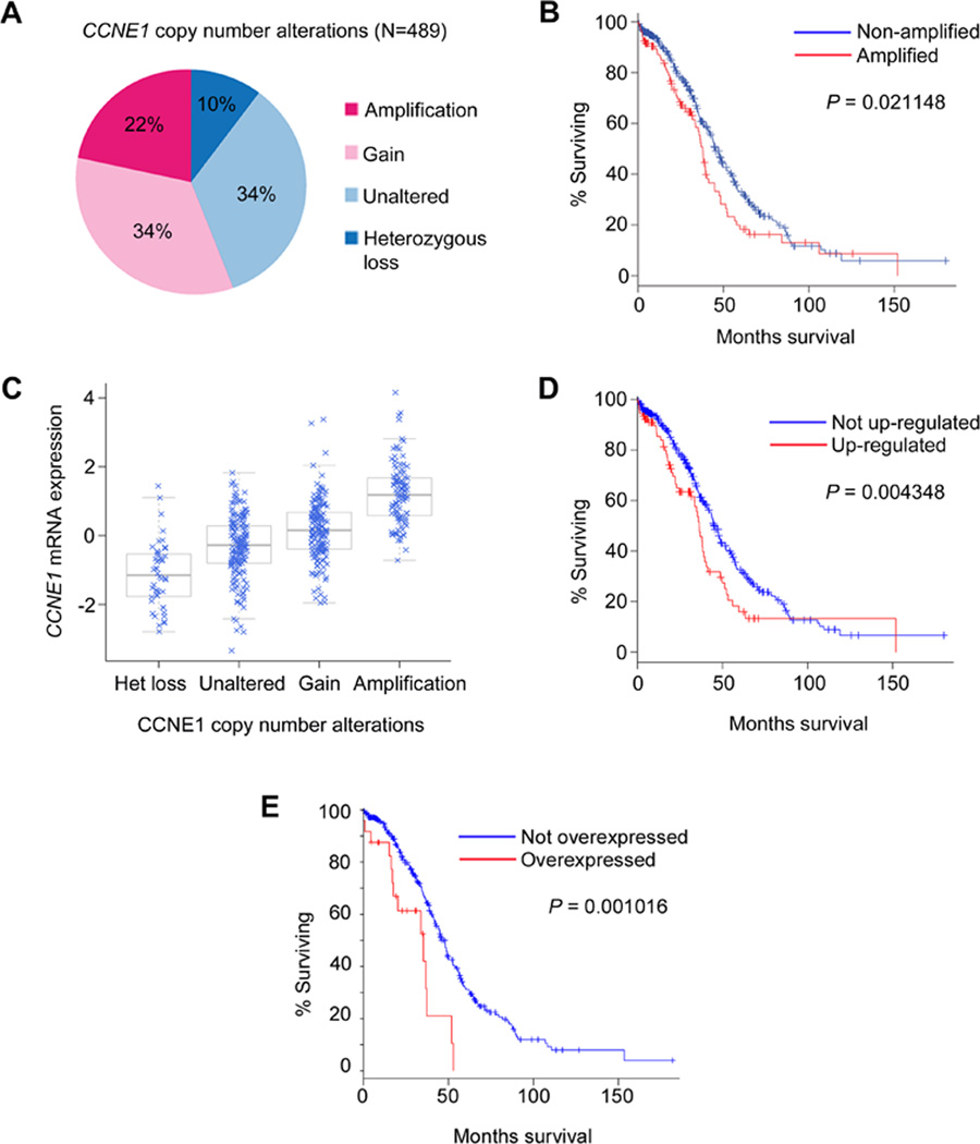
To determine the frequency of somatic CCNE1 amplifications in HGSOC, we queried The Cancer Genome Atlas (TCGA) database, which contains clinically annotated genomic data from 489 HGSOC samples (5). Datasets were analyzed using the Memorial Sloan-Kettering Cancer Center cBio Cancer Genomics Portal (http://www.cbioportal.org/public-portal/index.do) (22). Somatic copy number was determined by GISTIC analysis as previously described (22, 23). Our analysis revealed CCNE1 alterations in 319 of 489 cases, including 106 cases (21.7%) of amplification, 165 cases (33.7%) of copy number gain, and 48 cases (9.8%) of heterozygous loss (Fig. 1A). Consistent with previous reports (14, 24), CCNE1 amplifications were associated with reduced overall patient survival, determined by Kaplan-Meier analysis (P = 0.021148, log-rank test) (Fig. 1B). We next examined the mRNA data for these cases. As shown in Fig. 1C, CCNE1 mRNA levels tended to increase with copy number. Using a Z-score threshold of +1.5 to define up-regulation, we found that CCNE1 mRNA was up-regulated in 90/489 cases (18.4%) and associated with reduced overall survival (P = 0.004348, log-rank test) (Fig. 1D). Lastly, we examined the protein expression data, generated by reverse phase protein array (RPPA) analysis. We found that Cyclin E1 was overexpressed (Z-score >1.5) in 30/412 cases (7.1%) and, again, was strongly associated with reduced overall survival (P = 0.001016, log-rank test) (Fig. 1E).
Figure 1. CCNE1 alterations in high-grade serous ovarian carcinoma.
A, Distribution of somatic CCNE1 copy number alterations across 489 tumors. B, Reduced overall 5-year survival for patients with CCNE1 amplifications. C, Relationship between CCNE1 copy number and mRNA expression. D-E, Reduced overall 5-year survival for patients with elevated CCNE1 mRNA (D) or overexpression of Cyclin E1 protein (E) (Z-scores >1.5).
In just over half of amplified cases (56.6%; 60/106), amplification resulted in mRNA up-regulation, and conversely, two-thirds (66.7%; 60/90) of tumors with high mRNA levels had an underlying amplification. Correlation with protein expression, however, was lower. Although high Cyclin E1 protein levels could be attributed to CCNE1 amplification in 63.3% (19/30) of cases, only 24.4% (19/78) of amplified cases actually overexpressed Cyclin E1 protein. This seems surprisingly low and might be an underestimate due to the fact that RPPA data was available for only 78 amplified cases. Amplification without corresponding expression might indicate that amplified CCNE1 is not always constitutively transcribed or that transcription is countered by post-transcriptional repression, perhaps by microRNAs (25). Alternatively, low Cyclin E1 protein expression could reflect increased rates of ubiquitin-mediated degradation.
CCNE1 amplification is associated with increased Cyclin E1 protein expression
Given the discordance between amplification and protein expression in many TCGA samples, we sought to independently validate the data. To this end, we analyzed a TMA containing 140 primary HGSOC samples by FISH (n=87 informative cases) and IHC (n=138 informative cases)
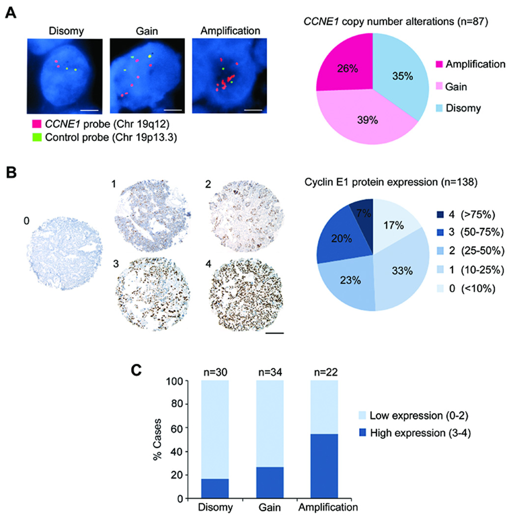
In the FISH analysis, 23 cases (26.4%) showed CCNE1 amplification, 34 cases (39.1%) showed relative CCNE1 gain, and 30 cases (34.5%) were disomic for chromosome 19 (Fig. 2A). FISH classification criteria are described in the Materials and Methods. In the IHC analysis, protein expression was scored on a scale of 0–4 according to the percentage of immunopositive tumor cells present (Fig. 2B). For statistical analysis, scores 0–2 (<50% positive cells) were considered “low expression” and scores 3–4 (>50% positive cells) were considered “high expression”. Using this scale, 38 cases (27.5%) were identified as high Cyclin E1 expressers (Fig. 2B). Integration of the FISH and IHC data revealed a significant difference in protein levels between cases with amplification, relative gain, or disomy (P = 0.0110, Chi-square test) (Fig. 2C). Twelve of 22 CCNE1-amplified cases (54.5%) were high expressers; a significantly higher proportion than in the TCGA dataset. Conversely, 12 of 26 high expressers (46.2%) were CCNE1-amplified. These data suggest that CCNE1 amplification is linked to high protein expression in 46–55% of HGSOC cases.
Figure 2. CCNE1 amplification is associated with increased Cyclin E1 protein expression.
A-B, A TMA containing 140 cases of primary high-grade serous ovarian carcinoma was analyzed by FISH and IHC to determine gene copy number and protein expression level, respectively. A, Dual-color FISH analysis using a CCNE1 probe (red) and chromosome 19 reference probe (green). Nuclei were counterstained with DAPI (blue). Graph represents distribution of CCNE1 copy number alterations across 87 cases. Scale bar = 5 µm. B, IHC analysis of Cyclin E1 expression. The percentage of immunoreactive tumor cells (brown) was scored on a scale of 0–4. Graph shows the distribution of expression scores across 138 cases. Scale bar = 100 µm. C, Integration of FISH and IHC data shows that Cyclin E1 expression is significantly higher in CCNE1-amplified cases (P = 0.0110, chi-square test).
Cyclin E1 overexpression occurs early in serous tumorigenesis
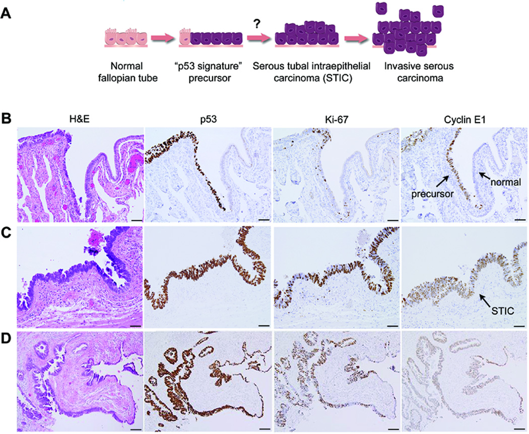
Although CCNE1 amplifications are present in late-stage HGSOC, it remains unclear whether CCNE1 deregulation contributes to early-stage tumor development. Given that tissue-targeted Cyclin E1 expression induces mammary and lung carcinogenesis in mice (26–28), we hypothesized that early CCNE1 aberrations may similarly drive HGSOC development. As previously mentioned, HGSOC likely arises from fallopian tube epithelium. We therefore asked whether Cyclin E1 is expressed in early serous lesions of the fallopian tube. We analyzed fallopian tube specimens from 14 patients clinically diagnosed with high-grade serous adenocarcinoma of the ovary or fallopian tube, containing lesions that spanned the morphologic continuum from normal epithelium to invasive carcinoma (Fig. 3A). This included putative precursor lesions called “p53 signatures” and non-invasive tumors termed “serous tubal intraepithelial carcinoma” (STIC). P53 signatures are stretches of secretory epithelial cells that exhibit intense p53 immunoreactivity but appear morphologically normal and have low proliferative activity (<10% Ki-67 positive nuclei) (3, 16) (Fig. 3B). P53 signatures have been shown to harbor DNA damage and somatic TP53 mutations that result in p53 nuclear accumulation (3). STICs are non-invasive lesions characterized by nuclear atypia, loss of polarity, increased nuclear/cytoplasmic ratio, and high proliferative activity (>50% Ki-67-positive nuclei) (29–32) (Fig. 3C,D). STICs also harbor somatic TP53 mutations and are usually p53-positive, although truncating mutations can result in loss of p53 immunoreactivity. It is hypothesized that p53 signatures are a normal physiologic entity possibly caused by damaging ovulatory factors, and that additional oncogenic events are required for p53 signatures to progress to STIC. P53-positive lesions exhibiting features intermediate between a p53 signature and STIC have been called “tubal intraepithelial lesions in transition” (TILT) (17). TILTs are also hypothesized to be precursors of STIC and we have grouped them with p53 signatures in our analysis. By immunostaining serial tissue sections for p53, Ki-67, and Cyclin E1, we identified precursor lesions (p53 signature/TILT) in 9 cases and STIC in 13 cases (Table 1). Normal tubal epithelium was present in 11 cases. Cyclin E1 expression in each lesion was scored as negative (<10% positive nuclei), low (10–50% positive nuclei), or high (>50% positive nuclei). We found that while Cyclin E1 was consistently absent in normal tubal epithelium, 3 out of 9 putative precursor lesions (33%) were Cyclin E1-positive (Table 1). As shown in Fig. 3B, Cyclin E1-positive cells outnumbered Ki-67-positive cells in the precursor lesions, indicating that Cyclin E1 expression was not simply a read out of proliferation. Our data show that Cyclin E1 overexpression can occur very early, even before STIC development, suggesting that deregulated CCNE1 might cooperate with mutant p53 to drive tumorigenesis. Several studies have demonstrated oncogenic synergism between Cyclin E1 and p53 defects, both in vitro and in vivo (10, 11, 28). Of the 13 STIC cases we analyzed, Cyclin E1 expression was high in 7 cases, (54%), low in 2 cases (15%), and absent in 4 cases (31%) (Fig. 3C,D and Table 1). This suggests that only a subset of STICs exhibit high-level Cyclin E1 expression and they may represent a distinct group of patients in which CCNE1 deregulation drives early-stage tumor progression.
Figure 3. Cyclin E1 expression occurs early during serous tumorigenesis.
A, An illustration of the proposed carcinogenic sequence for serous tumorigenesis in the fallopian tube epithelium. The “p53 signature” is hypothesized to precede STIC development. B, Example of high Cyclin E1 expression in a putative precursor lesion. Precursor cells are differentiated from adjacent normal cells by their nuclear p53 expression (brown), typically indicating somatic mutation of TP53. Note that Cyclin E1 is expressed in precursor cells but not in adjacent normal cells. The proliferation marker Ki-67 is absent in most precursor cells, indicating low proliferative activity. H&E (hematoxylin and eosin) staining highlights histologic features. C-D, Example of high Cyclin E1 expression in STIC, shown at high (C) and low (D) magnification. STICs are highly proliferative, indicated by high Ki-67 expression, and typically express mutant p53. STICs usually exhibit nuclear atypia and loss of polarity, evident by H&E staining. Scale bars = 20 µm (B-C), 50 µm (E).
Table 1.
Cyclin E1 expression in early-stage serous lesions of the fallopian tube
| Negative (<10% pos nuclei) (%) |
Low (10–50% pos nuclei) (%) |
High (>50% pos nuclei) (%) |
|
|---|---|---|---|
| Total cases, N=14 | |||
| Normal FTE (n=11) | 11/11(100) | 0/11 (0) | 0/11(0) |
| P53 signature or TILT (n=9) | 6/9 (66) | 0/9 (0) | 3/9 (33) |
| STIC (n=13) | 4/13 (31) | 2/13 (15) | 7/13 (54) |
FTE, fallopian tube epithelium; TILT, tubal intraepithelial lesion in transition; STIC, serous tubal intraepithelial carcinoma.
Constitutive Cyclin E1 expression drives over-proliferation of fallopian tube secretory epithelial cells (FTSECs)
To understand how Cyclin E1 might transform normal or p53-compromised secretory cells in the fallopian tube epithelium, we developed an in vitro model of Cyclin E1-mediated transformation using primary human cells. Secretory epithelial cells were enzymatically dissociated from the fimbrial region of a fallopian tube tissue sample and cultured in vitro using methods recently described (18, 19). The cells were promptly transduced with TERT (telomerase reverse transcriptase) to delay replicative senescence. Next, we tried transducing TERT-expressing cells with CCNE1, but it did not enhance their growth and they underwent senescence within 2 passages (data not shown), likely reflecting a p53-mediated response to excess Cyclin E1, as seen in other primary cell types (10, 33). To circumvent this outcome, we instead transduced TERT-expressing cells with mutant TP53R175H, one of the most common mutations found in p53 signatures, STIC, and HGSOC (3, 5). TP53R175H is a conformational mutant that exerts a dominant negative effect by hetero-oligomerizing with wild-type p53, thus compromising its ability to transactivate target genes (34). TP53R175H might also have gain-of-function oncogenic properties (34). Given that TP53 is often somatically mutated at the precursor stage, we reasoned that CCNE1 deregulation is likely to occur in the presence of mutant p53. Therefore, using TP53R175H to immortalize FTSECs is both physiologically relevant and chronologically accurate. FTSECs co-expressing TERT and p53R175H grew slowly but were proliferative enough to be expanded into a cell line, designated “FT282”.
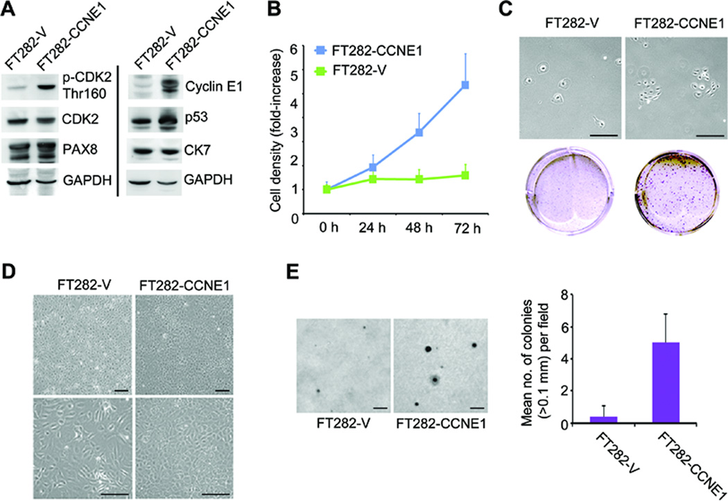
To characterize the effects of constitutive Cyclin E1 expression on p53-compromised but untransformed cells, we transduced FT282 cells with either CCNE1 or an empty vector control. The resulting cell lines were designated “FT282-CCNE1” and “FT282-V”, respectively. Western blot analysis was used to validate exogenous gene expression (Cyclin E1, p53) and to verify the expression of appropriate Müllerian lineage markers, namely PAX8 (paired box 8) and CK7 (cytokeratin-7) (Fig. 4A). Cyclin E1 overexpression triggered phosphorylation of its binding partner CDK2 (cyclin-dependent kinase 2) on Thr160, indicating CDK2 activation (35) (Fig. 4A). Accordingly, FT282-CCNE1 cells exhibited accelerated growth in SRB proliferation assays (Fig. 4B). FT282-CCNE1 cell density increased by 4.45-fold (± 0.96 s.d.) over 72 h whereas FT282-V cell density increased by only 1.61-fold (± 0.27 s.d.) (P =1.14^–11, Student’s t-test) (Fig. 4B). When seeded sparsely in clonogenic assays, FT282-CCNE1 cells exhibited clonal growth whereas FT282-V cells typically remained as single cells, often appearing senescent (Fig. 4C). When seeded at moderate density (~50%), FT282-V cells grew to 90–100% confluency and thereafter could be maintained as contact-inhibited monolayers for weeks. Conversely, FT282-CCNE1 cells continued to divide when confluent and quickly became overcrowded, indicating loss of contact inhibition (Fig. 4D). Tightly packed FT282-CCNE1 cultures remained viable, however, showing no evidence of stress-induced apoptosis. Lastly, Cyclin E1 overexpression induced a small but significant degree of anchorage–independent growth, evidenced by colony formation in soft agar. While FT282-V cells produced virtually no colonies (0.4 colonies/field ± 0.70 s.d.), FT282-CCNE1 cells formed small, slow-growing colonies (50 colonies/field ± 1.76 s.d.) (P = 4.47^–7, Student’s t-test) (Fig. 4E). Loss of contact inhibition and anchorage-independent growth by FT282-CCNE1 cells emerged at mid-passage number (~p20). FT282-V cells at equivalent passage number did not exhibit a similar phenotype. These data suggest that TP53 mutation alone does not drive FTSEC proliferation but rather impairs the G1/S checkpoint such that growth arrest cannot be executed in response to Cyclin E1 deregulation. Under these conditions, Cyclin E1 readily drives inappropriate cell growth.
Figure 4. Constitutive Cyclin E1 expression leads to uncontrolled growth of immortal FTSECs.
A, Primary human FTSECs immortalized with TERT and TP53R175H were transduced with CCNE1 (FT282-CCNE1) or empty vector (FT282-V). Western blot was used to validate expression of Cyclin E1, p53, and endogenous Müllerian lineage markers (PAX8, CK7). Phospho-CDK2-Thr160 indicates CDK2 activation. A composite blot is shown; samples were loaded in duplicate to accommodate numerous antibody incubations. Full length blots are presented in Supplementary Figure S1. B, Constitutive expression of Cyclin E1 increased cell proliferation rate (P = 1.14^–11 at t = 72 h, Student’s t-test). C, Cyclin E1 expression also promoted clonogenic growth. Top panel shows cells in culture; bottom panel shows cells fixed and stained with crystal violet. D, Cyclin E1 induced the loss of contact inhibition, resulting in overcrowded cell culture. Images depict confluent cells at low (upper panels) and high (lower panels) magnification. E, Cyclin E1 promoted subtle anchorage-independent colony formation in soft agar (P = 4.47^–7, Student’s t-test). Error bars represent s.d. Scale bars = 50 µm (B-C), 500 µm (E).
Constitutive Cyclin E1 expression causes DNA damage in FTSECs
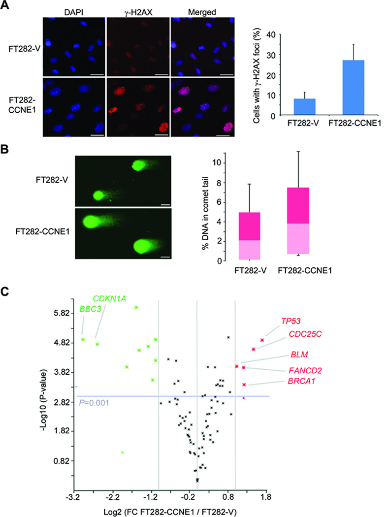
Recent studies suggest that deregulated Cyclin E1 induces genomic instability by generating DNA replication stress (36, 37). Replication stress, broadly defined as inefficient DNA replication, is caused by impediments to replication fork progression, for example, obstructive DNA lesions, replication inhibitors, or depletion of components required for DNA synthesis. Stalled replication forks are dangerous because they can collapse into double strand breaks. The mechanism by which Cyclin E1 generates replication stress is not completely clear. It was reported that deregulated Cyclin E1 forces S-phase entry without adequate nucleotide pools, resulting in incomplete DNA replication and fork collapse (36). Cyclin E1 overexpression has also been shown to cause excessive origin firing, which increases the number of active replication forks and creates spatial conflicts between replication and transcription machineries (37). To determine whether Cyclin E1 overexpression induced DNA damage in our model, we analyzed cells for the presence of γ-H2AX (p-H2A.X Ser139) nuclear foci by immunofluorescent staining. H2AX is a histone protein phosphorylated by ATM (ataxia telangiectasia mutated) or ATR (ataxia telangiectasia and Rad3 related) at sites of DNA damage. It is predominantly phosphorylated by ATM at double strand breaks (DSBs) (38). We detected prominent γ-H2AX foci in 27.2 % (± 7.6 % s.d.) of FT282-CCNE1 nuclei compared to only 8.0 % (± 3.1 % s.d.) of FT282-V nuclei, representing a ~3.4-fold increase in DSBs (P = 0.0035, Student’s t-test) (Fig. 5A). Since H2AX can also by phosphorylated by ATR at single strand breaks (SSBs) (38), we looked for evidence of SSBs using the Comet assay, in which migration of damaged DNA gives the appearance of a comet “tail” in single-cell gel electrophoresis (39). We found a significantly higher proportion of DNA in the tails of FT282-CCNE1 cells (5.09 % ± 5.09 % s.d.) compared to FT282-V cells (3.04% ± 3.37 % s.d.), representing a 1.67-fold increase in DNA damage (P = 0.000383, Student’s t-test) (Fig. 5B). It should be noted that active replication forks appear as SSBs in the Comet assay. Therefore, the increase in DNA damage could represent stalled or lagging replication forks, consistent with replication stress.
Figure 5. Cyclin E1 expression leads to DNA damage in immortal FTSECs.
A, Immunofluorescent staining of γ-H2AX nuclear foci at sites of DNA double strand breaks (red dots). Nuclei were counterstained with DAPI (blue). Cyclin E1-overexpressing cells had significantly more γ-H2AX foci than control cells (P = 0.0035, Student’s t-test). Graph shows quantification of foci (± s.d.). B, Comet assay for DNA damage. Comet “tails” represent DNA with single strand or double strand breaks. Cyclin E1-overexpressing cells had a greater proportion of DNA in “tails” than did controls cells, indicating increased DNA damage (P = 0.000383, Student’s t-test). Box plot shows the range of % tail DNA/cell. Scale bars = 50 µm (A,B). C, Expression of 84 DNA damage response genes in Cyclin E1-overexpressing cells compared to vector controls, measured by quantitative PCR array. Volcano plot shows genes up-regulated (red) or down-regulated (green) by >2-fold (P < 0.0005). Fold-change values are presented in Table 2 and Supplementary Table S2.
Finally, we asked whether Cyclin E1-induced DNA damage would alter the regulation of DNA damage response (DDR) genes. Using quantitative PCR array profiling, we analyzed the expression of 84 DDR-related genes and identified 14 genes differentially expressed in FT282-CCNE1 cells compared to FT282-V cells (> 2-fold difference, P<0.001, Student’s t-test) (Fig. 5C and Supplementary Table S2). Up-regulated genes included TP53, CDC25C, BRCA1, FANCD2, and BLM (Table 2). Down-regulated genes included BBC3, CDKN1A, ATM, MPG, B2M, DDB2, GADD45G, MAPK12, and BAX. Although TP53 was up-regulated (+3.3-fold), it failed to transactivate its transcriptional targets, CDKN1A (p21CIP1) and BBC3 (Puma), consistent with the exertion of a dominant negative effect by ectopic p53R175H. P21CIP1 and Puma are key effectors of p53-induced cell cycle arrest and apoptosis, respectively (34). CDKN1A and BBC3 were down-regulated (−6.0 and −7.8-fold), suggesting that constitutive Cyclin E1 expression might repress these genes. Other alterations that would promote cell viability and proliferation included up-regulation of CDC25C (+2.78), which promotes mitotic entry; down-regulation of pro-apoptotic BAX (−2.08), down-regulation of GADD45G (-2.20), which induces growth arrest after DNA damage, and down-regulation of MAPK12 (−2.09), a stress-activated negative regulator of cell cycle progression. These results are consistent with our observation that confluent FT282-CCNE1 cells continue to proliferate and exhibit no apoptosis (Fig. 4D). Most of the genes found up-regulated (BRCA1, FANCD2, BLM, XRCC2) are directly involved in DNA damage repair, suggesting that Cyclin E1 overexpression may prompt the cell to increase its DNA repair capacity by up-regulating key repair factors.
Table 2.
DNA damage response genes up- or down-regulated in FT282-CCNE1 cells relative to FT282-V cells
| Gene Symbol | Fold-Regulation | P-value* |
|---|---|---|
| TP53 | 3.2583 | 0.000012 |
| CDC25C | 2.7845 | 0.000025 |
| BRCA1 | 2.3415 | 0.000391 |
| FANCD2 | 2.3307 | 0.000101 |
| XRCC2 | 2.3307 | 0.001179 |
| BLM | 2.0150 | 0.000093 |
| BAX | −2.0838 | 0.000061 |
| MAPK12 | −2.0935 | 0.000012 |
| GADD45G | −2.2026 | 0.000285 |
| DDB2 | −2.4103 | 0.000020 |
| B2M | −2.8664 | 0.000027 |
| MPG | −2.9812 | 0.000001 |
| ATM | −3.5208 | 0.000101 |
| CDKN1A | −6.0317 | 0.000017 |
| BBC3 | −7.8312 | 0.000012 |
P-values were calculated based on Student’s t-tests of the triplicate 2^(-ΔCt) values for each target gene in the control (FT282-V) and treatment (FT282-CCNE1) groups.
Discussion
The molecular pathogenesis of HGSOC remains poorly understood and oncogenic drivers of this cancer must be identified in order to guide novel therapeutic approaches. In this study we have characterized the oncogenic role of CCNE1 in HGSOC development and provided evidence that CCNE1 deregulation may contribute to serous tumorigenesis and chromosomal instability in the fallopian tube.
First, we identified cases in The Cancer Genome Atlas database with somatic CCNE1 gene amplification, mRNA upregulation, and/or protein overexpression, and showed that each alteration type is associated with reduced overall survival. This finding suggests that deregulated CCNE1 is an oncogenic driver of late-stage HGSOC progression when amplified, transcriptionally up-regulated, or overexpressed, although reduced survival rates may also reflect resistance to platinum-based chemotherapy, previously found to be associated with the CCNE1 amplicon (13, 14). Using FISH and IHC analysis, we showed that CCNE1 amplifications lead to high-level Cyclin E1 protein expression in >50% of cases. In agreement with our results, two studies have reported correlations between CCNE1 amplification, Cyclin E1 expression, and reduced survival in ovarian cancer (14, 24). However, one of them included multiple tumor subtypes in their analysis. Since there is now strong evidence that HGSOC pathogenesis differs dramatically from that of other subtypes (40), we restricted our study to high-grade serous tumors only, thus eliminating the possibility of subtype-specific confounding factors.
Secondly, we showed that CCNE1 deregulation might promote serous tumorigenesis within the fallopian tube epithelium. IHC analysis of fallopian tubes from HGSOC patients revealed Cyclin E1 overexpression in a subset of putative precursors and non-invasive lesions, thus implicating Cyclin E1 at the earliest stages of tumor development. A recent study examining Cyclin E1 expression in the fallopian tubes of 23 HGSOC patients noted a Cyclin E1-positive precursor in one case (41). Our data bolsters that finding and provides additional evidence for CCNE1 deregulation at the precursor stage. It has been reported that Cyclin E1 is present in a majority of STICs, although expression levels varied considerably (3, 41). Sehdev et al found that Cyclin E1 was expressed in 24/35 STICs (69%) collected from 22 patients (41). However, only 16 (46%) of those STICs exhibited high expression (>50% positive cells). We propose that high-level Cyclin E1 expression could highlight specific cases in which tumor development is driven by CCNE1 deregulation as opposed to other oncogenic mechanisms. Given that TP53 mutations are present in nearly all STICs and many putative precursors (3, 31), p53 dysfunction is likely required for Cyclin E1-driven tumorigenesis in the fallopian tube. It has been shown that excess Cyclin E1 triggers a DNA damage response, causing p53 stabilization and activation(42). Activated p53 rapidly intervenes by transcriptionally up-regulating the Cyclin E1/CDK2 inhibitor CDKN1A (p21CIP1), which shuts down CDK2 activity and mediates cell cycle arrest. If p21 is disabled, p53 will instead activate apoptosis through alternate mediators in order to mitigate the effects of Cyclin E1. Thus, p53 must be mutated (or otherwise inactivated) for the cell to tolerate Cyclin E1 deregulation (42). While our data support the hypothesis that CCNE1 deregulation is an early event, the causes of CCNE1 deregulation in the fallopian tube epithelium remain ill defined. Gene amplification is one possibility; two studies reported recurrent CCNE1 copy number gains/amplifications in early-stage (FIGO Stage I-II) fallopian tube carcinomas (43, 44). However, given that >40% of our Cyclin E1-overexpressing TMA samples lacked amplification (Fig. 2C), additional mechanisms of CCNE1 up-regulation must be at play, possibly transcriptional activation by other oncogenes or decreased ubiquitin-mediated degradation (8).
Thirdly, after observing Cyclin E1 expression in early tubal lesions, we created a model of Cyclin E1-mediated transformation using primary human FTSECs. The cells were immortalized in a physiologically relevant manner by introducing a form of mutant p53 (TP53R175H) that is found in both p53 signatures and STICs (3, 31). Through a series of in vitro assays, we demonstrated that constitutive Cyclin E1 expression induces over-proliferation of p53-compromised FTSECs and the acquisition of malignant characteristics such as clonal growth ability, loss of contact inhibition, and colony formation in soft agar. It should be noted that Cyclin E1 overexpression did not result in an aggressively malignant phenotype, like that seen with H-RASV12 or c-MYC (19), suggesting that additional genetic events are required for full transformation. To determine what those might be, we are currently screening genes that are coamplified or co-expressed with CCNE1 in HGSOC (for example, TPX2 (14)) for their ability to transform FTSECs. Alternatively, Cyclin E1 might require cleavage to reach its full oncogenic potential. A number of studies have shown that low molecular weight forms of Cyclin E1, produced by elastase-mediated cleavage, cause genomic instability and promote aggressive tumor phenotypes in breast cancer (45, 46). It has also been reported that the chromatin remodeling protein Rsf-1 (remodeling and spacing factor 1) is co-expressed with Cyclin E1 in HGSOC and that Rsf-1/Cyclin E1 complexes promote the transformation of immortalized rat kidney cells expressing mutant p53R175H (47). Interestingly, Rsf-1/Cyclin E1 complexes required p53R175H to induce tumorigenicity and could not transform p53 wild-type cells, again underscoring the importance of mutant p53 in Cyclin E1-mediated tumorigenesis.
Lastly, we demonstrated that Cyclin E1-overexpression in FTSECs causes DNA damage and alters the expression of DNA damage response genes. Multiple negative cell cycle regulators that are normally activated by DNA damage were down-regulated. Meanwhile, specific DNA repair genes were up-regulated, including BRCA1, FANCD2, XRCC2, and BLM. Interestingly, these genes are central components of a “fork protection pathway” that protects stalled DNA replication forks from degradation and facilitates fork recovery. Schlacher et al has shown that monoubiquitinated FANCD2 (Fanconi anemia, complementation group D2) complexes with BRCA1 to stabilize stalled forks and that BLM (Bloom syndrome, RecQ helicase-like) is required for efficient replication recovery (48). Chaudhury et al additionally reported that FANCD2 is a critical regulator of BLM helicase activity and that the two proteins act in concert to restart stalled replication forks (49). XRCC2 is a RAD51 paralog that complexes with RAD51C, another key fork stabilization factor (48). Up-regulation of these genes possibly enables cells to manage high levels of replication stress brought on by CCNE1 deregulation. Up-regulation of BRCA1 is notable given the rarity of CCNE1 amplification in BRCA1-mutated HGSOC. CCNE1-amplified tumors might depend on BRCA1 to mitigate replication stress and thus maintain cell viability in the face of accumulating chromosomal instability.
In sum, our data support a model whereby CCNE1 deregulation drives uncontrolled growth of FTSECs harboring somatic p53 defects and causes DNA damage by inducing replication stress, thus generating chromosomal instability and promoting tumorigenesis.
Supplementary Material
Acknowledgements
The authors thank the BWH Department of Pathology faculty and staff for allocation of tissues, and members of the Drapkin and Bowtell laboratories for fruitful discussions.
Grant Support
This work was supported by grants from the National Cancer Institute at the NIH P50-CA105009 (RD), NIH U01 CA-152990 (RD), NIH R21 CA-156021 (RD); the Honorable Tina Brozman ‘Tina’s Wish’ Foundation (RD), the Dr. Miriam and Sheldon G. Adelson Medical Research Foundation (RD), The Robert and Debra First Fund (RD), The Gamel Family Fund (RD), a Canadian Institutes of Health Research Fellowship (AK), a Kaleidoscope of Hope Foundation Young Investigator Research Grant (AK), and a National Health and Medical Research Council (NHMRC) project grant (APP 1042358) (DB).
Footnotes
Conflicts of interest: No conflicts of interest declared.
Author contributions: AK, PJ, NV, AL and RD designed and/or performed experiments. JL, MH, DE, and DB provided critical experimental materials; AK wrote the manuscript; RD edited the manuscript.
References
- 1.Boyle PLB. International Agency for Research on Cancer, and World Health Organization, “World cancer report 2008”. WHO press; 2008. [Google Scholar]
- 2.Nik NN, Vang R, Shih IM, Kurman RJ. Origin and Pathogenesis of Pelvic (Ovarian, Tubal, and Primary Peritoneal) Serous Carcinoma. Annu Rev Pathol. 2013;16:27–45. doi: 10.1146/annurev-pathol-020712-163949. [DOI] [PubMed] [Google Scholar]
- 3.Lee Y, Miron A, Drapkin R, Nucci MR, Medeiros F, Saleemuddin A, et al. A candidate precursor to serous carcinoma that originates in the distal fallopian tube. J Pathol. 2007;211:26–35. doi: 10.1002/path.2091. [DOI] [PubMed] [Google Scholar]
- 4.Dubeau L, Drapkin R. Coming into focus: the nonovarian origins of ovarian cancer. Ann Oncol. 2013;24(Suppl 8):viii28–viii35. doi: 10.1093/annonc/mdt308. [DOI] [PMC free article] [PubMed] [Google Scholar]
- 5.TCGA. Integrated genomic analyses of ovarian carcinoma. Nature. 2011;474:609–615. doi: 10.1038/nature10166. [DOI] [PMC free article] [PubMed] [Google Scholar]
- 6.Gorringe KL, George J, Anglesio MS, Ramakrishna M, Etemadmoghadam D, Cowin P, et al. Copy number analysis identifies novel interactions between genomic loci in ovarian cancer. PLoS One. 2010;5:e11408. doi: 10.1371/journal.pone.0011408. [DOI] [PMC free article] [PubMed] [Google Scholar]
- 7.Bowtell DD. The genesis and evolution of high-grade serous ovarian cancer. Nat Rev Cancer. 2010;10:803–808. doi: 10.1038/nrc2946. [DOI] [PubMed] [Google Scholar]
- 8.Siu KT, Rosner MR, Minella AC. An integrated view of cyclin E function and regulation. Cell Cycle. 2012;11:57–64. doi: 10.4161/cc.11.1.18775. [DOI] [PMC free article] [PubMed] [Google Scholar]
- 9.Spruck CH, Won KA, Reed SI. Deregulated cyclin E induces chromosome instability. Nature. 1999;401:297–300. doi: 10.1038/45836. [DOI] [PubMed] [Google Scholar]
- 10.Minella AC, Swanger J, Bryant E, Welcker M, Hwang H, Clurman BE. p53 and p21 form an inducible barrier that protects cells against cyclin E-cdk2 deregulation. Curr Biol. 2002;12:1817–1827. doi: 10.1016/s0960-9822(02)01225-3. [DOI] [PubMed] [Google Scholar]
- 11.Loeb KR, Kostner H, Firpo E, Norwood T, K DT, Clurman BE, et al. A mouse model for cyclin E-dependent genetic instability and tumorigenesis. Cancer Cell. 2005;8:35–47. doi: 10.1016/j.ccr.2005.06.010. [DOI] [PubMed] [Google Scholar]
- 12.Engler DA, Gupta S, Growdon WB, Drapkin RI, Nitta M, Sergent PA, et al. Genome wide DNA copy number analysis of serous type ovarian carcinomas identifies genetic markers predictive of clinical outcome. PLoS ONE. 2012;7:e30996. doi: 10.1371/journal.pone.0030996. [DOI] [PMC free article] [PubMed] [Google Scholar]
- 13.Etemadmoghadam D, deFazio A, Beroukhim R, Mermel C, George J, Getz G, et al. Integrated genome-wide DNA copy number and expression analysis identifies distinct mechanisms of primary chemoresistance in ovarian carcinomas. Clin Cancer Res. 2009;15:1417–1427. doi: 10.1158/1078-0432.CCR-08-1564. [DOI] [PMC free article] [PubMed] [Google Scholar]
- 14.Etemadmoghadam D, George J, Cowin PA, Cullinane C, Kansara M, Gorringe KL, et al. Amplicon-dependent CCNE1 expression is critical for clonogenic survival after cisplatin treatment and is correlated with 20q11 gain in ovarian cancer. PLoS ONE. 2010;5:e15498. doi: 10.1371/journal.pone.0015498. [DOI] [PMC free article] [PubMed] [Google Scholar]
- 15.Liu JF, Hirsch MS, Lee H, Matulonis UA. Prognosis and hormone receptor status in older and younger patients with advanced-stage papillary serous ovarian carcinoma. Gynecol Oncol. 2009;115:401–406. doi: 10.1016/j.ygyno.2009.08.023. [DOI] [PubMed] [Google Scholar]
- 16.Mehrad M, Ning G, Chen EY, Mehra KK, Crum CP. A Pathologist’s Road Map to Benign, Precancerous, and Malignant Intraepithelial Proliferations in the Fallopian Tube. Adv Anat Pathol. 2010;17:293–302. doi: 10.1097/PAP.0b013e3181ecdee1. [DOI] [PubMed] [Google Scholar]
- 17.Jarboe E, Folkins A, Nucci MR, Kindelberger D, Drapkin R, Miron A, et al. Serous carcinogenesis in the fallopian tube: a descriptive classification. Int J Gynecol Pathol. 2008;27:1–9. doi: 10.1097/pgp.0b013e31814b191f. [DOI] [PubMed] [Google Scholar]
- 18.Karst AM, Drapkin R. Primary culture and immortalization of human fallopian tube secretory epithelial cells. Nat Protoc. 2012;7:1755–1764. doi: 10.1038/nprot.2012.097. [DOI] [PMC free article] [PubMed] [Google Scholar]
- 19.Karst AM, Levanon K, Drapkin R. Modeling high-grade serous ovarian carcinogenesis from the fallopian tube. Proc Natl Acad Sci U S A. 2011;108:7547–7552. doi: 10.1073/pnas.1017300108. [DOI] [PMC free article] [PubMed] [Google Scholar]
- 20.Counter CM, Hahn WC, Wei W, Caddle SD, Beijersbergen RL, Lansdorp PM, et al. Dissociation among in vitro telomerase activity, telomere maintenance, and cellular immortalization. Proc Natl Acad Sci U S A. 1998;95:14723–14728. doi: 10.1073/pnas.95.25.14723. [DOI] [PMC free article] [PubMed] [Google Scholar]
- 21.Junk DJ, Vrba L, Watts GS, Oshiro MM, Martinez JD, Futscher BW. Different mutant/wild-type p53 combinations cause a spectrum of increased invasive potential in nonmalignant immortalized human mammary epithelial cells. Neoplasia. 2008;10:450–461. doi: 10.1593/neo.08120. [DOI] [PMC free article] [PubMed] [Google Scholar]
- 22.Cerami E, Gao J, Dogrusoz U, Gross BE, Sumer SO, Aksoy BA, et al. The cBio cancer genomics portal: an open platform for exploring multidimensional cancer genomics data. Cancer Discov. 2012;2:401–404. doi: 10.1158/2159-8290.CD-12-0095. [DOI] [PMC free article] [PubMed] [Google Scholar]
- 23.Gao J, Aksoy BA, Dogrusoz U, Dresdner G, Gross B, Sumer SO, et al. Integrative analysis of complex cancer genomics and clinical profiles using the cBioPortal. Sci Signal. 2013;6:pl1. doi: 10.1126/scisignal.2004088. [DOI] [PMC free article] [PubMed] [Google Scholar]
- 24.Nakayama N, Nakayama K, Shamima Y, Ishikawa M, Katagiri A, Iida K, et al. Gene amplification CCNE1 is related to poor survival and potential therapeutic target in ovarian cancer. Cancer. 2010;116:2621–2634. doi: 10.1002/cncr.24987. [DOI] [PubMed] [Google Scholar]
- 25.Ofir M, Hacohen D, Ginsberg D. MiR-15 and miR-16 are direct transcriptional targets of E2F1 that limit E2F–induced proliferation by targeting cyclin E. Mol Cancer Res. 2011;9:440–447. doi: 10.1158/1541-7786.MCR-10-0344. [DOI] [PubMed] [Google Scholar]
- 26.Bortner DM, Rosenberg MP. Induction of mammary gland hyperplasia and carcinomas in transgenic mice expressing human cyclin E. Mol Cell Biol. 1997;17:453–459. doi: 10.1128/mcb.17.1.453. [DOI] [PMC free article] [PubMed] [Google Scholar]
- 27.Ma Y, Fiering S, Black C, Liu X, Yuan Z, Memoli VA, et al. Transgenic cyclin E triggers dysplasia and multiple pulmonary adenocarcinomas. Proc Natl Acad Sci U S A. 2007;104:4089–4094. doi: 10.1073/pnas.0606537104. [DOI] [PMC free article] [PubMed] [Google Scholar]
- 28.Smith AP, Henze M, Lee JA, Osborn KG, Keck JM, Tedesco D, et al. Deregulated cyclin E promotes p53 loss of heterozygosity and tumorigenesis in the mouse mammary gland. Oncogene. 2006;25:7245–7259. doi: 10.1038/sj.onc.1209713. [DOI] [PubMed] [Google Scholar]
- 29.Piek JM, van Diest PJ, Zweemer RP, Jansen JW, Poort-Keesom RJ, Menko FH, et al. Dysplastic changes in prophylactically removed Fallopian tubes of women predisposed to developing ovarian cancer. J Pathol. 2001;195:451–456. doi: 10.1002/path.1000. [DOI] [PubMed] [Google Scholar]
- 30.Paley PJ, Swisher EM, Garcia RL, Agoff SN, Greer BE, Peters KL, et al. Occult cancer of the fallopian tube in BRCA-1 germline mutation carriers at prophylactic oophorectomy: a case for recommending hysterectomy at surgical prophylaxis. Gynecol Oncol. 2001;80:176–180. doi: 10.1006/gyno.2000.6071. [DOI] [PubMed] [Google Scholar]
- 31.Kindelberger DW, Lee Y, Miron A, Hirsch MS, Feltmate C, Medeiros F, et al. Intraepithelial carcinoma of the fimbria and pelvic serous carcinoma: Evidence for a causal relationship. Am J Surg Pathol. 2007;31:161–169. doi: 10.1097/01.pas.0000213335.40358.47. [DOI] [PubMed] [Google Scholar]
- 32.Medeiros F, Muto MG, Lee Y, Elvin JA, Callahan MJ, Feltmate C, et al. The tubal fimbria is a preferred site for early adenocarcinoma in women with familial ovarian cancer syndrome. Am J Surg Pathol. 2006;30:230–236. doi: 10.1097/01.pas.0000180854.28831.77. [DOI] [PubMed] [Google Scholar]
- 33.Bartkova J, Rezaei N, Liontos M, Karakaidos P, Kletsas D, Issaeva N, et al. Oncogene-induced senescence is part of the tumorigenesis barrier imposed by DNA damage checkpoints. Nature. 2006;444:633–637. doi: 10.1038/nature05268. [DOI] [PubMed] [Google Scholar]
- 34.Brosh R, Rotter V. When mutants gain new powers: news from the mutant p53 field. Nat Rev Cancer. 2009;9:701–713. doi: 10.1038/nrc2693. [DOI] [PubMed] [Google Scholar]
- 35.Brown NR, Noble ME, Lawrie AM, Morris MC, Tunnah P, Divita G, et al. Effects of phosphorylation of threonine 160 on cyclin-dependent kinase 2 structure and activity. J Biol Chem. 1999;274:8746–8756. doi: 10.1074/jbc.274.13.8746. [DOI] [PubMed] [Google Scholar]
- 36.Bester AC, Roniger M, Oren YS, Im MM, Sarni D, Chaoat M, et al. Nucleotide deficiency promotes genomic instability in early stages of cancer development. Cell. 2011;145:435–446. doi: 10.1016/j.cell.2011.03.044. [DOI] [PMC free article] [PubMed] [Google Scholar]
- 37.Jones RM, Mortusewicz O, Afzal I, Lorvellec M, Garcia P, Helleday T, et al. Increased replication initiation and conflicts with transcription underlie Cyclin E-induced replication stress. Oncogene. 2013;32:3744–3753. doi: 10.1038/onc.2012.387. [DOI] [PubMed] [Google Scholar]
- 38.Podhorecka M, Skladanowski A, Bozko P. H2AX Phosphorylation: Its Role in DNA Damage Response and Cancer Therapy. J Nucleic Acids. 2010 doi: 10.4061/2010/920161. 2010. [DOI] [PMC free article] [PubMed] [Google Scholar]
- 39.Olive PL, Banath JP. The comet assay: a method to measure DNA damage in individual cells. Nat Protoc. 2006;1:23–29. doi: 10.1038/nprot.2006.5. [DOI] [PubMed] [Google Scholar]
- 40.Karst AM, Drapkin R. Ovarian cancer pathogenesis: a model in evolution. J Oncol. 2010:932371. doi: 10.1155/2010/932371. 2010. [DOI] [PMC free article] [PubMed] [Google Scholar]
- 41.Sehdev AS, Kurman RJ, Kuhn E, Shih Ie M. Serous tubal intraepithelial carcinoma upregulates markers associated with high-grade serous carcinomas including Rsf-1 (HBXAP), cyclin E and fatty acid synthase. Mod Pathol. 2010;23:844–855. doi: 10.1038/modpathol.2010.60. [DOI] [PMC free article] [PubMed] [Google Scholar]
- 42.Minella AC, Grim JE, Welcker M, Clurman BE. p53 and SCFFbw7 cooperatively restrain cyclin E-associated genome instability. Oncogene. 2007;26:6948–6953. doi: 10.1038/sj.onc.1210518. [DOI] [PubMed] [Google Scholar]
- 43.Nowee ME, Snijders AM, Rockx DA, de Wit RM, Kosma VM, Hamalainen K, et al. DNA profiling of primary serous ovarian and fallopian tube carcinomas with array comparative genomic hybridization and multiplex ligation-dependent probe amplification. J Pathol. 2007;213:46–55. doi: 10.1002/path.2217. [DOI] [PubMed] [Google Scholar]
- 44.Snijders AM, Nowee ME, Fridlyand J, Piek JM, Dorsman JC, Jain AN, et al. Genome-wide-array-based comparative genomic hybridization reveals genetic homogeneity and frequent copy number increases encompassing CCNE1 in fallopian tube carcinoma. Oncogene. 2003;22:4281–4286. doi: 10.1038/sj.onc.1206621. [DOI] [PubMed] [Google Scholar]
- 45.Bagheri-Yarmand R, Biernacka A, Hunt KK, Keyomarsi K. Low molecular weight cyclin E overexpression shortens mitosis, leading to chromosome missegregation and centrosome amplification. Cancer Res. 2010;70:5074–5084. doi: 10.1158/0008-5472.CAN-09-4094. [DOI] [PMC free article] [PubMed] [Google Scholar]
- 46.Duong MT, Akli S, Wei C, Wingate HF, Liu W, Lu Y, et al. LMW-E/CDK2 deregulates acinar morphogenesis, induces tumorigenesis, and associates with the activated b-Raf-ERK1/2-mTOR pathway in breast cancer patients. PLoS Genet. 2012;8:e1002538. doi: 10.1371/journal.pgen.1002538. [DOI] [PMC free article] [PubMed] [Google Scholar]
- 47.Sheu JJ, Choi JH, Guan B, Tsai FJ, Hua CH, Lai MT, et al. Rsf-1, a chromatin remodelling protein, interacts with cyclin E1 and promotes tumour development. J Pathol. 2013;229:559–568. doi: 10.1002/path.4147. [DOI] [PMC free article] [PubMed] [Google Scholar]
- 48.Schlacher K, Wu H, Jasin M. A distinct replication fork protection pathway connects Fanconi anemia tumor suppressors to RAD51-BRCA1/2. Cancer Cell. 2012;22:106–116. doi: 10.1016/j.ccr.2012.05.015. [DOI] [PMC free article] [PubMed] [Google Scholar]
- 49.Chaudhury I, Sareen A, Raghunandan M, Sobeck A. FANCD2 regulates BLM complex functions independently of FANCI to promote replication fork recovery. Nucleic Acids Res. 2013;41:6444–6459. doi: 10.1093/nar/gkt348. [DOI] [PMC free article] [PubMed] [Google Scholar]
Associated Data
This section collects any data citations, data availability statements, or supplementary materials included in this article.


