Abstract
Additional cuneiform bones of the foot have been described in reference to the medial bipartite cuneiform or as small accessory ossicles. An additional middle cuneiform has not been previously documented. We present the case of a patient with an additional ossicle that has the appearance and location of an additional middle cuneiform. Recognizing such an anatomical anomaly is essential for ruling out second metatarsal base or middle cuneiform fractures and for the preoperative planning of arthrodesis or open reduction and internal fixation procedures in this anatomical location.
INTRODUCTION
The middle cuneiform is the smallest of the three cuneiform bones and articulates proximally with the navicular and distally with the base of the second metatarsal. Medial and lateral surfaces are partly articular with the medial and lateral cuneiforms, respectively. The ossification centre of the middle cuneiform appears during the second to fourth years of life and is the last of the cuneiforms to ossify. It completely ossifies by the sixth year of life [1].
The most recognized developmental anomaly of the cuneiform bones is a bipartite medial cuneiform. This is a rare, segmentation defect that is commonly asymptomatic and mistaken for a fracture. It occurs in 0.3% of the population, more commonly in males and bilaterally. Segmentation may be complete or incomplete and occurs horizontally (axially) into a smaller dorsal (os cuneiform-I dorsale) and larger planter (os cuneiform-I plantare) segment. Both segments articulate with the first metatarsal distally, proximally with the navicular and medially with the middle cuneiform [2].
Middle and lateral cuneiform anomalies are rare. However, there are several recognized accessory ossicles in proximity that are described. The os intercuneiforme occurs in 0.03% population and is present on the dorsum of the mid-foot in an interval between the medial and middle cuneiforms just distal to the navicular. It is triangular in appearance and thought to belong to the medial as opposed to the middle cuneiform [3]. Very rarely an accessory bone element called the os cuneo-I metatarsale-II dorsale is found interposed dorsally between the middle cuneiform and the base of the second metatarsal [4]. The os cuneo-I metatarsale-I plantare is similar to the previous accessory bone but occurs on the planter aspect of the foot at the base of the first metatarsal and articulates with the planter base of the first metatarsal and the first cuneiform [5]. Both were described as being peppercorn and cherrystone in size, respectively. The uncinate process of the lateral cuneiform is also considered a normal variant, which is located on its planter side by the base of the third metatarsal. If this occurs in isolation, it can also be termed the os unci [6]. All these accessory ossicles are much inferior to the size of their neighbouring tarsal bones and do not form articulating joint surfaces.
There has been no documented occurrence of an additional middle or lateral cuneiform. We present the case of a patient who has an additional bone identical in size to the middle cuneiform, being located between the second metatarsal base and the middle cuneiform. We postulate that this is an additional middle cuneiform.
CASE REPORT
A 63-year-old male complained of ongoing pain in the tarsometatarsal joints (TMTJs) and first metatarsophalangeal joint (MTPJ) of his left foot for 6 years. He denied any previous history of trauma or congenital abnormality, and there was no relevant family history.
Examination revealed hallux valgus and hammering of the second toe. There was swelling of the foot dorsum and first MTPJ with maximal tenderness over the first, second and third TMTJs and the first MTPJ. Passive motion of these joints was stiff and painful. The foot had normal neurovascular examination.
Dorsoplanter, lateral and oblique radiographs demonstrated an additional bone immediately distal to the middle cuneiform articulating with the second metatarsal base and middle cuneiform. Second and third metatarsals were shorßt in comparison with normal (Figs 1–3). Osteoarthritis was seen in all TMTJs and the first MTPJ. A computed tomography (CT) scan confirmed the presence of the additional bone and degenerative joint disease (Figs 4 and 5).
Figure 2:
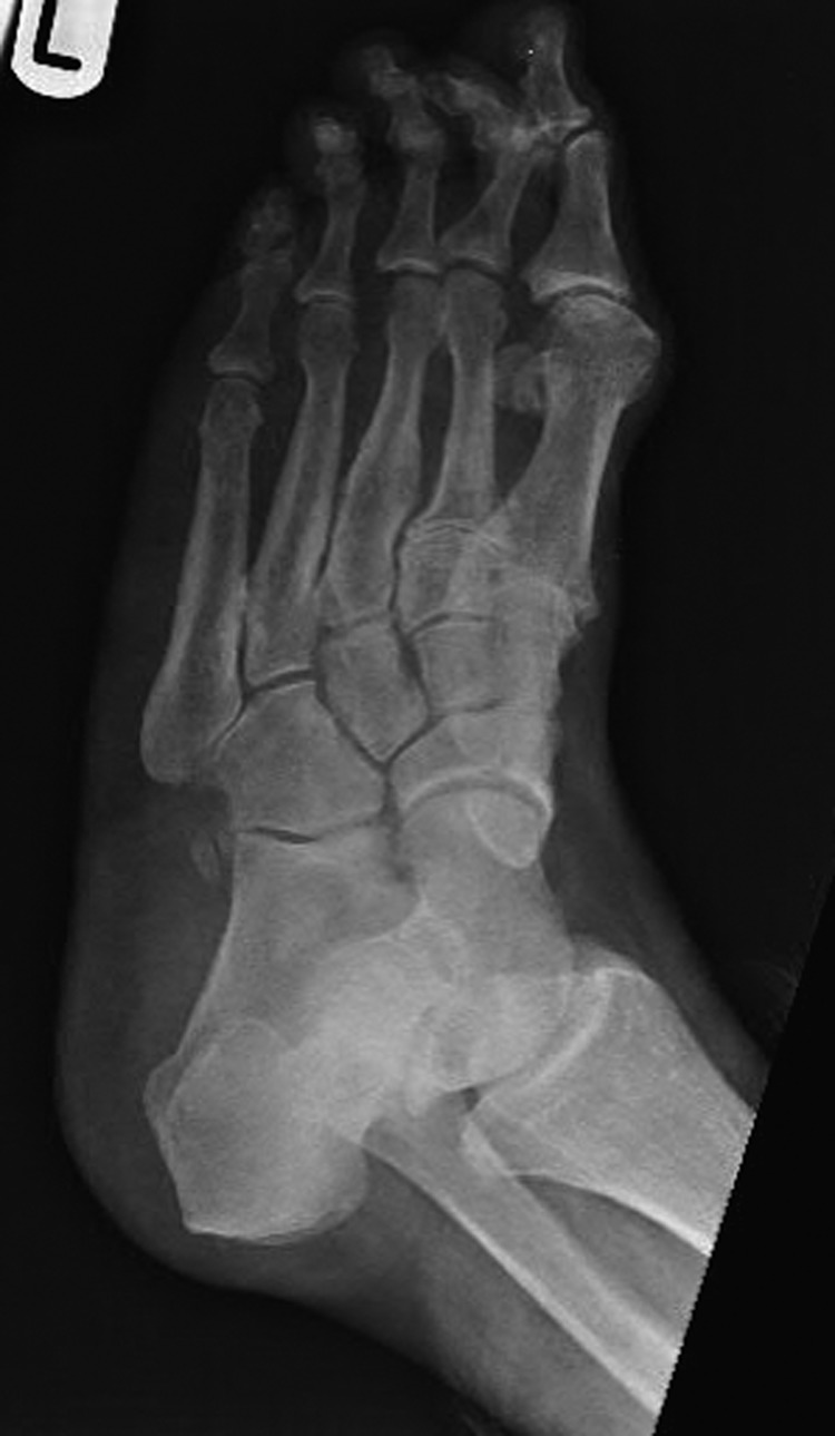
Oblique radiograph of the left foot with the clearly visible additional middle cuneiform.
Figure 1:
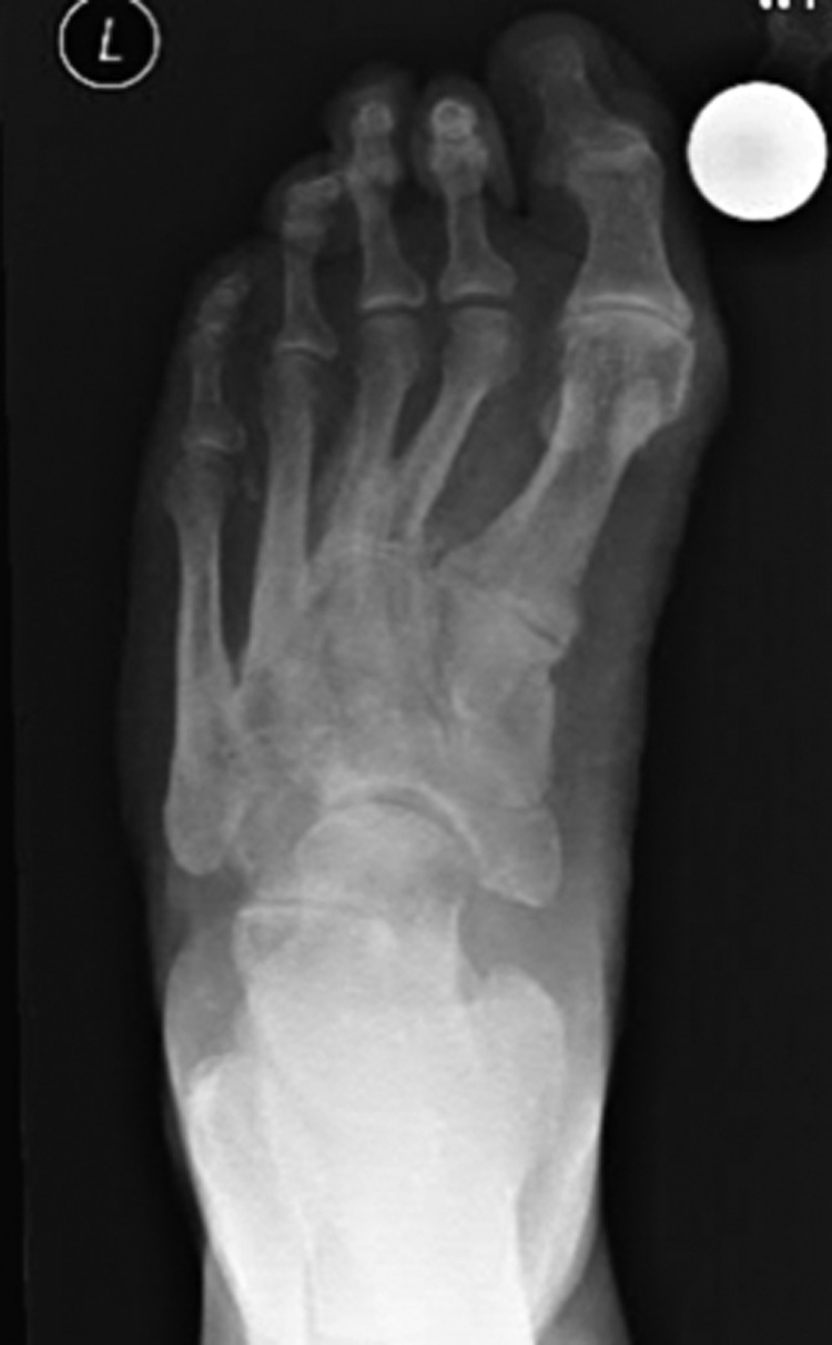
Dorsoplanter radiograph of the left foot with the clearly visible additional middle cuneiform.
Figure 3:
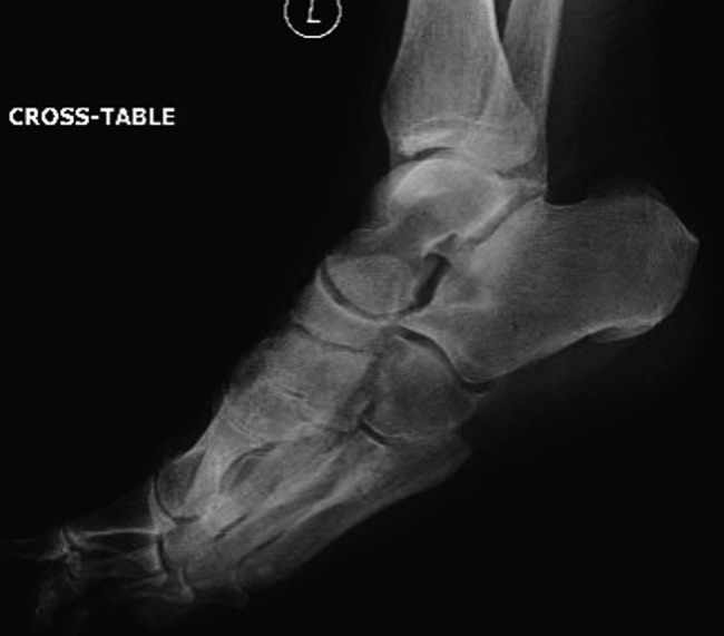
Lateral radiograph of the left foot with the clearly visible additional middle cuneiform.
Figure 4:
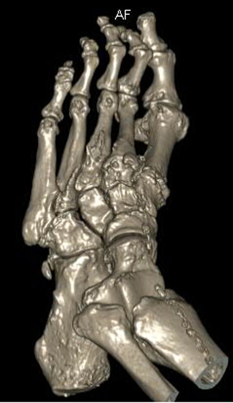
Three-dimensional CT reconstruction of the left foot in the dorsoplanter orientation.
Figure 5:
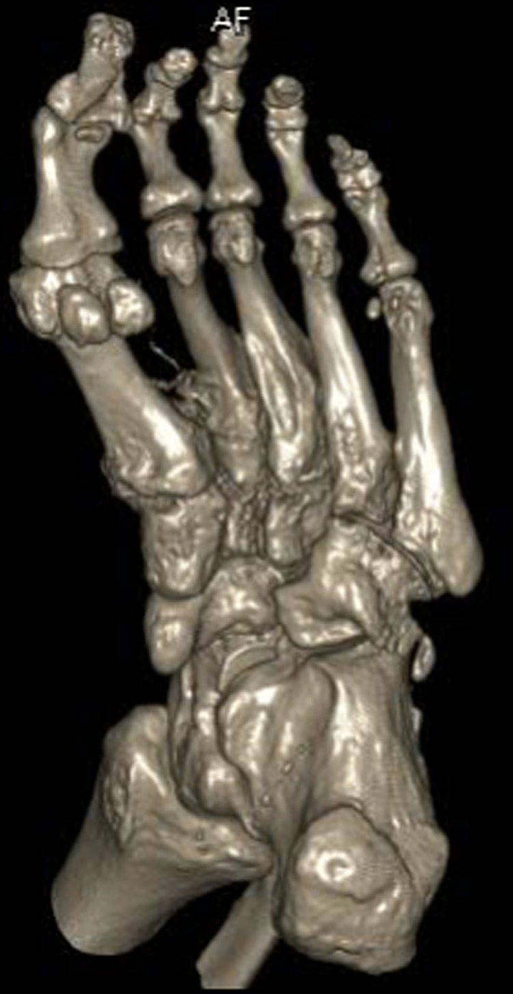
Three-dimensional CT reconstruction of the left foot in the planterdorsal orientation.
The patient had undergone previous diagnostic and therapeutic corticosteroid injections into his first, second and third TMTJs, temporarily improving the mid-foot pain. The patient wanted complete pain resolution and so underwent arthrodesis of these joints and the first MTPJ (Figs 6 and 7). Operative findings confirmed radiographic findings with the presence of an additional middle cuneiform covered with degenerate cartilage proximally and distally.
Figure 6:
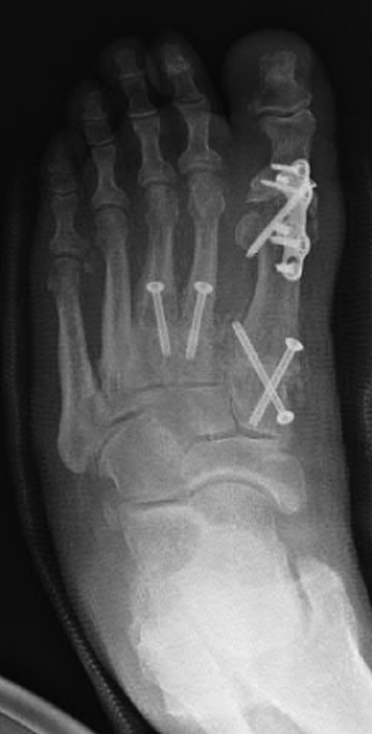
Oblique radiograph of the left foot following first MTPJ and first, second and third TMTJ fusions.
Figure 7:
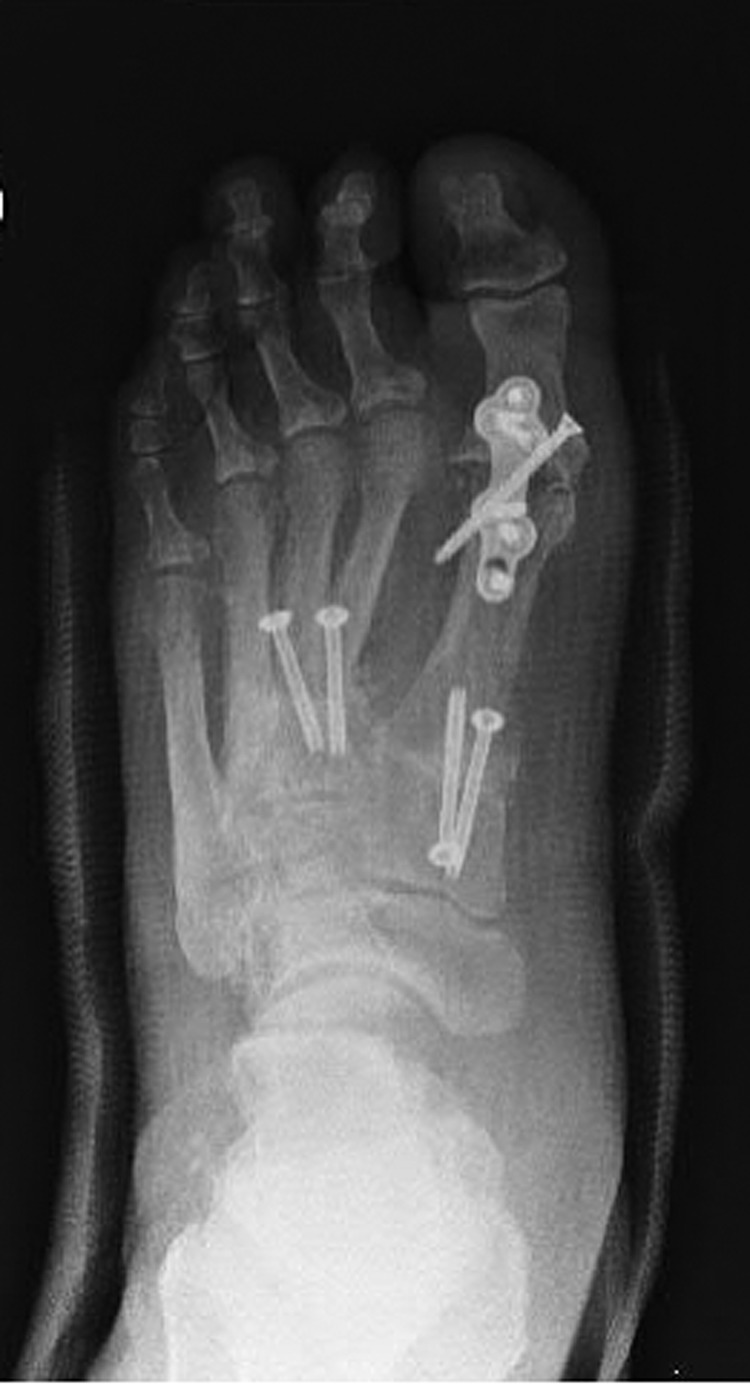
Dorsoplanter radiograph of the left foot following first MTPJ and first, second and third TMTJ fusions.
DISCUSSION
The additional bone of this patients foot appears to exist as a completely separate entity to the regular middle cuneiform proximally and the second metatarsal distally. It has developed the characteristic cuneiform wedge shape and size and articulates with the second metatarsal. Given the clinical and radiological conclusions, this extra ossicle represents an interesting finding of a potential additional middle cuneiform.
Accessory ossicles around the mid-foot are multiple and well documented. They are all generally small, having characteristic shapes and locations, all of which differ to the additional bone in our patient's foot. The bipartite medial cuneiform is the only other additional foot bone that has a similar shape. The location of this variant is more medial. The axial orientation in relation to the medial cuneiform compared with the location of our additional excludes the presence of a bipartite medial cuneiform.
If our additional ossicle does represent a duplicate middle cuneiform and should others substantiate its occurrence, then the clinical significance of its presence arises when one should distinguish it from an acute or a chronic non-united fracture of the second metatarsal base or middle cuneiform. Recognition of a fracture in this area is of upmost importance to ensure that there is no associated mid-foot subluxation or dislocation, which unrecognized, can lead to degenerate osteoarthritis. In the presence of pain in this area, magnetic resonance imaging would help differentiate it from an acute fracture and CT scanning would topographically map it.
Another clinical significance could arise in the case of a second TMTJ arthrodesis. The dilemma is presented as to whether or not fusion be performed between the second metatarsal base and the additional ossicle (Figs 6 and 7), or to span the additional ossicle fusing the second metatarsal to the middle cuneiform. Either way, careful assessment of the anatomy must be performed preoperatively to ensure that all joint surfaces are prepared and arthrodesed appropriately.
In conclusion, we report an interesting finding of an ossicle that has the appearance and location of a potential additional middle cuneiform, a finding that has yet to be described but could have clinical implications should its existence be further substantiated.
CONFLICT OF INTEREST STATEMENT
None declared.
REFERENCES
- 1.Scheuer L, Black S. Developmental Juvenile Osteology. London: Academic Press, 2000, 287–8. [Google Scholar]
- 2.Burnett SE, Case DT. Bipartite medial cuneiform: new frequencies from skeletal collections and meta analysis of previous cases. J Comp Hum Biol 2011;62:109–25. [DOI] [PubMed] [Google Scholar]
- 3.Dwight T. Os intercuneiforme tarsi, os paracuneiforme tarsi, calcaneus secundarius. Anat Anz 1902;20:465–72. [Google Scholar]
- 4.Schoen W. Seltenere akzessorische Knochen am Fussrucken. Rontgenpraxis 1935;7:775–6. [Google Scholar]
- 5.Pfitzner W. Beirage zur Kenntmiss des Menschlichen Extremitatenskelets: VII. Die Variationen in Aufbau des Fusskelets. Morphologische Arbeiten, Germany: Gustav Fischer, 1896, 245–527. [Google Scholar]
- 6.Freyschmidt J, Brossman J, Wiens J, Sternberg A. Borderlands of Normal and Early Pathological Findings in Skeletal Radiography. 5th edn New York: Thieme, 2003, 426–7. [Google Scholar]


