

Abstract
Purpose
To evaluate the efficacy of low level laser therapy (LLLT) in the treatment of temporomandibular disorders (TMD) in relation to pain intensity, tender points, joint sounds and jaw movements.
Materials and Methods
Twenty patients received 6 sessions of LLLT (3 times a week for 2 weeks) with semiconductive diode laser (gallium arsenide; 904 nm, 0.6 W, 60 s, 4 J/cm2). Pain intensity, number of tender points, joint sounds and active range of motion were assessed before and immediately after each session and after 1, 2 weeks, 1, 3 and 6 months.
Results
Statistically significant results were achieved in all study parameters.
Conclusion
LLLT promoted satisfactory results in reducing the pain intensity, number of tender points, joint sounds and improvement in the range of jaw motion. Hence it is an effective and efficient treatment method for TMDs.
Keywords: Temporomandibular disorders, Low level laser therapy, Semiconductive diode laser
Introduction
Temporomandibular disorders (TMD) is a collective term embracing a number of clinical problems that involve the masticatory musculature, the temporomandibular joint (TMJ) and associated structures, or both. Epidemiological studies show that about 75 % of the population presents one sign of TMD and 33 % present at least one symptom [1].
Non-surgical treatment of TMDs continues to be the most effective way of managing over 80 % of patients [2], which include psychotherapy or behavioral therapy, pharmacotherapy, occlusal splint therapy and various physical therapies like thermal therapy, acupuncture, electrical stimulation, ultrasound therapy, physiotherapy and low intensity laser therapy.
Low level laser therapy (LLLT) has been investigated and used clinically in the treatment of a variety of acute and chronic musculoskeletal injuries, degenerative conditions and wound healing for about 20 years. The basic effects of LLLT are bio-stimulative, regenerative, analgesic and anti-inflammatory [3].
The relative clinical efficacy of LLLT for the treatment of TMDs is controversial. Some authors reported the efficacy of LLLT to be superior to placebo therapy [4–19] and other physical therapies [2–23], while, others found no significant differences between LLLT and placebo for the measures of TMJ pain [24–26]. However there is an advantage of using LLLT in the treatment of TMDs as it is non-invasive, cost effective and does not have any known side effects.
In view of the fact that outcomes in LLLT studies may depend on patient samples, treatment protocols and procedure and study design, the purpose of this study was to evaluate the effectiveness and the outcome of LLLT in the treatment of TMDs.
Materials and Methods
Patients who reported to the Department of Oral and Maxillofacial Surgery, SRM Dental College, Chennai, with orofacial pain, clicking of the joint, limited mouth opening, or jaw stiffness were examined. Cases with congenital abnormality, neoplastic conditions and those with a recent history of acute trauma or any form of treatment within the last month were excluded. Orthopantomographic radiographs were taken for all the patients to rule out any gross anatomical deformity in relation to the TMJ.
After evaluation 28 patients were selected for the study. Three patients declined the treatment protocol due to multiple outpatient visits and for 5 patients follow-up could not be completed due to poor patient compliance. Thereby, 20 patients (11 males, 9 females) 19–47 years of age (mean: 28.55 years) were included in the study.
Based on the findings elicited, corresponding diagnosis was made. There were twelve patients of mainly myogenic origin and eight patients of mainly arthrogenic origin. Three of the patients, who were treated 2 years earlier by arthrocentesis, reported with relapse of symptoms were also included in the study. None of the other patients had been subjected to any type of treatment earlier.
All the patients were explained about the procedure in detail and a signed consent was taken. They were examined by the first investigator. Pain intensity, number of tender points, joint sounds, maximal pain-free and maximal possible mouth opening and right and left lateral jaw motion were assessed before and immediately after every session and after 1, 2 weeks, 1, 3 and 6 months from the first session.
Pain intensity was recorded in millimetres on a 100 mm Visual Analogue Scale (VAS). Number of tender points (minimum: 0, maximum: 20) were assessed by palpation of the following 10 points on both sides: preauricular (mouth closed–mouth open–through external auditory meatus), masseter, temporalis, medial and lateral pterygoid, sternocleidomastoid, trapezius and back of the neck.
Joint sounds were assessed by auscultation of TMJ during mouth opening and closing, listening for the presence of opening and closing clicks as well as fine and coarse crepitation. The total number of sounds on both sides was recorded.
Both maximum pain free mouth opening and maximum possible mouth opening were recorded. The vertical inter-incisal distance between the midpoints of upper and lower central incisors was measured by a ruler and recorded in millimetres. Lateral jaw motion was assessed by measurement of the horizontal distance between the midpoints of upper and lower central incisors in millimetres.
All patients were treated with six sessions of LLLT. CDHC DenLase 980/7 Diode Laser Therapy System (Fig. 1), a Class IV laser product, producing semi-conductive (diodic) gallium arsenide (GaAs) laser (input: 5 V–14 A, visible output: 1 mW max @ 630–670 nm, invisible output: 7 W max @ 800–990 nm manufactured by China Daheng Group Inc.) was utilized in the study was used. LLLT (wavelength: 904 nm, mean output power: 0.6 W, duration: 60 s, dosage: 4 J/cm2) was applied to all the tender points selected during examination. The subjects and the clinician used protective eyewear.
Fig. 1.
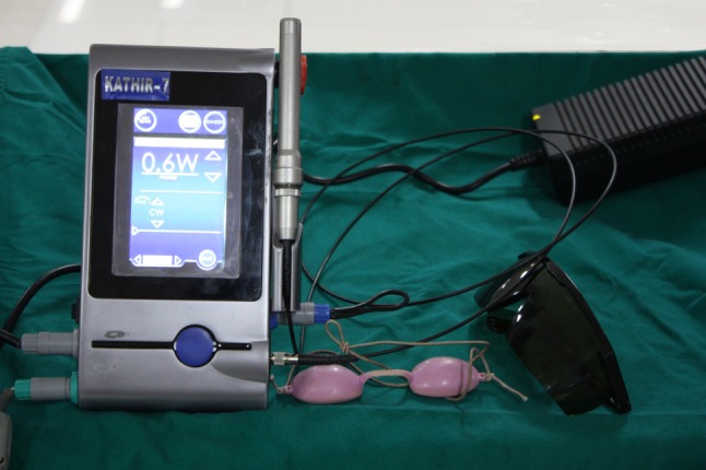
Laser dispensing system
The therapeutic LLLT application was achieved through direct contact of the probe on the skin. The laser beam was delivered through a handheld single laser probe. The probe was placed perpendicular directly on the skin behind, in front of, and above the joint area (Fig. 2), and into the external acoustic meatus (Fig. 3). It was also applied over the painful muscle spots (tender points) like masseter, temporalis, sternocleidomastoid and trapezius and over the back of the neck (Fig. 4). Intra-orally the LLLT was applied on to the masseter, anterior border of the ramus of the mandible—attachment of temporalis (Fig. 5), posterior and superior to the molars in the buccal vestibule—lateral pterygoid muscle and on the lingual aspect of the posterior mandible—medial pterygoid muscle. Each tender point was exposed to 60 s of LLLT.
Fig. 2.
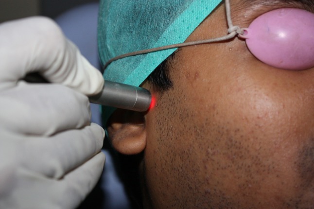
LLLT around temporomandibular joint area
Fig. 3.
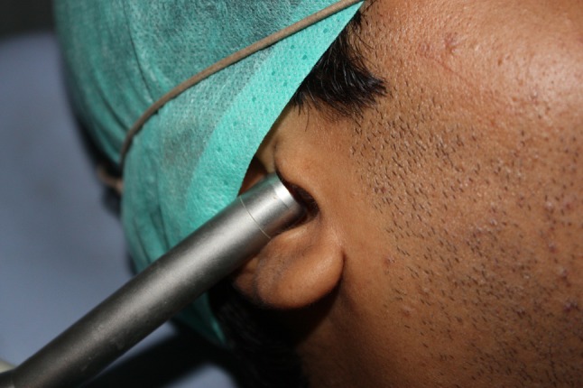
LLLT behind the joint area through the external acoustic meatus
Fig. 4.
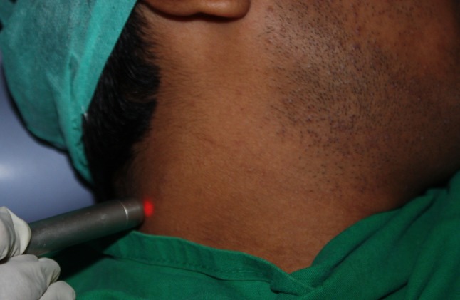
LLLT to the back of the neck
Fig. 5.
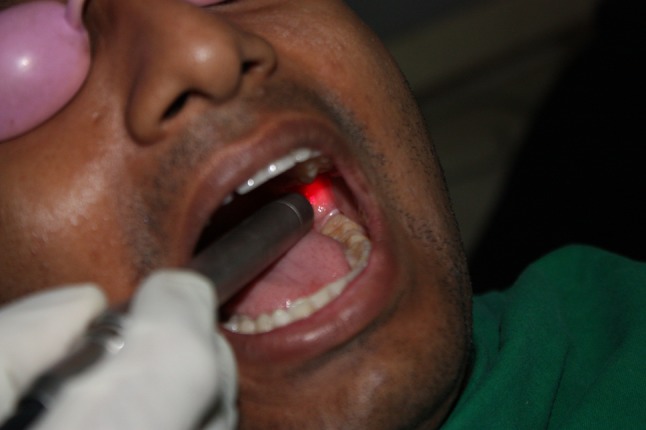
LLLT to the anterior border of the ramus of the mandible
Statistical analysis of the parameters obtained was conducted using SPSS for Windows, Version 15 (SPSS Inc., Chicago, IL, USA). Differences were analyzed by paired samples t test, Wilcoxon signed-ranks test and Mann–Whitney test. Any p value <.05 was considered significant.
Results
The descriptive scores of the parameters evaluated before the commencement of LLLT, immediately after the first session, after 1, 2 weeks, 1, 3 and 6 months are given in Table 1.
Table 1.
Study parameters at different intervals
| Pre | A1 | W1 | W2 | M1 | M2 | M3 | |
|---|---|---|---|---|---|---|---|
| Pain intensity | 66.45 ± 15.53 | 49.05 ± 15.29 | 33.70 ± 16.84 | 13.85 ± 12.06 | 5.50 ± 9.58 | 4.13 ± 10.99 | 3.42 ± 9.79 |
| Number of tender points | 9.75 ± 3.59 | 7.20 ± 3.96 | 4.45 ± 3.23 | 2.70 ± 3.37 | 1.60 ± 2.87 | 1.20 ± 2.91 | 1.05 ± 2.23 |
| Number of joint sounds | 2.58 ± 1.64 | 2.47 ± 1.61 | 2.11 ± 1.69 | 1.63 ± 1.42 | 1.16 ± 1.21 | 1.16 ± 1.25 | 1.13 ± 1.30 |
| Maximum pain free mouth opening | 37.30 ± 8.189 | 38.05 ± 7.96 | 40.25 ± 6.80 | 42.35 ± 5.779 | 43.45 ± 5.28 | 43.40 ± 5.46 | 43.65 ± 5.32 |
| Maximum possible mouth opening | 40.60 ± 7.03 | 41.50 ± 6.20 | 42.60 ± 6.01 | 43.80 ± 4.73 | 44.30 ± 4.54 | 44.35 ± 4.54 | 44.40 ± 4.52 |
| Right lateral excursion | 7.00 ± 2.36 | 7.25 ± 2.09 | 8.05 ± 1.82 | 8.95 ± 1.43 | 8.85 ± 1.43 | 8.95 ± 1.43 | 8.95 ± 1.41 |
| Left lateral excursion | 7.45 ± 1.84 | 8.35 ± 1.63 | 8.75 ± 1.71 | 9.40 ± 1.27 | 9.40 ± 1.27 | 9.30 ± 1.38 | 9.35 ± 1.36 |
Pre—baseline, A1—after 1st session, W1—1 week, W2—2 weeks, M1—1 month, M2—3 months, M3—6 months
Values: mean ± SD
Pain Intensity and Tender Points
The pain intensity was measured on a 100 mm VAS which showed 23.19 % reduction after the first session with LLLT, 49.29 % after 3 sessions/1 week, 79.16 % after 6 session/2 weeks, 91.72 % after 1 month, 93.78 % after 3 months and 94.86 % after 6 months (Fig. 6). The reduction in pain intensity was statistically highly significant (p < .001). Similar results were achieved in relation to the number of tender points (p < .001). There was a reduction of 26.15, 54.35, 72.30, 83.59, 87.69 and 89.23 % after the first session, 1, 2 weeks, 1, 3 and 6 months respectively (Fig. 7).
Fig. 6.

Pain intensity
Fig. 7.

Number of tender points
Joint Sounds
There was no significant reduction in the joint sounds after the first session with LLLT, but there was a gradual improvement in this parameter as the treatment progressed. There was a decrease in joint sound of 18.21 % after 1 week, 36.82 % after 2 weeks, 55.04 % after 1 month, 55.42 % after 3 months and 56.19 % after 6 months (Fig. 8). The reduction in the number of joint sounds was statistically significant only after 1 week of treatment (p < .05).
Fig. 8.

Number of joint sounds
Active Range of Motion
Maximum mouth opening parameters were statistically significant in all follow-up periods. There was significant mean increment of 6.35 mm (Fig. 9) in pain free mouth opening and 3.8 mm (Fig. 10) in maximum possible mouth opening during the 6 month follow-up period (p < .001). The right and left lateral jaw movements increased significantly (p < .001) by a mean increment of 1.95 and 1.9 mm correspondingly (Figs. 11, 12).
Fig. 9.

Maximum pain free mouth opening
Fig. 10.

Maximum possible mouth opening
Fig. 11.

Right lateral excursion
Fig. 12.

Left lateral excursion
Myogenic and Arthrogenic Groups
The comparison of the mean changes in clinical parameters at 6 months follow-up period (Table 2) revealed that the reduction in number of joint sound was highly significant in the myogenic group (p < .001). However, there were no significant differences in other parameters in both the groups.
Table 2.
Comparison of the mean change in clinical parameters at 6 months between myogenic and arthrogenic groups
| Myogenic | Arthrogenic | p value | |
|---|---|---|---|
| Pain intensity | 1.83 ± 4.30 | 12.25 ± 14.99 | .098a |
| Number of tender points | 0.25 ± 0.62 | 2.63 ± 4.30 | .181a |
| Number of joint sounds | 0.25 ± 0.45 | 2.38 ± 0.91 | .000a |
| Maximum pain free mouth opening | 44.75 ± 3.91 | 41.38 ± 7.00 | .216b |
| Maximum possible mouth opening | 45.33 ± 4.45 | 42.88 ± 4.54 | .750b |
| Right lateral excursion | 8.83 ± 1.33 | 9.13 ± 1.64 | .259b |
| Left lateral excursion | 9.67 ± 1.43 | 8.75 ± 1.16 | .643b |
Values: mean ± SD
aMann–Whitney test
bIndependent samples test
Male and Female Groups
There was no significant difference between the outcomes of the LLLT in both the groups as shown in Table 3 (p < .05). However, both the groups showed good response to the treatment.
Table 3.
Comparison of the mean change in clinical parameters at 6 months between male and female groups
| Male | Female | p value | |
|---|---|---|---|
| Pain intensity | 1.82 ± 4.04 | 11.11 ± 14.60 | .131a |
| Number of tender points | 0.18 ± 0.60 | 2.44 ± 4.06 | .080a |
| Number of joint sounds | 0.64 ± 0.92 | 1.67 ± 1.41 | .095a |
| Maximum pain free mouth opening | 45.55 ± 3.04 | 40.78 ± 6.72 | .052b |
| Maximum possible mouth opening | 46.00 ± 3.74 | 42.33 ± 4.82 | .287b |
| Right lateral excursion | 9.00 ± 1.34 | 8.89 ± 1.61 | .413b |
| Left lateral excursion | 9.73 ± 1.34 | 8.78 ± 1.30 | .434b |
Values: mean ± SD
aMann–Whitney test
bIndependent samples test
All the study parameters showed significant improvement at 6 months follow-up to LLLT.
Discussion
The American Academy of Craniomandibular Disorders has cited physical therapy as an important treatment modality in the management of TMDs. Physical therapy is intended to relieve musculoskeletal pain, reduce inflammation, and restore oral motor function [1]. Numerous physical therapy interventions are potentially effective in managing TMD, including exercise and manual therapy techniques, thermal therapies by application of cold or heat, electrophysical modalities like transcutaneous electric neural stimulation, acupuncture and LLLT.
Low Level Laser Therapy
Since the first postulation of the principle governing the emission by stimulation by Albert Einstein in 1917, the laser light technology has flared in leaps and bounds.
LLLT, first published by Andre Mester, is the application of light, usually a low power laser to a pathology to promote tissue regeneration, reduce inflammation and relieve pain. The light is typically of narrow spectral width in the red or near infrared spectrum (600–1,000 nm), with a power density (irradiance) between 1 mW–5 W/cm2. It is typically applied to the injury for a minute or so, a few times a week for several weeks. Unlike other medical laser procedures, LLLT is not an ablative or thermal mechanism, but rather a photochemical effect comparable to photosynthesis in plants whereby the light is absorbed and exerts a chemical change [3].
Mechanisms of Low Level Laser Therapy
The consensus about the mechanism of LLLT effects inevitably involves mitochondria. The effects of HeNe laser and other illumination on mitochondria isolated from rat liver have included increased proton electrochemical potential, more ATP synthesis, increased RNA and protein synthesis and increases in oxygen consumption, membrane potential, and enhanced synthesis of NADH and ATP [3].
This photostimulatory effect in mitochondria processes enhanced growth factor release and ultimately led to cell proliferation.
Low Level Laser Therapy and Temporomandibular Disorders
Considerable work has focussed on determining the effects of laser on pain and click management with varying results [4–26]. Kulekcioglu et al. [11] showed that, after 15 sessions of LLLT, TMD of both myogenic and arthrogenic causes responded to therapy with a significant reduction in pain, improvement in mouth opening and lateral motion, and a diminished number of trigger points. Çetiner et al. [12] expressed similar results in 24 patients after 10 sessions of LLLT. Improved outcome of laser therapy was documented by Santos et al. [19] by applying LLLT at a comparatively higher dose.
In this study the treatment was carried out on alternate days and the regimen was completed in 2 weeks (3 sessions/week). After every session recordings of the parameters were taken. In 5 of the patients follow-up could not be completed hence were not included in the study, however those patients had relief of symptoms during the treatment regimen. The main drawback of the procedure was that it had to be done at multiple sessions, because of this reason three patients declined the treatment protocol as they found it difficult to commit for treatment.
Most of the earlier studies have used lower power than the one used in this study. Wertz reported that in many of studies related to the efficacy of LLLT, analysis uncovered one or more reasons for the negative findings reported, the most common being the use of extremely low doses [27]. He also recommended the use of higher dosage of 600–700 mW.
The number of treatment sessions is another parameter which has no consensus drawn. Most of the studies report 10–20 sessions of treatment, while in this study the sessions were restricted to six keeping in mind the comparatively higher dosage of 700 mW delivered.
Improvement in all the study parameters were seen in all cases except in two. Both the patients were females, had TMD of mainly arthrogenic origin, both had been treated with arthrocentesis 2 years earlier. It was noticed that in these cases there was a temporary relief in pain intensity; betterment in joint sounds i.e., the joint sounds were of crepitus type, which after LLLT had been reduced to clicking sound. However there was no significant decrease in the number of joint sounds during the treatment as well as in the follow-up period; the mouth opening increased and remained so even in the follow-up period.
This study does not include a control group due to the limited sample size hence it is open for further research using a larger sample size and a control group to assess and compare the effectiveness of LLLT.
Conclusion
This study supports the use of LLLT as an alternative to other conventional treatment modalities in TMDs by producing positive outcomes. The subjects were comfortable with the treatment and satisfied by the outcome and had a better lifestyle following treatment. There was a rapid decrease in the pain intensity; the number of tender points and joint sounds. The active range of motion was also increased in all the patients. During the follow-up period there was no relapse of the disease except in two cases which we believe is due to the complexity of the disorder and in such cases there is a demand for a more complex treatment.
Thus, it can be suggested as an efficient treatment method, with the dose level tested, as a primary modality in early and less complex conditions and as an adjunctive procedure in more advanced and complex disease conditions.
The findings of this study are restricted to a specific set of parameters. However, optimal treatment parameters (e.g., wavelength, dosage, number of treatment sessions) have not been agreed on and are still debatable. Further research should focus on optimal treatment parameters such as the intensity and duration with double-blind, randomized controlled trials. Moreover, comparison of the effectiveness of different modalities in myogenic and arthrogenic TMD deserves further investigation.
Contributor Information
Nabeel Sayed, Email: dr_nabeels@yahoo.com.
C. Murugavel, Email: dr_mrgvl@yahoo.co.in
A. Gnanam, Email: drgnanam@hotmail.com
References
- 1.Okeson JP. Management of temporomandibular disorders and occlusion. 6. Philadelphia: Mosby; 2008. [Google Scholar]
- 2.Dimitroulis G. Temporomandibular disorders: a clinical update. Br Med J. 1998;317:190. doi: 10.1136/bmj.317.7152.190. [DOI] [PMC free article] [PubMed] [Google Scholar]
- 3.Karu T. The science of low power laser therapy. Amsterdam: Gordon and Beach Science; 1998. [Google Scholar]
- 4.Conti PC. Low level laser therapy in the treatment of temporomandibular disorders (TMD): a double-blind pilot study. Cranio. 1997;15:144. doi: 10.1080/08869634.1997.11746005. [DOI] [PubMed] [Google Scholar]
- 5.Bezuur NJ, Habets LL, Hansson TL. The effect of therapeutic laser treatment in patients with craniomandibular disorders. J Craniomandib Disord. 1988;2:83. [PubMed] [Google Scholar]
- 6.Hatano Y. Laser in diagnosis of the TMJ problems. Laser Dent. 1989;1:169. [Google Scholar]
- 7.Bertolucci LE, Grey T. Clinical comparative study of microcurrent electrical stimulation to mid-laser and placebo treatment in degenerative joint disease of the temporomandibular joint. J Craniomandib Pract. 1995;13:116. doi: 10.1080/08869634.1995.11678054. [DOI] [PubMed] [Google Scholar]
- 8.Eckerdal A, Bastian L. Can low reactive-level laser therapy be used in the treatment of neurogenic facial pain? A double-blind, placebo controlled investigation of patients with trigeminal neuralgia. Laser Ther. 1996;8:247. doi: 10.5978/islsm.8.247. [DOI] [Google Scholar]
- 9.Bradley PF. A review of the use of the neodymium YAG laser in oral and maxillofacial surgery. Br J Oral Maxillofac Surg. 1997;35:26. doi: 10.1016/S0266-4356(97)90005-X. [DOI] [PubMed] [Google Scholar]
- 10.Al Pinherio, Cavalcanti ET, Pinherio TI. Low level laser therapy is an important tool to treat disorders of the maxillofacial region. J Clin Laser Med Surg. 1998;16:223. doi: 10.1089/clm.1998.16.223. [DOI] [PubMed] [Google Scholar]
- 11.Kulekcioglu S, Sivrioglu K, Ozcan O, Parlak M. Effectiveness of low-level laser therapy in temporomandibular disorder. Scand J Rheumatol. 2003;32:114. doi: 10.1080/03009740310000139. [DOI] [PubMed] [Google Scholar]
- 12.Çetiner S, Kahraman S, Yücetas SU. Evaluation of low-level laser therapy in the treatment of temporomandibular disorders. Photomed Laser Surg. 2006;24:5. doi: 10.1089/pho.2006.24.637. [DOI] [PubMed] [Google Scholar]
- 13.Fikácková H, Dostálová T, Navrátil L, Klaschka J. Effectiveness of low-level laser therapy in temporomandibular joint disorders: a placebo-controlled study. Photomed Laser Surg. 2007;25:297. doi: 10.1089/pho.2007.2053. [DOI] [PubMed] [Google Scholar]
- 14.Mazzetto MO, Carrasco TG, Bidinelo EF, de Andrade Pizzo RC, Mazzetto RG. Low intensity laser application in temporomandibular disorders: a phase I double-blind study. Cranio. 2007;25:186. doi: 10.1179/crn.2007.029. [DOI] [PubMed] [Google Scholar]
- 15.Frare JC, Nicolau RA. Clinical analysis of the effect of laser photobiomodulation (GaAs-904 nm) on temporomandibular joint dysfunction. Rev Bras Fisioter. 2008;12:37. doi: 10.1590/S1413-35552008000100008. [DOI] [Google Scholar]
- 16.Carrasco TG, Mazzetto MO, Mazzetto RG, Mestriner W., Jr Low intensity laser therapy in temporomandibular disorder: a phase II double-blind study. Cranio. 2008;26:274. doi: 10.1179/crn.2008.037. [DOI] [PubMed] [Google Scholar]
- 17.Lassemi E, Jafari SM, Motamedi MH, Navi F, Lasemi R. Low-level laser therapy in the management of temporomandibular joint disorder. J Oral Laser Appl. 2008;8:83. [Google Scholar]
- 18.Shirani AM, Gutknecht N, Taghizadeh M, Mir M. Low-level laser therapy and myofacial pain dysfunction syndrome: a randomized controlled clinical trial. Laser Med Sci. 2009;24(5):715. doi: 10.1007/s10103-008-0624-5. [DOI] [PubMed] [Google Scholar]
- 19.Santos TS, Piva MR, Ribeiro MH, Antunes AA, Melo AR, Silva ED. Laser therapy efficacy in temporomandibular disorders: control study. Braz J Otorhinolaryngol. 2010;76:294. doi: 10.1590/S1808-86942010000300004. [DOI] [PMC free article] [PubMed] [Google Scholar]
- 20.Taha AR, Taftazani EA. Effect of soft laser and occlusal splints on temporomandibular joint disorders (electromyographic study) Cairo Dent J. 2003;19:179. [Google Scholar]
- 21.Kogawa EM, Kato MT, Santos CN, Conti PC. Evaluation of the efficacy of low-level laser therapy (LLLT) and the microelectric neurostimulation (MENS) in the treatment of myogenic temporomandibular disorders: a randomized clinical trial. J Appl Oral Sci. 2005;13:280. doi: 10.1590/S1678-77572005000300015. [DOI] [PubMed] [Google Scholar]
- 22.Núñez SC, Garcez AS, Suzuki SS, Ribeiro MS. Management of mouth opening in patients with temporomandibular disorders through low-level laser therapy and transcutaneous electrical neural stimulation. Photomed Laser Surg. 2006;24:1. doi: 10.1089/pho.2006.24.45. [DOI] [PubMed] [Google Scholar]
- 23.Kato MT, Kogawa EM, Santos CN, Conti PC. Tens and low-level laser therapy in the management of temporomandibular disorders. J Appl Oral Sci. 2006;14:130. doi: 10.1590/S1678-77572006000200012. [DOI] [PMC free article] [PubMed] [Google Scholar]
- 24.Hansen HJ, Thoroe U. Low power laser biostimulation of chronic oro-facial pain. A double-blind placebo controlled cross-over study in 40 patients. Pain. 1990;43:169. doi: 10.1016/0304-3959(90)91070-Y. [DOI] [PubMed] [Google Scholar]
- 25.Venancio DA, Camparis CM, Lizarelli FZ. Low intensity laser therapy in the treatment of temporomandibular disorders: a double-blind study. J Oral Rehabil. 2005;32:800. doi: 10.1111/j.1365-2842.2005.01516.x. [DOI] [PubMed] [Google Scholar]
- 26.Emshoff R, Bösch R, Pümpel E, Schöning H, Strobl H. Low-level laser therapy for treatment of temporomandibular joint pain: a double-blind and placebo-controlled trial. Oral Surg Oral Med Oral Pathol Oral Radiol Endod. 2008;105:452. doi: 10.1016/j.tripleo.2007.09.012. [DOI] [PubMed] [Google Scholar]
- 27.Wertz RL. Class IV high power laser therapy in physical medicine and rehabilitation. Dyn Chiropr. 2006;24(23):8. [Google Scholar]