

Abstract
Introduction
Malignancies of the tongue represent one of the greatest management challenges for the maxillofacial surgeons as well as oncologists, because of the adverse effects of treatment on oral and pharyngeal function, the eventual quality of life, and the poor prognosis of advanced disease. Therefore, it is important to use judgment and experience in determining the best method of treatment.
Material and methods
We reviewed forty cases of oral tongue cancer patients admitted in the Dental and Facio-Maxillary Surgical Oncology department in National Institute of Cancer Research and Hospital, and department of Oral and Maxillofacial Surgery, Dhaka Dental College and Hospital, Dhaka, Bangladesh, during the past four years and followed till the lesion healed or recurred and followed later on upto two years. All cases were thoroughly examined, investigated with routine blood examinations and radiography of the involved region. Preoperative biopsy of the lesion and staging was done in each and every case. Postoperative biopsy was taken where there was a doubt about the possibility of recurrence.
Results
Squamous cell carcinoma (well differentiated) is by far the most common malignancy of the oral tongue. Generally a correlation is recognized between tumor size, nodal presence, metastasis, and eventual prognosis. When surgeons detect oral tongue cancer at an early stage, they can often treat it with surgery or can, often treat it with surgery or radiation. In later stages the cancer may require a combination of surgery, radiation and chemotherapy.
Conclusion
Standard and uniform protocol has not been explored till now for the practice in our country. So current management strategies of oral tongue cancer cannot be underestimated.
Keywords: Tongue Cancer, Management, Oral Care, Prevention, Bangladesh
Introduction
One of the most common site for intraoral carcinoma is the tongue. Though there is no recognized survey on tongue cancer in Bangladesh, but 30 % of head and neck cancers in nearby country like India constitutes of tongue cancers [1]. These tumors most frequently arise from the lateral aspect (85 %) of the oral tongue. The majority of tongue cancers are squamous cell carcinoma and are well differentiated, while poorly differentiated varieties are rare [2]. These arise from the lining that covers the muscles of the tongue. Studies shows that tongue cancer usually occurs after the age of 40, with men affected more than women (10 %) [3–5]. The condition may appear as a lump, white or red spot or ulcer. Pain is a relatively late occurrence as a rule [3]. Though exact etiology of tongue cancer is still unknown, but several risk factors have been identified. Chewing tobacco, pan, betal nuts or quid is very common in parts of Asia and is known to be a main cause of oral cancer including tongue [5, 6]. The etiological factors of oral squamous cell carcinoma (OSCC) are different in different areas of the world [7]. The common site of oral cancer also differs due to some habit. Due to some geographic distribution and socio-economic condition, the rate of oral cancer is high in our country than the developed countries [8, 9]. So current management strategies of oral tongue cancer cannot be underestimated.
The tongue is divided into two parts for the purpose of diagnosis and treatment of tongue tumors. The first is the oral or mobile tongue (front two-thirds of tongue), cancers that develop in this part of the tongue come under a group of cancers called mouth (oral) cancer. The other is the base of the tongue (back third of tongue), cancers that develop in this part are called oropharyngeal cancers. Tumors of each of these areas present slightly differently and are treated differently [5]. Predictors of outcome of tongue cancer still remain unknown. Standard and uniform protocol is not explored till now for the practice in our country. This guideline has been written to assist doctors and other health care providers in the recognition, diagnosis and management of oral tongue cancer patients.
Initial Evaluation
Examination of the Oral Cavity
Hold the tip of the tongue with a piece of gauze by the left hand and pull it out to inspect and palpate the dorsal surface of the tongue as well as lateral borders. Then turn it upwards to allow examination of the ventral surface of the tongue and floor of the oral cavity. The doctor will ask the patient specific questions about symptoms and make initial evaluation by medical and physical examination (cancer screening exam) (Fig. 1).
Know the risk factors associated with tongue cancer e.g. men over the age of 40, tobacco and betel nut users, people who consume excessive amounts of alcoholic beverages.
Look for red, white or red and white patches or any sores that do not heal for long time or lumps in the tongue or a lump on the neck.
Complain of pain on swallowing or of any other constant numbness or pain or difficulty in moving the tongue or jaw.
Check for any unexplained bleeding, bad breath.
Feel for loose or painful teeth or dentures that may not fit well.
Fig. 1.
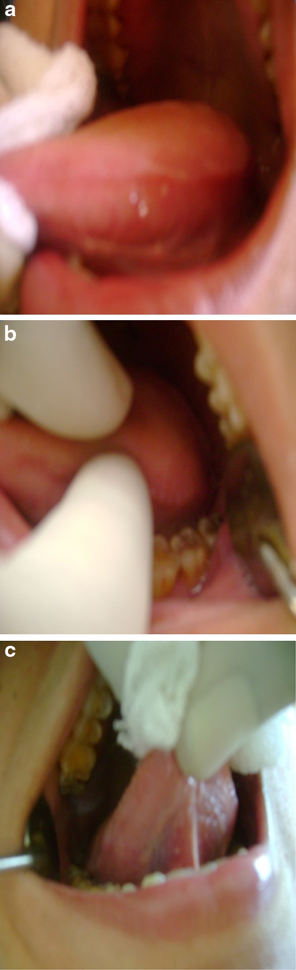
Technique of examination of the oral tongue and common site of tongue cancer
Diagnosis
Doctors use a variety of diagnostic tools to confirm the disease and to determine the extent of involvement, along with initial clinical evaluation before choosing a treatment. These include:
Tongue biopsy—Incisional, Punch biopsy, Fine Needle Aspiration (FNA) biopsy and microscopic examination
FNAC of neck lymphnode or biopsy and microscopic examination
X-rays (OPG, Chest X-ray)
CT scan (CAT scan)
MRI (Magnetic Resonance Imaging)
Ultrasound scan
Bone scan
Radionuclide neck scanning (SPECT, PET)
Assessment of disease by using TNM, Broder’s and Anneroth’s multifactorial classification (staging system).
Goals of Treatment
The primary goal of treatment is to treat primary tumor in the oral tongue, control neck disease (nodal metastasis) and preserve function of the tongue as much as possible [10]. The tongue has the reputation of being a site from which metastasis to the neck nodes occurs early and frequently [3]. The incidence of lymph node metastases is proportional to both the tumor T-stage and its depth, and there is also correlation between certain histological grades of the tumour and lymph node metastases [2]. Different treatment protocols and technologies are used to treat oral tongue cancer, which often includes surgery, chemotherapy (newer chemotherapy drugs), radiotherapy (IMRT, CHART), others—biological therapies, gene therapy, REOLYSIN (in advanced cancer), Light therapy (PDT or photodynamic therapy)[11]. The ultimate long term goal of treatment is to prevent recurrence (local or distant), to reduce or prevent complications and to prolong survival.
Principles of Treatment of the Oral Tongue
When surgeons detect oral tongue cancer at an early stage, then can often treat it with surgery or radiation. In later stages the cancer may require a combination of surgery, radiation and chemotherapy [10] (Fig. 2).
Fig. 2.

Common tongue pathology, surgical management and postoperative recurrence
Standard Treatment Options [2]
In Bangladesh in different specialized Maxillofacial Surgery centers the most common protocol of treatment of oral tongue cancers are same with negligible modifications being wide surgical resection, resection of floor of mouth musculature, removal of bone and associated muscle attachments and the sacrifice of both sensory and motor cranial nerves along with or without neck dissection.
Carcinoma of the Tip of Tongue
Stage I: Mode of treatment is surgery (wide excision): Tip of the tongue is excised perorally in a V-shaped fashion with a margin of at least 1 (one) centimeter and a new tongue tip made by primary reconstruction.
Stage II or larger T1 lesions: Wide surgical excision or radiotherapy of the primary site and consideration should be given to irradiating the neck also.
Carcinoma of the Dorsum of the Tongue
Stage I: Mode of treatment is surgery (wide excision) or irradiation. If the tumor is less than 2 cm in diameter excision would be transoral in an ellipse and with a margin of at least 2 cm.
Stage II: For larger tumors treatment of choice is, an extensive surgery or radiotherapy or a combination of both, along with neck dissection. But in order to preserve speech and swallowing function of the tongue, most of the cases radiation therapy is preferred.
Stage III: External beam radiotherapy with or without interstitial implant or in advanced cases palliative irradiation is given.
Carcinoma of the Lateral Border of the Tongue
Stage I: Partial glossectomy or interstitial irradiation
Stage II: Hemiglossectomy to subtotal glossectomy or external beam radiotherapy (in selected cases).
Stage III: Subtotal glossectomy, here only a stump of tongue above the vallecula is spared, along with base of the tongue on the contra lateral side or Total glossectomy and postoperative radiotherapy.
Principles of Treatment of the Neck
Treatment of the neck is a highly controversial point. But based on the high incidence of occult neck metastases in the clinically No neck 50 % [2] prophylactic or elective neck dissection (usually supraomohyoid neck dissection) is advocated even in T1No and T2No cases. There is less controversy concerning the management of stage III and stage IV (massive primary or neck), combined surgery and radiation are most often employed [12]. Tumors that have spread to the lymph nodes on one or both sides may need selective or radical neck dissection. It lowers the risk of cancer recurrence. Then a course of radiotherapy help to get rid of any cancer cells left behind [5].
Multidisciplinary Approach
Multidisciplinary specialists can develop a unique treatment plan for each patient, with different modalities of treatment combined with or without surgery. Team members are dental hygienist, dentist, oral and maxillofacial surgeon, oncologists (surgeon, radiation specialist, chemotherapist), nutritionist, psychiatrist or psychologist and social worker.
Surgery
Surgeons use the latest techniques to minimize the loss of function of the tongue and surrounding structures. Surgery for tongue cancers depends on the tumor size, type, location and depth [2]. Clinical evaluation of stage of disease is often difficult in cancers of the tongue and therefore, extent of resection is best decided at the time of operation [1]. In case of early cancers that, denotes cancers which have not spread beyond the primary site, it can be removed transorally, adequate surgical margins are crucial at the initial excision [12]. If the tumor extends deeply to involve the underlying muscles, the surgeon may remove the nearby neck nodes (prophylactic neck dissection) with moderate excisions of tongue, occasionally hemiglossectomy is needed [13]. However larger lesions (T3 and resectable T4) will require mandibulotomy for access and resection. The lesions which encroach the gingiva or superficially extend over it, should be treated with marginal mandibulectomy for oncologic clearance. In cases of gross involvement of mandible, composite resection will be required in conjunction with selective or radical neck dissection. The extent of mandibular resection will depend on extent of bone involvement [1].
Reconstruction and Rehabilitation
Depending upon the size, location and spread of the cancer, the treatment team can determine whether reconstructive and plastic surgery is necessary or not. Usually oral and maxillofacial surgeons, reconstructive and plastic surgeons, otorhinolaryngologists restore physical appearance and function of the tongue and surrounding structures. Usually a peroral large wedge resection to hemiglossectomy defect can be closed primarily. However, subtotal glossectomy for advance (T3 or T4) will require repair of the defect by myocutaneous flap in order to achieve the closure [1]. Patients who need rehabilitation after surgery or radiation therapy will find help from dietitian, speech therapist, swallowing therapist, physical therapist, occupational therapist and psychotherapist.
Radiation Therapy
Radiation therapy involves the use of external beam radiotherapy (electron beams), or interstitial brachytherapy (radioactive isotope)[2, 13]. Radiation therapy is an alternate to surgery in early and intermediate size tumours of the anterior tongue. Radiation therapy is usually started 4–6 weeks after the surgery. The postoperative radiotherapy dose commonly ranges between 55 and 65 Gy [1]. To minimize loss of function of the tongue and normal surrounding structures such as mucous membrane and salivary glands [11] IMRT (Intensity modulated radiotherapy) and CHART (continuous hyperfractionated accelerated radiation therapy) irradiation is advocated.
Brachytherapy
Brachytherapy is a technique of radiotherapy selectively used in the treatment of mainly T1 and T2 lesions. Low-dose-rate (LDR) brachytherapy is frequently chosen for the treatment of stage II mobile tongue cancer [14]. However little is known about the biological mechanism underlying this therapy. LDR may overcome the radioresistence of non-cycling and hypoxic cells [15]. French studies at Creteil and Nancy have shown that interstitial brachytherapy alone at a dose of 65 to 70 Gy is preferable (for T1–T2 lesions) and the neck can be observed for surgical salvage in case of nodal recurrences. The commonly used radio-isotopes are Ir-192, cs-137, I-125, Pd-203. There are different ways of implanting the radioactive sources. The Paris system is most widely used in head and neck cancers [1].
Chemotherapy
Medical oncologists administer chemotherapy when the oral tongue cancer has spread to lymph nodes or other organs in the body, usually in stage II cancers with surgery and in stage III and IV cancers with radiotherapy. Chemotherapy is commonly administered intravenously and/or intramuscularly or orally. Some drugs are used for injecting directly into the arteries [16] that supply the cancer or in intralesions. These cytotoxic drugs destroy cancer cells. To date, various regimens have been reported with variable response rates and duration of response [1]. Few commonly used protocols used in Bangladesh are given in Table 1.
Table 1.
Commonly used protocols of cytotoxic drug combinations
|
Protocol I
Cisplatin 25–30 mg i.v. (1 h inf) d 1–3 5-Fluorouracil 500–750 mg i.v. (cont.inf) d 1–3 Bleomycin 15 mg i.v. (bolus) d 1 followed by 16 mg/m2 (cont.inf) d 1–3 To be repeated every 3 weeks |
Protocol II
Paclitaxel 260 mg/m2 i.v. (3 h inf) d 1 Caboplatin 450 AUC i.v. (30 min inf) d 2 To be repeated every 3 weeks |
|
Protocol III
5-Fluorouracil 750 mg/m2 i.v. (cont inf) d 1–4 To be repeated every week up to 12 weeks then review |
Protocol IV
Ifosfamide 1,500 mg/m2 i.v. (30 min inf) d 1–5 with mesna uroprotection Cisplatin 10 mg/ms2 i.v. (30 min inf) d 1–5 To be repeated every 4 weeks |
Biological Therapies
Biological therapies are treatments made from naturally occurring body substances. There are many different types of biological therapies like monoclonal antibodies (cetuximab and zalutumumab) and cancer growth blockers. Both monoclonal antibodies block the epidermal growth factor receptors (EGFR) on cancer cells. Scientists hope that blocking these receptors will stop the signals that tell the cancers to grow. Now cetuximab is used with radiotherapy, for locally advanced squamous cell head and neck cancer, if platinum-based chemotherapy (such as cisplatin or carboplatin) is not working or could not be used. Zalutumumab is also under trial. Cancer growth factor blockers like tyrosine kinase inhibitors (TKIS) e.g. Iressa [11] and lidocaine which also act by blocking epidermal growth factor receptors, are also under trial [17].
Gene Therapy
OncoVEX, ONYX-15 are genetically engineered viruses, which are able to kill cancer cells, but not normal healthy cells. Now researchers are using it in combination with radiotherapy and chemotherapy drugs like 5FU and cisplatin [11].
Reolysin (in Advanced Cancer)
Currently a virus called Reovirus is being used, which is able to kill cancer cells when injected directly into the tumour. Reovirus in combination with radiotherapy or chemotherapy like paclitaxel or carboplatin is under trial [11].
Light Therapy (Photodynamic Therapy or PDT)
Using light treatment to kill cancer cells helps to shrink or slow the growth of the cancer and relieve symptoms. So it may be useful, as a palliative treatment in patients with advanced head and neck cancer who are not able to have any more standard treatment [11].
Complication and Management
Problems usually may be related to surgical treatment of the primary tumor or radiation treatment or chemotherapy. The major functional consideration with tongue resection results in significant mobility problems and dramatic changes in articulation, as well as aesthetics, swallowing, nutritional status and psychosocial function. Radiation treatment causes acute mucosal changes, erythema and edema to frank necrosis and breakdown (mucositis), candidiasis, loss of taste or altered taste, dysphagia. Late mucosal changes include dryness, atrophy telangiectasia, sclerosis and chronic edema. Late salivary gland changes range from hyposalivation to asilorrhoea (xerostomia). Trismus is yet another manifestation of progressive muscle fibrosis secondary to radiation therapy of the facial region. It is most commonly a late complication of therapy, often observed a year or more after completion of treatment. Further it can cause rampant dental caries, osteoradionecrosis. In addition, complications associated with chemotherapy are nausea, vomiting, diarrhoea, stomatitis haematologic toxicity, renal toxicity, shortness of breath, hypertension. Secondary effects include dehydration and malnutrition [18]. Recent research suggests that Inj. Amifostine a few minutes before radiotherapy helps to protect against the harmful side effects of chemotherapy and radiotherapy like xerostomia. Some studies also suggest that acupuncture appears to help with dry mouth, following radiotherapy and for pain, following a neck dissection. Further, acupuncture and moxibustion help to relieve the symptoms of lymphoedema following surgery or radiotherapy treatment but these are under trial [11, 13].
Oral Care Considerations
In patients with mucositis, irrigation or rinsing with bland rinses: 0.9 % saline solution (1 tsp of table salt to 32 oz of water) or sodium bicarbonate solution (1 tsp in 8 oz of water) or salt and soda (½ tsp of salt and 2 tbsp of sodium bicarbonate in 32 oz of warm water) should be performed every 2 h for 1–2 days maximum, since chronic use may impair timely healing of stomatitis. Moisturing can be achieved with water-soluble lubricating jelly, cellulose film-forming agents for covering localized ulcerative lesions (e.g. hydroxypropyl cellulose, coating agents such as milk of magnesia, sucralfate suspension combined with topical anesthetic agents may provide topical analgesia [19]. To relieve pain, topical anesthetics, systemic analgesics (including opioids) are used. Intraoral use of capsaicin topical lotions and creams (0.025 and 0.075 %) is under trial [20]. Chlorhexidine gluconate 0.12 % oral rinse has been used in combination with fluoride gel to control cariogenic flora [21]. Management of xerostomia includes the use of saliva substitutes (oral rinses containing hydroxyethyl-, hydroxypropyl-, or carboxymethyl-cellulose) which are temporary palliative agents used with oral care protocols.
Prevention
Specific Measure
Avoid using tobacco in any form.
Be sure that dentures fit properly and edges of teeth are smooth.
Avoid heavy use of alcoholic beverages.
General Measure
A diet high in fresh fruit and vegetables reduce the risk of mouth cancer.
Some scientists believe that antioxidant vitamins A, C and E and minerals supplement may help to prevent cancer.
Chemoprevention: There are two groups of drugs in clinical trials i.e. cyclooxygenase-2 inhibitors (COX-2) and retinoids (vitamin A) which help to prevent tongue cancer.
- Established tongue cancer is prevented from spreading or recurring by:
- Completing the full course of surgical, radiation, and/or chemotherapy treatments as ordered by the doctor.
- Maintaining good oral hygiene, taking good care of teeth and gums, and having regular dental checkups, particularly if the patient wears dentures.
- Visit the doctor immediately if a return of any previous symptoms, such as a lump or ulcer on the tongue that does not heal, but worsens and spreads is noticed.
Conclusion
To increase public awareness continuing educational campaigns are needed at community and national levels and routine dental check up should be instituted. Dental practitioner can play a vital role in cancer prevention and early diagnosis. Early diagnosis is critical if patients are to be cured. Further treatment modality should be such as to decrease the morbidity to the patient as well as to decrease or perhaps obviate the chances of any recurrence [22].
References
- 1.Mohanti BK, Bahadur S, Lal P, Gairola M, Rath GK. Cancers of the head and neck. In: Rath GK, Mohanti BK, editors. Textbook of radiation oncology: principles and practice. 1. India: Elsevier, Reed Elsevier India Private Limited; 2002. pp. 131–199. [Google Scholar]
- 2.Watkinson JC, Gaze MN, Wilson JA (2000) Stell and Marans head and neck surgery, 4th edn. Reed Educational and Professional Publishing Ltd., Oxford, pp 288–301, 68–73
- 3.Mc Gregor IA, Mc Gregor FM (1986) Tongue. In: Cancers of the face and mouth: pathology and management for surgeons, 1st edn. Longman Group Limited, New York, Churchill Livingstone, pp 474, 477
- 4.Disease-Tongue Cancer (2009) M.D. Anderson cancer Center. (Internet). The University of Texas M.D. Anderson Cancer Center [updated 2009] Web. http://www.mdanderson.org/Departments/headandneck/display.cfm?-52k. Accessed 20 Feb 2009
- 5.Tongue cancer or Cancer of the Tongue (2008) Its various causes along with symptoms and Treatments. (Internet). Cancer-Info-Guide.com/ (updated 2008) Web. http://www.cancer-infoguide.com/tongue-cancer.Html. Accessed 20 Feb 2009
- 6.Benoit JG (2006) Malignant tumors of the mobile tongue. (Internet). (Last updated 2006 May 4) Web. http://www.emedicine.com/ent/topic256.htm. Accessed 7 Aug 2006
- 7.Shaheed I, Hossain A, Molla MR (1995) Histological and causative factor of oral cancer in Bangladesh. Oral Oncology, Applied. In: Proceedings of the 4th international congress on oral cancer. Ogaki City, Japan, pp 27–30
- 8.Khan Z (2012) An overview of oral cancer in indian subcontinent and recommendations to decrease its incidence. WebmedCentral CANCER 3(8):WMC003626
- 9.Syam Sundar B, Nageswara Rao R, Faheem MdK. Epidemiological and clinico pathological study of oral cancers in a tertiary care hospital. Int J Biol Med Res. 2012;3(4):2376–2380. [Google Scholar]
- 10.Dhingra PL (2004) Diseases of ear, nose and throat, 3rd edn. Reed Elsevier India Private Ltd., p 274
- 11.What’s new in head and neck cancer (2002) (Internet). Cancer Research UK: Cancer Help UK. Cancer Research UK Charity Number 1089464 (2002) (updated 2009 January 09). Web. http://www.cancerhelp.org.uk/help/default.asp?page=13112. Accessed 07 March 2009
- 12.Oral Tongue Cancer Treatment (2009) Mayo Clinic in Florida. (Internet). Mayo Foundation for Medical Education and Research 2001–2009. Web. http://www.mayoclinic.org/oraltonguecancer/treatment.html. Accessed 19 Feb 2009
- 13.Brock M (1991 July 27) Squamous carcinoma of the oral tongue. Grand Rounds presentation from the Bobby R. Alford Department of Otolaryngology—Head and Neck Surgery. Baylor College of Medicine. Houston, Texas
- 14.Oota S, Shibuya H, Yoshimura R, Watanabe H, Miura M. Brachytherapy of stage II mobile tongue carcinoma. Prediction of local control and QOL. Radiat Oncol. 2006;1:21. doi: 10.1186/1748-717X-1-21. [DOI] [PMC free article] [PubMed] [Google Scholar]
- 15.Sakata K, Someya M, Nagakura H, Nakata K, Oouchi A, Takagi M, et al. Brachytherapy for oral tongue cancer: an analysis of treatment results with various biological markers. Jpn J Clin Oncol. 2008;38(6):402–407. doi: 10.1093/jjco/hyn050. [DOI] [PubMed] [Google Scholar]
- 16.Nishioka T, Homma A, Furuta Y, Aoyama H, Suzuki F, Ohmori K, et al. A novel approach to advanced carcinoma of the tongue: cases successfully treated with combination of superselective intra-arterial chemotherapy and external/high-dose-rate interstitial radiotherapy. Jpn J Clin Oncol. 2006;36(12):822–826. doi: 10.1093/jjco/hyl111. [DOI] [PubMed] [Google Scholar]
- 17.Sakaguchi M, Kuroda Y, Hirose M. The antiproliferative effect of lidocaine on human tongue cancer cells with inhibition of the activity of epidermal growth factor receptor. Anesth Analg. 2006;102:1103–1107. doi: 10.1213/01.ane.0000198330.84341.35. [DOI] [PubMed] [Google Scholar]
- 18.August M. Complications associated with treatment of head and neck cancer. In: Kaban LB, Pogrel MA, Perrott DH, editors. Complications in oral and maxillofacial surgery. Philadelphia: W.B. Saunders Company; 1997. pp. 180–191. [Google Scholar]
- 19.Oral Complications of Chemotherapy and Head/Neck Radiation. (Internet). National Cancer Institute. (Last modified: 11/06/2008). Web. http://www.cancer.gov/cancertopics/pdq/supportivecare/oralcomplicaions/healthprofessional. Accessed 05 March 2009
- 20.Hawk RJ, Millikan LE. Treatment of oral postherpetic neuralgia with topical capsaicin. Int J Dermatol. 1988;27:336. doi: 10.1111/j.1365-4362.1988.tb02364.x. [DOI] [PubMed] [Google Scholar]
- 21.Epstein JB, McBride BC, Stevenson-Moore P, et al. The efficacy of chlorhexidine gel in reduction of streptococcus mutans and lactobacillus species in patients treated with radiation therapy. Oral Surg Oral Med Oral Pathol. 1991;71(2):172–178. doi: 10.1016/0030-4220(91)90461-K. [DOI] [PubMed] [Google Scholar]
- 22.Cawson RA, Odell EW, Porter S. Oral cancer. In: Michael P, editor. Cawson’s essentials of oral pathology and oral medicine. 7. Edinburgh: Elsevier Science Limited, Churchill Livingstone; 2002. pp. 247–252. [Google Scholar]