Introduction
Melanin pigmentation in the human cochlea may have an important role in preventing oxidative stress by limiting free-radical formation.1 Melanocytes are distributed in the stria vascularis and spiral ligament regions and recent evidence illustrates the distribution of melanin in the human cochlea with lower relative amount of melanin observed in the basilar turns compared to apical turns.2 In this important report, African American (AA) individuals had greater cochlear melanin levels compared to Caucasian individuals and these results are hypothesized to account for decreased rates of presbycusis in AA patients.
This temporal bone histopathology case illustrates the melanin distribution in the adult cochlea in a patient with a history of presbycusis and noise-induced hearing loss with a preponderance of melanin in the mid turns compared to basal turns.
Case Report
This temporal bone is from an 80-year-old male, obtained with IRB approval (St. Vincent Medical Center IRB No. 06-030) who presented with presbycusis and a history of noise exposure relating to military service in World War II. There was a bilateral symmetric downsloping sensorineural hearing loss at 43-dB in the right ear and a 45-dB loss in the left ear. He had a bilateral 90–100 db loss at 3000–8000 Hz with a noise notch at 4 kHz. He was treated with hearing aids prior to his death of cardiac arrest.
Histopathology
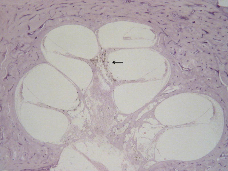
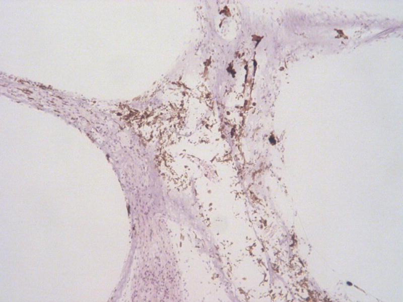
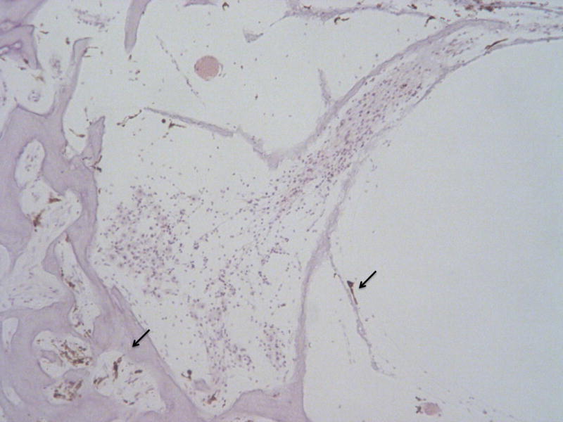
Microscopic examination revealed an abundant accumulation of melanin in the middle segment of the modiolus (Figs. 1 and 2), and scant in the anterior basal segment (Fig. 3). The organ of Corti was also missing in the anterior basal segment.
Fig. 1.

Image of cochlea showing copious melanin pigment in the middle segment (arrow) but little in the base. (Hematoxyline and eosin [H&E] × 20).
Fig. 2.

Higher power (100×) of the middle segment of the cochlea with abundant melanin in the modiolus. (H&E)
Fig. 3.

Anterior basal segment of the showing a portion of the modiolus and part of the osseous spiral lamina with little melanin (arrows), ×100. (H&E)
Discussion
This clinical case illustrates the concept that melanin distribution is disproportionally present in the cochlear mid-turns and apex compared to basal turns. Sun et al., hypothesize that the distribution of melanin in the cochlea may account for lower levels of oxidative protection at the cochlear base in presbycusis.2 With the exception of hydrops, sensorineural hearing loss caused by presbycusis, maternal-fetal rubella infection, aminoglycosides, and noise exposure disproportionally impact the basal turn with variable losses of spiral ganglion and hair cells reflected in a down-sloping audiogram.3 The location of melanin and known function as a free radical scavenger may explain some patterns of sensorineural hearing loss and highlights the need for further investigation of melanocyte function in the cochlea.2
Acknowledgments
Supported by NIDCD.NIH U24 DC 011962.
Footnotes
The authors disclose no conflicts of interest.
References
- 1.Ohlemiller KK, Rybak Rice ME, Lett JM, et al. Absence of strial melanin coincides with age-associated marginal cell loss and endocochlear potential decline. Hear Res. 2009;249:1Y14. doi: 10.1016/j.heares.2008.12.005. [DOI] [PubMed] [Google Scholar]
- 2.Sun DQ, Zhou X, Lin FR, Francis HW, Carey JP, Chien WW. Racial difference in cochlear pigmentation is associated with hearing loss risk. Otol Neurotol. 2014 Oct;35(9):1509–14. doi: 10.1097/MAO.0000000000000564. [DOI] [PubMed] [Google Scholar]
- 3.Rizk HG, Linthicum FH., Jr Histopathologic categorization of presbycusis. Otol Neurotol. 2012 Apr;33(3):e23–4. doi: 10.1097/MAO.0b013e31821f84ee. [DOI] [PubMed] [Google Scholar]


