

Abstract
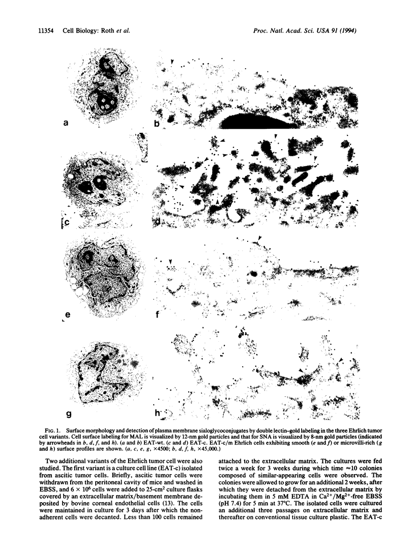
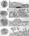
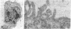
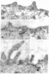
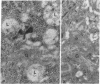
Three variants of the classical Ehrlich ascites tumor (EAT) cell have been studied by quantitative, sialic acid-specific, lectin-gold ultrastructural cytochemistry. Electron microscopic examination revealed pronounced differences in the surface morphology of the three cell variants. The wild-type Ehrlich cells (EAT-wt), grown in the peritoneal cavity of mice, exhibited a smooth surface profile. A variant form selected for growth as monolayer on basement membrane (EAT-c) showed a complex surface profile with numerous microvilli. The third variant (EAT-c/m), the cultured cells reinoculated into mice and passaged 20-25 times as ascites, presented a smooth surface profile similar to the EAT-wt cells. Quantitative single as well as double lectin-gold labeling revealed significant differences in the nature of cell surface sialoglycoproteins. The most significant finding was the presence of cell surface Neu5Ac alpha 2-6Gal residues as detected with the Sambucus nigra lectin on EAT-c and EAT-c/m cells, whereas EAT-wt cells contained little or none of such carbohydrate sequences. On the contrary, labeling by Maackia amurensis lectin, which recognizes the Neu5Ac alpha 2-3Gal beta 1-4GlcNAc sequence, was intense on all three Ehrlich cell variants; it was 20-60 times greater than alpha-2,6-linked sialic acid-containing glycoconjugates. Specific cell surface lectin binding combined with morphologic study appears to have identified a small subpopulation of cells within the ascites tumor that are capable of attaching to and growing on a basement membrane.
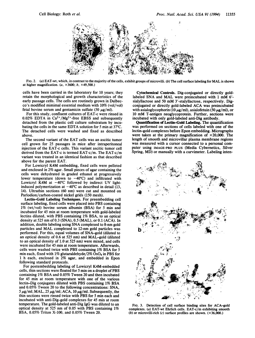
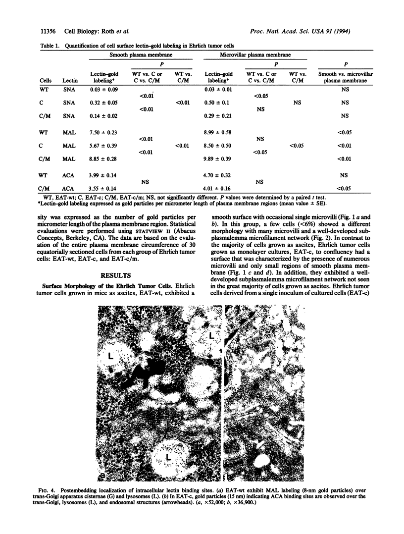
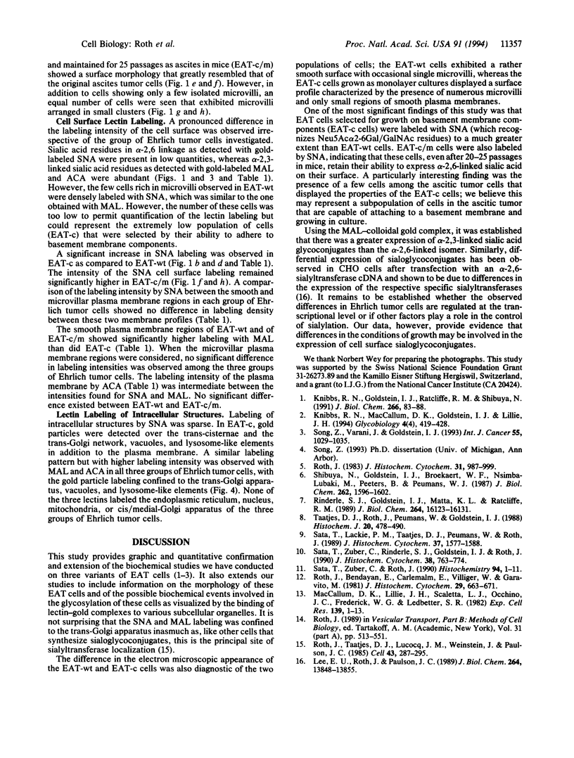
Full text
PDF
Images in this article
Selected References
These references are in PubMed. This may not be the complete list of references from this article.
- Knibbs R. N., Goldstein I. J., Ratcliffe R. M., Shibuya N. Characterization of the carbohydrate binding specificity of the leukoagglutinating lectin from Maackia amurensis. Comparison with other sialic acid-specific lectins. J Biol Chem. 1991 Jan 5;266(1):83–88. [PubMed] [Google Scholar]
- Knibbs R. N., MacCallum D. K., Lillie J. H., Goldstein I. J. Wild-type and cultured Ehrlich ascites tumour cells differ in tumorigenicity, lectin binding patterns and binding to basement membranes. Glycobiology. 1994 Aug;4(4):419–428. doi: 10.1093/glycob/4.4.419. [DOI] [PubMed] [Google Scholar]
- Lee E. U., Roth J., Paulson J. C. Alteration of terminal glycosylation sequences on N-linked oligosaccharides of Chinese hamster ovary cells by expression of beta-galactoside alpha 2,6-sialyltransferase. J Biol Chem. 1989 Aug 15;264(23):13848–13855. [PubMed] [Google Scholar]
- MacCallum D. K., Lillie J. H., Scaletta L. J., Occhino J. C., Frederick W. G., Ledbetter S. R. Bovine corneal endothelium in vitro. Elaboration and organization and of a basement membrane. Exp Cell Res. 1982 May;139(1):1–13. doi: 10.1016/0014-4827(82)90313-5. [DOI] [PubMed] [Google Scholar]
- Rinderle S. J., Goldstein I. J., Matta K. L., Ratcliffe R. M. Isolation and characterization of amaranthin, a lectin present in the seeds of Amaranthus caudatus, that recognizes the T- (or cryptic T)-antigen. J Biol Chem. 1989 Sep 25;264(27):16123–16131. [PubMed] [Google Scholar]
- Roth J. Application of lectin--gold complexes for electron microscopic localization of glycoconjugates on thin sections. J Histochem Cytochem. 1983 Aug;31(8):987–999. doi: 10.1177/31.8.6190857. [DOI] [PubMed] [Google Scholar]
- Roth J., Bendayan M., Carlemalm E., Villiger W., Garavito M. Enhancement of structural preservation and immunocytochemical staining in low temperature embedded pancreatic tissue. J Histochem Cytochem. 1981 May;29(5):663–671. doi: 10.1177/29.5.6166664. [DOI] [PubMed] [Google Scholar]
- Roth J., Taatjes D. J., Lucocq J. M., Weinstein J., Paulson J. C. Demonstration of an extensive trans-tubular network continuous with the Golgi apparatus stack that may function in glycosylation. Cell. 1985 Nov;43(1):287–295. doi: 10.1016/0092-8674(85)90034-0. [DOI] [PubMed] [Google Scholar]
- Sata T., Lackie P. M., Taatjes D. J., Peumans W., Roth J. Detection of the Neu5 Ac (alpha 2,3) Gal (beta 1,4) GlcNAc sequence with the leukoagglutinin from Maackia amurensis: light and electron microscopic demonstration of differential tissue expression of terminal sialic acid in alpha 2,3- and alpha 2,6-linkage. J Histochem Cytochem. 1989 Nov;37(11):1577–1588. doi: 10.1177/37.11.2478613. [DOI] [PubMed] [Google Scholar]
- Sata T., Zuber C., Rinderle S. J., Goldstein I. J., Roth J. Expression patterns of the T antigen and the cryptic T antigen in rat fetuses: detection with the lectin amaranthin. J Histochem Cytochem. 1990 Jun;38(6):763–774. doi: 10.1177/38.6.2335739. [DOI] [PubMed] [Google Scholar]
- Sata T., Zuber C., Roth J. Lectin--digoxigenin conjugates: a new hapten system for glycoconjugate cytochemistry. Histochemistry. 1990;94(1):1–11. doi: 10.1007/BF00266783. [DOI] [PubMed] [Google Scholar]
- Shibuya N., Goldstein I. J., Broekaert W. F., Nsimba-Lubaki M., Peeters B., Peumans W. J. The elderberry (Sambucus nigra L.) bark lectin recognizes the Neu5Ac(alpha 2-6)Gal/GalNAc sequence. J Biol Chem. 1987 Feb 5;262(4):1596–1601. [PubMed] [Google Scholar]
- Song Z., Varani J., Goldstein I. J. Differences in cell surface carbohydrates, and in laminin and fibronectin synthesis, between adherent and non-adherent Ehrlich ascites tumor cells. Int J Cancer. 1993 Dec 2;55(6):1029–1035. doi: 10.1002/ijc.2910550625. [DOI] [PubMed] [Google Scholar]
- Taatjes D. J., Roth J., Peumans W., Goldstein I. J. Elderberry bark lectin--gold techniques for the detection of Neu5Ac (alpha 2,6) Gal/GalNAc sequences: applications and limitations. Histochem J. 1988 Sep;20(9):478–490. doi: 10.1007/BF01002646. [DOI] [PubMed] [Google Scholar]