

Abstract
A unique strain of Clostridium botulinum (IBCA10-7060) was recently discovered which produces two toxins; botulinum neurotoxin (BoNT) serotype B and a novel BoNT reported as serotype H. Previous molecular assessment showed that the light chain (LC) of the novel BoNT most resembled the bont of the light chain of known subtype F5 while the C-terminus of the heavy chain (HC) most resembled the binding domain of serotype A. We evaluated the functionality of both toxins produced in culture by first incorporating an immunoaffinity step using monoclonal antibodies to purify BoNT from culture supernatants and tested each immune-captured neurotoxin with full-length substrates vesicle-associated membrane protein 2 (VAMP-2), synaptosomal-associated protein 25 (SNAP-25), syntaxin, and shortened peptides representing the substrates. The BoNT/B produced by this strain behaved as a typical BoNT/B, having immunoaffinity for anti-B monoclonal antibodies, and cleaving both full length VAMP-2 and a peptide based on the sequence of VAMP-2 in the expected location. As expected, there was no activity towards SNAP-25 or syntaxin. The novel BoNT demonstrated immunoaffinity for anti-A monoclonal antibodies, but did not cleave SNAP-25 as expected for BoNT/A. Instead, the novel BoNT cleaved VAMP-2 and VAMP-2-based peptides in the same location as BoNT/F5. This is the first discovery of a single botulinum neurotoxin with BoNT/A antigenicity and BoNT/F light chain function. This work suggests that the newly reported serotype H may actually be a hybrid of previously known BoNT serotype A and serotype F, specifically subtype F5.
Introduction
Botulism, a lethal disease if untreated, is caused by intoxication by highly toxic proteins known as botulinum neurotoxins (BoNTs), which are produced by various species of clostridia. BoNT, of which there are 7 confirmed serotypes (A through G), are each comprised of a heavy chain (HC), which binds to receptors on the neuron and facilitates translocation across the cell membrane, and a light chain (LC) which is a protease, cleaving specific proteins needed for nerve signal transmission. The descending flaccid paralysis that is characteristic of botulism is the result of this enzymatic cleavage of one or more of the proteins that make up the Soluble NSF Attachment Protein Receptor (SNARE) complex. BoNT/A, /C, and /E cleave synaptosomal-associated protein 25 (SNAP-25)1–6 whereas BoNT/B, /D, /F, and /G cleave vesicle-associated membrane protein 2 (VAMP-2), also known as synaptobrevin-27–12. BoNT/C is unique in that it also cleaves syntaxin13,14.
Typically C. botulinum produces only one serotype of toxin; however, some strains of C. botulinum produce more than one serotype of BoNT. These are referred to as bivalent toxin producers and are notated as Ab, Ba, Af, or Bf, with the capital letter representing the major BoNT produced by the strain. Most serotypes of BoNT also possess genetic and amino acid variance within each serotype. This variance is referred to as subtype, with the addition of a number following the letter to designate the subtype, i.e. subtype BoNT/A1. Currently, all subtypes within each serotype are known to cleave their protein target in the same location, with the exception of BoNT/F512.
The California Department of Public Health (CDPH) described a unique strain of Clostridium botulinum (IBCA10-7060) that they isolated in 2010 from the stool of an infant botulism case15. Although the authors did not state the origin of the sample, based on US surveillance reports, the origin is international16. This bivalent strain, identified as C. botulinum Bh, produced two toxins; the major component being serotype B, specifically subtype B2. The second minor toxin was reported as serotype H15; however, the designation of this novel toxin as a new serotype has since been questioned17. The novel bont gene (GenBank accession number KGO15617.1) can be divided into three sections with the N-terminal portion, corresponding to the LC, most similar to bont/F5 (86% identical at the nucleotide level), the C-terminal portion consisting of the binding domain of the HC nearly identical (96–97%) to bont/A1, and the middle portion similar to bont/F (77% identical)18. The authors demonstrated that the novel toxin causes botulism in animals; however, they did not report the exact functional mechanism by which it does so; namely, which of the SNARE complex protein or proteins is involved and where that protein(s) is cleaved.
Our laboratory previously reported that BoNT/F5 cleaved VAMP-2 in a different location than the other BoNT/F subtypes12. This discovery was accomplished through the use of immunoaffinity purification of BoNT/F5 from a culture supernatant followed by incubation of the BoNT/F5 with SNARE complex proteins SNAP-25, syntaxin, and VAMP-2 and by mass spectrometry of the resultant cleavage products. Using a similar approach, we analyzed the novel toxin reported as serotype H by CDPH. Our studies show that this novel toxin cleaves VAMP-2 in the same location as BoNT/F5. However, the novel toxin is unique from BoNT/F5 in that it can be immunopurified using antibodies directed toward the HC of BoNT/A. This is the first discovery of a botulinum neurotoxin with BoNT/A antigenicity and BoNT/F light chain function. This suggests that the secondary toxin produced by IBCA10-7060 may be a hybrid of known serotypes F and A (BoNT F/A) rather than a new unrecognized serotype.
Materials and Methods
Materials
Botulinum neurotoxin is highly toxic and thus requires stringent safety measures. All neurotoxins were handled in a Class II biosafety cabinet with HEPA filters. Monoclonal antibodies (mAbs) to BoNT/A (CR2 and RAZ1) and /B (2B18.2 and B12.1) were acquired from Dr. James Marks at the University of California at San Francisco. Dynabeads® (M-280/Streptavidin) were purchased from Invitrogen (Carlsbad, CA). BoNT/A, /B, and /F complexes were purchased from Metabiologics (Madison, WI). All chemicals were from Sigma-Aldrich (St. Louis, MO) except where indicated. SNAP-25 and syntaxin were purchased from Genway (San Diego, CA) and VAMP-2 was purchased from AtGen (Sampyeong-dong, South Korea). Peptide substrates LSELDDRADALQAGASQFESSAAKLKRKYWWKNLK (abbreviated as SubB) for measurement of BoNT/B activity, and TSNRRLQQTQAQVDEVVDIMRVNVDKVLERDQKLSELDDRADAL (abbreviated as SubF) for evaluation of BoNT/F activity were synthesized by Midwest Bio-tech Inc. (Fishers, IN). Sulfo-NHS-Biotin was purchased from Thermo Fisher Scientific (Waltham, MA).
Preparation of culture supernatants
IBCA10-7060 (CDPH) was streaked for isolation on Egg Yolk Agar (EYA) plates. A single colony was selected, assigned the designation of CDC69016, and further cultured in chopped meat glucose starch (CMGS) medium. Single colonies of CDC69016 were selected from EYA plates and incubated into trypticase peptone glucose yeast (TPGY) and/or CMGS media. Cultures were incubated for 5 days at 35°C under anaerobic conditions.
Preparation of mAb-coated beads
The mAbs were biotinylated in a fresh solution of 300 μM sulfo-NHS-biotin in water. A ratio of 4 μL of the 300 μM biotin was added to 20 μg of mAb for each serotype. The mixtures were incubated overnight at room temperature with no mixing. The biotinylated mAbs were bound to streptavidin beads as previously described19.
Extraction of BoNT from culture supernatants
An aliquot of the culture supernatant between 100 μL and 500 μL was placed into a deep well plate with phosphate buffered saline with tween (PBST) (1X or 10X) added to achieve a final volume between 500 μL and 550 μL with a final concentration of 1X PBST. To extract BoNT/B, 20 μL of beads coated with 2B18.2 and B12.1 mAbs were added to the culture supernatant. The deep well plate was capped and placed on a plate shaker for 1 hr at the minimal speed necessary to keep the beads in solution. The deep well plate was then uncapped and placed into a KingFisher Flex magnetic particle processor (Thermo Fisher Scientific, Waltham, MA) for automated bead washing, which included two washes with 1 mL each of 2M NaCl followed by two washes with 1 mL each of 1X PBST. The beads were eluted from the KingFisher Flex into 80 μL of water and removed from the KingFisher Flex.
The process was the same for extraction of the novel toxin with the exception that beads coated with CR2 and RAZ1 mAbs were used. Negative controls consisted of culture supernatant medium with no organism or toxin, with the remainder of the extraction protocol as above. Positive controls consisted of culture supernatant medium spiked with varying levels of BoNT/A, /B, or /F complexes, diluted in PBST to appropriate levels immediately prior to spiking with the remainder of the extraction protocol as above.
Endopep-MS reaction with MALDI-TOF analysis
The aqueous extract was removed from the beads, which contain the bound toxin, and they were reconstituted in reaction buffer consisting of 0.05 M Hepes (pH 7.3), 25 mM dithiothreitol, and 20 μM ZnCl2 and protein or peptide substrate as indicated in Table 1. All samples then were incubated at 37°C for 4 hr with no agitation. A 2-μL aliquot of each reaction supernatant was mixed with 18 μL of matrix solution consisting of α-cyano-4-hydroxy cinnamic acid (CHCA) at 5 mg/mL in 50% acetonitrile, 0.1% trifluoroacetic acid (TFA), and 1 mM ammonium citrate. A 0.5-μL aliquot of this mixture was pipeted onto one spot of a 384-spot matrix-assisted laser desorption/ionization (MALDI) plate (Applied Biosystems, Framingham, MA). Mass spectra of each spot were obtained by scanning from 900 to 5500 m/z in MS-positive ion reflector mode (for peptide analysis) or by scanning from 900 to 30,000 m/z in MS-positive linear mode (for protein analysis) on an Applied Biosystems 4800 Proteomics Analyzer (Framingham, MA). The instrument uses an Nd-YAG laser at 355 nm, and each spectrum is an average of 2400 laser shots.
Table 1.
Volume and concentration of reagents for incubation with BoNT.
| Substrate | Volume of buffer | Volume of substrate | Concentration of substrate |
|---|---|---|---|
| VAMP-2 | 15 uL | 5 uL | 1 mg/mL |
| SNAP-25 | 15 uL | 5 uL | 1 mg/mL |
| Syntaxin | 15 uL | 5 uL | 1 mg/mL |
| SubB | 18 uL | 2 uL | 1 mM |
| SubF | 18 uL | 2 uL | 1 mM |
Digestion and LC-MS/MS Analysis of BoNT
To positively identify the novel botulinum neurotoxin extracted from the culture supernatant of CDC69016, the toxin was digested with trypsin and chymotrypsin following the same protocol previously used19. Peptides from the toxin were identified by LC-MS/MS with database searching as described previously19.
Results
Characterization of BoNT/B in CDC69016
BoNT/B is known to cleave VAMP-2 between 76Q and 77F7. To test the light chain enzymatic function of the BoNT/B component in the culture supernatant of strain CDC69016, we began by immunoextraction of the BoNT/B using beads coated with mAbs to BoNT/B. The beads were then incubated with VAMP-2 and Figure 1 shows that this resulted in two peaks in the mass spectrometer which correspond to cleavage of VAMP-2 by BoNT/B. The peak at m/z 13825 corresponds to intact VAMP-2, the peak at m/z 12215 in Figure 1B corresponds to the N-terminal cleavage product, and the peak at m/z 1628 in Figure 1B corresponds to the C-terminal cleavage product. Cleavage products with these masses can only be generated from cleavage of VAMP-2 between 76Q and 77F, and these cleavage products were present in supernatants produced in both TPGY and CMGS media.
Figure 1.
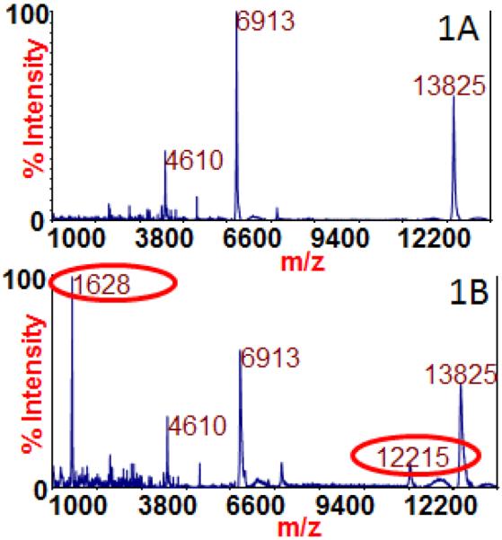
Mass spectra of VAMP-2 incubated with no toxin (1A) and BoNT/B immunopurified from the culture supernatant of CDC69016 (2B). The peaks at m/z 13825 and 6913 correspond to singly charged and doubly charged intact VAMP-2 in both 1A and 1B and the cleavage products are circled in red in 1B. The peak at m/z 12215 in 1B corresponds to the N-terminal cleavage product of VAMP-2 by BoNT/B, and the peak at m/z 1628 in 2B corresponds to the C-terminal cleavage product.
To further demonstrate the function of the BoNT/B extracted from the culture supernatant of strain CDC69016, we also tested the immunoextracted material with a peptide substrate (SubB) which is a shortened version of VAMP-2 containing the BoNT/B cleavage site. BoNT/B has been reported to cleave this peptide in the location that corresponds to the region of VAMP-2 between 76Q and 77F20, generating an N-terminal cleavage product of 1760 m/z and a C-terminal cleavage product of m/z 2284. The peaks at 4025 and 2013 m/z in Figure 2A correspond to singly charged and doubly charged intact SubB, the peak at 1760 m/z in Figure 2B is the N-terminal cleavage product of SubB after being cleaved by BoNT/B, and the peak at m/z 2284 in Figure 2B is the C-terminal cleavage product. The BoNT/B immunoextracted from the culture supernatant of CDC69016 was also tested with SNAP-25 and syntaxin, the other SNARE complex proteins, and the toxin did not cleave these proteins (data not shown).
Figure 2.

Mass spectra of SubB incubated with no toxin (2A) and BoNT/B immunopurified from the culture supernatant of CDC69016 (2B). The peaks at m/z 4025 and 2013 correspond to singly charged and doubly charged intact SubB in 2A and the cleavage products are circled in red in 2B. The peak at m/z 2284 in 2B corresponds to the N-terminal cleavage product of SubB by BoNT/B, and the peak at m/z 1760 in 2B corresponds to the C-terminal cleavage product.
Characterization of Novel Toxin in CDC69016
Gene sequencing of the novel toxin showed that the C-terminal portion, particularly the binding domain, had a high similarity to BoNT/A118. Therefore, we theorized that an antibody designed for the C-terminal portion of BoNT/A1 might immunoextract the novel toxin from the CDC69016 culture supernatant. Our laboratory previously reported that mAbs CR2 and RAZ1, designed for the C-terminal portion of BoNT/A, were successful at extraction of multiple subtypes of BoNT/A21. Additionally, one reported region of BoNT/A critical for binding to mAb CR2 is 1056KLDGCRDTHR1065 in BoNT/A1 and 1056KLDGCRDPRR1065 in BoNT/A222. According to the gene sequence, the novel bont gene is >85% similar to bont genes A1 and A2 in this region. Therefore, mAbs CR2 and RAZ1 were used to immunoextract the novel BoNT from the CDC69016 culture supernatant.
Digestion of the immunoextracted toxin and analysis of the subsequent peptides by LC-MS/MS yielded accurate identification of the novel BoNT with 61% sequence coverage. Twenty-five peptides identified by MS/MS are unique to this toxin and are listed in Table 2 as evidence of the immunocapture of this unique toxin. MS/MS of one of these unique peptides, IYSFTEFNLAYEFK is depicted in Figure 3A alongside the MS/MS of a very similar peptide from BoNT/F5, IYSFTEFNLAHEFK, in Figure 3B. The MS/MS of both peptides are quite similar, particularly among the y8-y12 fragment ions; yet differences in the spectra are observed due to the mass difference between the tyrosine and histidine residues. The MS/MS of the unique peptide YFNLFDKELNKK from BoNT F/A is even more similar to the MS/MS of the peptide YFNLFDKELNEK from BoNT/A1 in Figure 4, and in this case, the mass difference is harder to distinguish as lysine and glutamic acid are separated in mass by only one Dalton. This does not pose an analytical challenge for the instrumentation used in this study as the resolution of this instrument is able to resolve peptides separated by as little as 0.03 Da. These MS/MS and the twenty-three additional MS/MS not shown here prove the successful immunocapture of this novel toxin.
Table 2.
Peptides identified by LC-MS/MS which are unique to the novel BoNT in CDC69016. Specific residues which are unique to the novel BoNT are underlined, and the closest BoNT neighbor is listed for each peptide.
| Amino acid sequence | Nearest BoNT |
|---|---|
| 2PVVINSFNYDDPVNDNTIIYIRPPYYETSNTYFK35 | BoNT/F5 |
| 36AFQIMDNVWIIPER49 | BoNT/F7 |
| 52LGIDPSLFNPPVSLK66 | BoNT/F5 |
| 98INSKPAGQILLEEIK112 | BoNT/F7 |
| 113NAIPYLGNSYTQEEQFTTNNR133 | BoNT/F5 |
| 141LANGNIVQQMANLIIWGPGPDLTTNK166 | BoNT/F5 |
| 168TGGIIYSPYQSMEATPYK185 | BoNT/F5 |
| 274NIVPQSIQSQLYNK287 | BoNT/F5 |
| 302VNTATALINIDEFK315 | BoNT/F5 |
| 346IYSFTEFNLAHEFK359 | BoNT/F5 |
| 445DLFFIASQESYGENTINTYK464 | BoNT/F5 |
| 465EIDDTTTLDPSFEDILDK482 | BoNT/F5 |
| 483VILNFNEQVIPQMPNR498 | BoNT/F5 |
| 499NVSTDIQKDNYIPK512 | BoNT/F5 |
| 628TDIIDSYEVGR638 | None |
| 639NYNTFFYLNAQK650 | BoNT/F |
| 680TLDNALNVR688 | None |
| 748IDSSYNINEIER759 | BoNT/CD |
| 771NIEQFITESSIAYLINIINNETIQK795 | BoNT/F5 |
| 813NHSSILGNSVEELNSK828 | None |
| 829VNNHLDNGIPFELSSYTNDSLLIR852 | BoNT/F1 |
| 896FSEIDKNQVQLSNLESSK913 | BoNT/A1 |
| 1026LINEESISDLGNIHASNNIMFK1047 | BoNT/A1 |
| 1063YFNLFDKELNKK1074 | BoNT/A1 |
| 1114YLDVNNVGIR1123 | BoNT/A1 |
Figure 3.

MS/MS of the peptide IYSFTEFNLAYEFK unique to novel BoNT (3A) and the similar peptide IYSFTEFNLAHEFK from BoNT/F5 (3B).
Figure 4.

MS/MS of the peptide YFNLFDKELNKK unique to novel BoNT (4A) and the similar peptide YFNLFDKELNEK from BoNT/A1 (4B).
Once it was determined that the novel BoNT could be immunopurified from the culture supernatant of CDC69016 using the RAZ1 and CR2 mAbs, we proceeded to characterize the functional activity of the toxin. VAMP-2 was incubated in the absence and presence of the type A immunoextracted toxin. The peaks at m/z 13825 and 6913 correspond to singly charged and doubly charged intact VAMP-2 in both the negative control (Figure 5A) and the experimental with the novel BoNT (Figure 5B). However, the dominant peak in the experimental sample (Figure 5B) is m/z 4025, which corresponds to the C-terminal cleavage product of cleavage of VAMP-2 between 54L and 55E. The N-terminal cleavage product is larger and thus does not ionize well and is not visible. This C-terminal cleavage product was present in supernatants produced in both TPGY and CMGS media.
Figure 5.

Mass spectra of VAMP-2 incubated with no toxin (5A) and the novel toxin immunopurified from the culture supernatant of CDC69016 (5B). The peaks at m/z 13825 and 6913 correspond to singly charged and doubly charged intact VAMP-2 in both 5A and 5B and the cleavage products are circled in red in 5B. The peak at m/z 4025 in 5B corresponds to the C-terminal cleavage product.
The immunoextracted novel toxin was also tested with a peptide substrate (SubF) which is a shortened version of VAMP-2. BoNT/F5 has been reported to cleave this peptide in the location corresponding to the region of VAMP-2 between 54L and 55E12, producing an N-terminal cleavage product of m/z 3255 and a C-terminal cleavage product of 1874 m/z. The peak at 2556 m/z in Figure 6A and 6B corresponds to doubly charged intact SubF, the peak at 1874 m/z in Figure 6B is the C-terminal cleavage product of SubF after being cleaved by the novel toxin, and the peak at m/z 3255 in Figure 6B is the N-terminal cleavage product. The novel toxin immunoextracted from the culture supernatant of CDC69016 was also tested with SNAP-25 and syntaxin, the other SNARE complex proteins, and the toxin did not cleave any of these proteins (data not shown).
Figure 6.

Mass spectra of SubF incubated with no toxin (6A) and the novel toxin immunopurified from the culture supernatant of CDC69016 (6B). The peak at m/z 2556 corresponds to doubly charged intact SubF, and the cleavage products are circled in red in 6B. The peak at m/z 3255 in 6B corresponds to the N-terminal cleavage product of SubF by the novel toxin, and the peak at m/z 1874 in 6B corresponds to the C-terminal cleavage product.
Since both the toxins produced in this bivalent strain cleaved the same protein substrate, but at different sites, the question was posed as to whether both toxins could act upon the same substrate at the same time. There are 22 amino acids between the two specific cleavage sites as seen in Figure 7A. A combination of RAZ1, CR2, 2B18.2, and B12.1 mAbs were used to extract both the BoNT/B and the novel BoNT from the CDC69016 culture supernatant. The toxins were incubated with VAMP-2, and the resultant spectrum is presented in Figure 7C. The peak at m/z 1628 corresponds to the C-terminal cleavage product of BoNT/B activity upon VAMP-2, and the peak at m/z 12215 corresponds to the N-terminal cleavage product of BoNT/B activity upon VAMP-2. The peak at m/z 4026 corresponds to the C-terminal cleavage product of the novel toxin activity upon VAMP-2. However, the peak at m/z 2416 corresponds to the mass of the peptide produced when both BoNT/B and the novel toxin cleave the substrate simultaneously. Therefore, it was demonstrated that both toxins could act on the same substrate at the same time.
Figure 7.

Sequence of VAMP-2 with cleavage locations and expected masses (7A) and mass spectra of VAMP-2 incubated with no toxin (7B) and a combination of BoNT/B and the novel toxin (7C). The peaks at m/z 13825 and 6913 correspond to singly charged and doubly charged intact VAMP-2 in both 7B and 7C, and the cleavage products are circled in red in 7C. The peak at m/z 1628 corresponds to the C-terminal cleavage product of BoNT/B cleavage, the peak at m/z 4026 corresponds to the C-terminal cleavage product of the novel toxin cleavage, the peak at m/z 12215 corresponds to the N-terminal cleavage product of BoNT/B, and the peak at m/z 2416 corresponds to the peptide produced by cleavage of VAMP-2 by both BoNT/B and the novel toxin.
Discussion
Previously, CDPH reported on a strain of C. botulinum which produces two BoNTs; one serotype B and the other a previously unreported toxin. The type B neurotoxin was reported to be the major neurotoxin produced by this C. botulinum strain15 and this finding was confirmed in the present study by the amount of enzymatic activity observed for the two toxins. The BoNT/B was extracted using monoclonal antibodies that bind to all known BoNT B subtypes (2B18.2 and B12.1) and the enzymatic activity on both full length VAMP-2 and a peptide mimic were found to be the same as all other BoNT/Bs with cleavage between 76Q and 77F. As previously reported, the BoNT from this dual toxin producing stain of C. botulinum is very similar to other BoNT B with a sequence that most closely resembles BoNT/B2; only 1 amino acid variance was observed between this toxin and BoNT/B2 ABM 7397523. Based on its similarity with other BoNT/B, it is not surprising that this BoNT/B cleaves VAMP-2 at the same site as all other BoNT/B.
The second toxin produced by the IBCA10-7060 was very different than any single known toxin. A comparison of the overall amino acid sequence of the novel toxin showed that BoNT F5 was the closest match (64% similarity) suggesting a potential new serotype. The C-terminal binding domain of the toxin most closely resembles BoNT/A1 (96% amino acid similarity), the N terminal portion (LC) resembled BoNT/F5 (80% amino acid similarity) and the middle sections most closely resembled other BoNT/F (64% amino acid similarity). BoNT/Fs have the highest degree of amino acid diversity among the BoNT serotypes. For example, all BoNT/B are 93% or more identical and BoNT/A are ≥84% identical, while BoNT/Fs differ by up to 30% in the case of BoNT/F1 and /F524. The lack of sequence similarity (47.3% identity) of the enzymatically-active light chain between BoNT/F5 and BoNT/F1 is even more pronounced. Differing serotypes with different sites of enzymatic activity have a higher sequence similarity than what was displayed in the light chain of BoNT/F5 and BoNT/F1. For instance, BoNT/E and BoNT/F have 56% similarity and cleave entirely different proteins5,9. Our previous discovery that BoNT/F5 cleaves VAMP-2 in a different location than other BoNT/F supports the understanding that drastic changes in amino acid sequence in critical locations can have radical changes in enzyme function12.
The novel toxin in CDC69016 has BoNT/A antigenicity based upon its response to mAbs RAZ1 and CR2, which are antibodies known to react with BoNT/A21. This is expected considering the high amino acid sequence homology (96–97%) reported between the C-terminal binding domain of the novel toxin and the C-terminal binding domain of BoNT/A. The novel toxin does not appear to have BoNT/F antigenicity, as attempts to isolate this toxin with polyclonal antibodies to BoNT/F and several monoclonal antibodies directed toward the HC of BoNT/F were unsuccessful (data not shown). Despite the BoNT/A antigenicity, the novel toxin was determined to have BoNT/F5 enzymatic function. This is not surprising considering the amino acid similarity between the light chain of BoNT/F5 and this toxin (80%)18. An amino acid identity of 80% such as exists between the light chain of the novel toxin and /F5 is greater than the 63% amino acid identity between the light chains of BoNT/F1 and /F7. BoNT/F1 and /F7 are known to have the same enzymatic function, but do have some differences in substrate recognition25. Based on the novel toxin's antigenicity toward type A antibody and the demonstration of a type F5 cleavage of substrate, we believe this novel toxin represents a novel hybrid between known serotypes of A and F.
The existence of a hybrid BoNT is not new. BoNT/C and /D are known to form mosaics, consisting of the light chain with high sequence homology to BoNT/C and the heavy chain resembling BoNT/D, and vice versa26. Such mosaics are notated with two capital letters with the first letter indicating the homology with the light chain and the second letter indicating the homology with the HC. One such example is the case of BoNT/DC. This mosaic toxin has 96% amino acid homology in the LC with that of BoNT/D yet the HC has 74% homology with BoNT/C126. The BoNT/DC mosaic has been reported to be immunoaffinity extracted with polyclonal antibodies to BoNT/C yet has BoNT/D enzymatic function, cleaving VAMP-2 rather than SNAP-25 or syntaxin, and cleaving VAMP-2 in the same location as BoNT/D27.
The BoNT/B produced by CDC69016 has the antigenicity and functionality expected of BoNT/B since it cleaved VAMP-2 between 76Q and 77F and was immunoaffinity purified using mAbs 2B18.2 and B12.1. The other neurotoxin produced by CDC69016 has the antigenicity of BoNT/A as it was immunopurified with mAbs RAZ1 and CR2 yet has the functionality of BoNT/F5 as it cleaved VAMP-2 between 54L and 55E. We have shown that both neurotoxins produced by CDC69016 have the same substrate (VAMP-2) and both toxins can cleave this protein at the same time. It remains to be seen if the multiple actions upon VAMP-2 effect botulism and/or its recovery.
Previously, a cocktail of type A monoclonal antibodies (including clonal relatives of RAZ1 and CR2) demonstrated protective capacity in an animal model28. CDPH previously reported that when sufficient levels of a heptavalent therapeutic product were provided, mice were protected against the effects of toxins produced by this unique strain15. The presence of the previously known serotype BoNT produced in excess by this strain makes final determination of the uniqueness of the novel toxin difficult. However, based on this study showing the toxin's antigenicity towards type A antibody, it's enzymatic function toward VAMP-2, and the previous report of neutralization with existing antibody, we suggest an alternative nomenclature for this novel toxin as BoNT F/A similar to those proposed for hybrids or mosaics between serotype D and C BoNTs.
Acknowledgements
The findings and conclusions in this report are those of the authors and do not necessarily represent the official position of the Centers for Disease Control and Prevention.
IBCA10-7060 was kindly provided to CDC by the California Department of Public Health. The authors greatly appreciate the deposition of GenBank sequence KGO15617.1 by Dr. Shashi Sharma of the FDA.
Abbreviations
- BoNT
(Botulinum Neurotoxin)
- SNAP
(synaptosomal-associated protein)
- VAMP
(vesicle-associated membrane protein)
- SNARE
(Soluble NSF Attachment Protein Receptor)
- CHCA
(α-cyano-4-hydroxy cinnamic acid)
- TFA
(trifluoroacetic acid)
- MALDI
(matrix-assisted laser desorption ionization)
- HC
(heavy chain)
- (Hc)
C-terminus of the heavy chain
- (LC)
light chain
- EYA
(egg yolk agar)
- (TPGY)
trypticase peptone glucose yeast
- (CMGS)
chopped meat glucose starch
- (PBST)
phosphate buffered saline with tween
- (mAb)
monoclonal antibody
- (CDPH)
California Department of Public Health
References
- (1).Binz T, Blasi J, Yamasaki S, Baumeister A, Link E, Sudhof TC, Jahn R, Niemann H. J Biol Chem. 1994;269:1617. [PubMed] [Google Scholar]
- (2).Blasi J, Chapman ER, Link E, Binz T, Yamasaki S, De Camilli P, Sudhof TC, Niemann H, Jahn R. Nature. 1993;365:160. doi: 10.1038/365160a0. [DOI] [PubMed] [Google Scholar]
- (3).Foran P, Lawrence GW, Shone CC, Foster KA, Dolly JO. Biochemistry. 1996;35:2630. doi: 10.1021/bi9519009. [DOI] [PubMed] [Google Scholar]
- (4).Schiavo G, Rossetto O, Catsicas S, Polverino de Laureto P, DasGupta BR, Benfenati F, Montecucco C. J Biol Chem. 1993;268:23784. [PubMed] [Google Scholar]
- (5).Schiavo G, Santucci A, Dasgupta BR, Mehta PP, Jontes J, Benfenati F, Wilson MC, Montecucco C. FEBS letters. 1993;335:99. doi: 10.1016/0014-5793(93)80448-4. [DOI] [PubMed] [Google Scholar]
- (6).Williamson LC, Halpern JL, Montecucco C, Brown JE, Neale EA. J Biol Chem. 1996;271:7694. doi: 10.1074/jbc.271.13.7694. [DOI] [PubMed] [Google Scholar]
- (7).Schiavo G, Benfenati F, Poulain B, Rossetto O, Polverino de Laureto P, DasGupta BR, Montecucco C. Nature. 1992;359:832. doi: 10.1038/359832a0. [DOI] [PubMed] [Google Scholar]
- (8).Schiavo G, Malizio C, Trimble WS, Polverino de Laureto P, Milan G, Sugiyama H, Johnson EA, Montecucco C. J Biol Chem. 1994;269:20213. [PubMed] [Google Scholar]
- (9).Schiavo G, Shone CC, Rossetto O, Alexander FC, Montecucco C. J Biol Chem. 1993;268:11516. [PubMed] [Google Scholar]
- (10).Yamasaki S, Baumeister A, Binz T, Blasi J, Link E, Cornille F, Roques B, Fykse EM, Sudhof TC, Jahn R, et al. J Biol Chem. 1994;269:12764. [PubMed] [Google Scholar]
- (11).Yamasaki S, Binz T, Hayashi T, Szabo E, Yamasaki N, Eklund M, Jahn R, Niemann H. Biochemical and biophysical research communications. 1994;200:829. doi: 10.1006/bbrc.1994.1526. [DOI] [PubMed] [Google Scholar]
- (12).Kalb SR, Baudys J, Webb RP, Wright P, Smith TJ, Smith LA, Fernandez R, Raphael BH, Maslanka SE, Pirkle JL, Barr JR. FEBS letters. 2012;586:109. doi: 10.1016/j.febslet.2011.11.033. [DOI] [PMC free article] [PubMed] [Google Scholar]
- (13).Blasi J, Chapman ER, Yamasaki S, Binz T, Niemann H, Jahn R. The EMBO journal. 1993;12:4821. doi: 10.1002/j.1460-2075.1993.tb06171.x. [DOI] [PMC free article] [PubMed] [Google Scholar]
- (14).Schiavo G, Shone CC, Bennett MK, Scheller RH, Montecucco C. J Biol Chem. 1995;270:10566. doi: 10.1074/jbc.270.18.10566. [DOI] [PubMed] [Google Scholar]
- (15).Barash JR, Arnon SS. The Journal of infectious diseases. 2014;209:183. doi: 10.1093/infdis/jit449. [DOI] [PubMed] [Google Scholar]
- (16).Centers for Disease Control and Prevention . US Department of Health and Human Services, CDC. 2011. [Google Scholar]
- (17).Johnson EA. The Journal of infectious diseases. 2014 [Google Scholar]
- (18).Gonzalez-Escalona N, Thirunavukkarasu N, Singh A, Toro M, Brown EW, Zink D, Rummel A, Sharma SK. Genome announcements. 2014;2 doi: 10.1128/genomeA.01275-14. [DOI] [PMC free article] [PubMed] [Google Scholar]
- (19).Kalb SR, Baudys J, Smith TJ, Smith LA, Barr JR. Analytical chemistry. 2014;86:3254. doi: 10.1021/ac5001509. [DOI] [PMC free article] [PubMed] [Google Scholar]
- (20).Barr JR, Moura H, Boyer AE, Woolfitt AR, Kalb SR, Pavlopoulos A, McWilliams LG, Schmidt JG, Martinez RA, Ashley DL. Emerging infectious diseases. 2005;11:1578. doi: 10.3201/eid1110.041279. [DOI] [PMC free article] [PubMed] [Google Scholar]
- (21).Kalb SR, Lou J, Garcia-Rodriguez C, Geren IN, Smith TJ, Moura H, Marks JD, Smith LA, Pirkle JL, Barr JR. PLoS ONE. 2009;4:e5355. doi: 10.1371/journal.pone.0005355. [DOI] [PMC free article] [PubMed] [Google Scholar]
- (22).Garcia-Rodriguez C, Levy R, Arndt JW, Forsyth CM, Razai A, Lou J, Geren I, Stevens RC, Marks JD. Nature biotechnology. 2007;25:107. doi: 10.1038/nbt1269. [DOI] [PubMed] [Google Scholar]
- (23).Hill KK, Smith TJ, Helma CH, Ticknor LO, Foley BT, Svensson RT, Brown JL, Johnson EA, Smith LA, Okinaka RT, Jackson PJ, Marks JD. Journal of bacteriology. 2007;189:818. doi: 10.1128/JB.01180-06. [DOI] [PMC free article] [PubMed] [Google Scholar]
- (24).Raphael BH, Choudoir MJ, Luquez C, Fernandez R, Maslanka SE. Applied and environmental microbiology. 2010;76:4805. doi: 10.1128/AEM.03109-09. [DOI] [PMC free article] [PubMed] [Google Scholar]
- (25).Kalb SR, Baudys J, Egan C, Smith TJ, Smith LA, Pirkle JL, Barr JR. Applied and environmental microbiology. 2011;77:1301. doi: 10.1128/AEM.01662-10. [DOI] [PMC free article] [PubMed] [Google Scholar]
- (26).Moriishi K, Koura M, Abe N, Fujii N, Fujinaga Y, Inoue K, Ogumad K. Biochimica et biophysica acta. 1996;1307:123. doi: 10.1016/0167-4781(96)00006-1. [DOI] [PubMed] [Google Scholar]
- (27).Moura H, Terilli RR, Woolfitt AR, Gallegos-Candela M, McWilliams LG, Solano MI, Pirkle JL, Barr JR. FEMS immunology and medical microbiology. 2011;61:288. doi: 10.1111/j.1574-695X.2010.00774.x. [DOI] [PubMed] [Google Scholar]
- (28).Nowakowski A, Wang C, Powers DB, Amersdorfer P, Smith TJ, Montgomery VA, Sheridan R, Blake R, Smith LA, Marks JD. Proceedings of the National Academy of Sciences of the United States of America. 2002;99:11346. doi: 10.1073/pnas.172229899. [DOI] [PMC free article] [PubMed] [Google Scholar]