

Abstract
Ellis Van Creveld syndrome (EVC) is a rare genetic disorder having autosomal recessive inheritance affecting the Amish population of Pennsylvania in USA with incidence of 1:244,000 for the general population. This syndrome consists of characteristic features such as bilateral postaxial polydactyly, chondroectodermal dysplasia, congenital heart defects and hypoplastic nails and teeth. There are few case reports of this syndrome reported in dental literature. We report a case of a 17 year old female presenting typical features of this syndrome and the oral findings of this patient which are the key diagnostic features.
Keywords: Ellis Van Creveld syndrome, Polydactyly, Chondroectodermal dysplasia
1. Introduction
Syndrome Ellis Van Creveld (EVC) or dysplasia chondroectodermal was described in 1940 by Richard W.B. Ellis and Simon Van Creveld as an rare autosomal recessive disorder due to a genetic defect located in chromosome 4p16.1–3 It is caused by mutation in the EVC gene as well as by a mutation in a non homologous gene EV2, located close to the EVC gene in a head to head configuration with parental consanguinity in about 30% of cases.2,4,5 In 1964, McKusick et al., reported a large number of cases in the Amish community of Lancaster country, Pennsylvania USA and also in Aboriginal kindred in Australia.6 The birth prevalence in Amish population is 1/5000 live births and in Non Amish population is 7/1,000,000.4,5 Around 150 cases are described in literature.3,4,7 Not more than 25 cases have been reported in India. EVC presents a characteristic tetrad 1. disproportionate dwarfism 2. bilateral postaxial polydactyly 3. ectodermal dysplasia 4. congenital heart malformation.1,5
2. Case report
A 17 year old female [Fig. 1] patient visited our institution with a complaint of decayed teeth and missing lower teeth. Patient gives history of exfoliation of deciduous teeth and no eruption of permanent teeth. There is no history of consanguineous marriage between parents. Antenatal, natal and neonatal histories were non contributory. No significant family history. On general examination patient had a short stature with bilateral postaxial polydactyly of hands and feet [Fig. 2] with bimanual hexadactyly noted on the ulnar side [Fig. 3]. She also had hypoplastic fingers and nails with wide gap between big toe and other two toes. A comprehensive examination was completed by a team of multidisciplinary specialists. The patient's intellectual ability was within the normal range. Chest X ray and echocardiography was performed to rule out any chest and cardiac defects.
Fig. 1.

Facial photograph.
Fig. 2.

Polydactyly of the lower limbs with dystrophic nails.
Fig. 3.

Hexadactyly noted on the ulnar side.
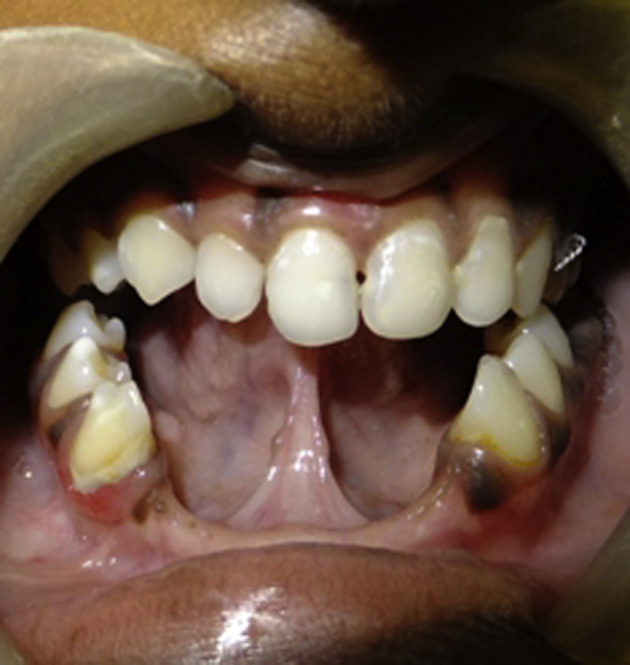
Extraoral examination revealed a concave facial profile, low set ears and malar hypoplasia. Intraorally soft tissue examination revealed multiple accessory labiogingival frenula with absence of mucobuccal fold in maxillary and mandibular anterior region [Fig. 4] and ankyloglossia [Fig. 5]. Hard tissue examination revealed class III malocclusion, congenitally missing lower central and lateral incisors, enamel hypoplasia and conical teeth [Fig. 5]. Panoramic radiography confirmed oligodontia of permanent teeth and multiple carious teeth. Hand wrist radiograph revealed incomplete duplication of left 5th metacarpal and phalanges seen in radial aspect with synostosis of the phalangeal joints suggestive of postaxial polydactyly. Right hand there is radio opaque density with soft tissue component noted in the ulnar aspect of 5th digit suggestive of poorly differentiated bone formation suggestive of incomplete axial polydactyly [Fig. 6]. Based on the clinical and radiographic findings of the dental and medical examination, the patient was diagnosed to have EVC syndrome. A team of oral and maxillofacial surgeon, orthodontist and prosthodontist were required to correct the craniofacial and dental defects. Therefore the patient was referred to the subsequent specialists for further treatment and has been on a regular follow up.
Fig. 4.

Multiple accessory upper labiogingival frenula with absence of mucobuccal fold.
Fig. 5.
Ankyloglossia, anodontia of the lower incisors and enamel hypoplasia.
Fig. 6.

Hand wrist radiograph.
3. Discussion
Ellis Van Creveld Syndrome also known as chondroectodermal dysplasia is genetic disorder with autosomal recessive inheritance and parental consanguinity.2,3 seen commonly in Amish population of Pennsylvania USA which does not show any gender predilection.2 Our patient is the eldest daughter of non consanguineous and normally developed parents. The diagnosis can be made as early as 18th week of gestation by ultrasonography when the increased nuchal translucency is evident.4,7,8 and later by clinical examination after birth.1,3,4
The most consistent clinical feature is chondrodystrophy due to defect in ossification affecting tubular bones resulting in shortened long bones of the limbs especially in distal and middle segments resulting in acromesomelic dwarfism.3,4,8 The other features include polydactyly usually bilateral postaxial hexadactyly most often seen in upper limbs on ulnar side and involves lower limb in 10% of cases. They also have wide hands and feet, sausage shaped fingers and dysplastic fingernails.2,8 Our patient had hexadactyly in both the upper and lower limbs with no syndactyly. Other features include genu valga, curvature of the humerus, talipes equinovarus, talipes calcaneovalgus4 and pectus carinatum with a long narrow chest.2–4,8 Congenital heart malformations are described in a 50–60% of patients. The anomalies include defects of the mitral and tricuspid valves, patent ductus arteriosus, ventricular septal defect, atrial septal defect and hypoplastic left heart syndrome which are the principal causes of decreased life-expectancy in these patients.3,8,9 The disease has characteristic oral manifestations that help early diagnosis at birth or during early childhood.5 The most common among them include fusion of the upper lip to the gingival margin resulting in the absence of mucobuccal fold, broad maxillary labial frenum described as partial harelip, multiple small accessory frenula, ankyloglossia, malocclusion, conical, microdontia teeth, hypodontia, anodontia (commonly the absence of permanent mandibular central and lateral incisors) and enamel hypoplasia.5,7,8,10 Our patient had all these findings. Other minor or variable manifestations include retarded eruption, supernumerary teeth, dental fusion, dysmorphic roots, taurodontism, abnormal occlusal anatomy with wide grooves and atypical cusps.5
Genitourinary abnormalities are seen in about 22% of the cases and include vulvar atresia, megaureters, nephrocalcinosis and renal agenesis.3,8 Several inconstant additional clinical findings are described, including strabismus, epi- and hypospadias, cryptorchidism and thoracic wall and pulmonary malformations.3,7 Exceptionally, hematological anomalies such as dyserythropoiesis and perinatal myeloblastic leukemia have been reported.3,7 The cognitive and motor development are normal with occasional CNS anomalies and hydrocephaly.8,9
The definitive diagnosis is molecular based on the homozygosity for a mutation in the EVC 1 and/or EVC 2 genes by direct sequencing. However the genetic mutations are seldom required for the clinical diagnosis as gene mutations is positive in only 2/3rd of patients. Due to the lack of availability of genetic studies the diagnosis was achieved clinically based on the observation of the symptoms and manifestations as described and with the aid of additional tests such as radiology, laboratory and cardiac function.1,3,8
Differential diagnosis includes other short rib polydactyly syndromes like Weyers acrodental dysostosis (Curry-Hall syndrome), asphyxiating thoracic dystrophy (Jeune syndrome), achondroplasia, chondroplasia punctata, orofaciodigital syndromes and Morquio's syndrome.3,4,7,8
EVC syndrome and Weyer's acrodental dysostosis (Curry-Hall syndrome) are allelic conditions caused by loss of function mutation in EVC and EVC2. These are separated by 2–6 kb of genomeric sequence on chromosome 4p16.10.2 Clinical features in Curry-Hall syndrome are similar to Ellis Van Creveld syndrome, which includes normal stature and cardiac defects; however thoracic dysplasia is generally absent. Additional features of Curry-Hall syndrome include presence of osseous cleft of symphysis of the mandible, while fifth carpal in distal row of wrist and multiple ossification centres in hamate is absent.4
Asphyxiating thoracic dystrophy or Jeune syndrome is a rare, potentially lethal, autosomal recessive disease; characterized by thoracic dystrophy, short limbs which is rhizomelic rather than mesomelic, small stature, polydactyly and generalized bony dysplasia. There are anomalies in pigmentation of the retina, renal involvement and hypoplastic lungs2,3 and absence of nail dystrophy and abnormal frenula.4 The orofaciodigital syndromes result from dominant sex-linked inheritance, they are limited to women and clinically characterized by multiple gingivolabial frenula, hypoplasia of the nasal cartilages, moderate mental retardation, fissured tongue and in a third of the cases ankyloglossia.2,3
In achondroplasia, rhizomelic shortening of the limbs along with large calvarial bones and small cranial base and facial bones is seen. Chondroplasia punctata presents with severe and symmetrical rhizomelic micromelia, punctate calcifications and alterations to the ossification in metaphyses and epiphyses of the long bones, microcephaly, micrognathia and flattened nasal bridge. Morquio's syndrome shows short neck, lumbar kyphosis, hypermobility of metacarpal joints, general osteoporosis, short trunk with proportionately long limbs and coxa valga. The hands show shortening of the metacarpals, inclination of the distal portions of the radius and ulna toward each other and a prominent maxilla along with broad mouth.4
To conclude a multidisciplinary team approach is always advised which includes pedodontist, oral and maxillofacial surgeon, prosthodontist, clinical geneticist, cardiologist, pulmonologist, orthopaedician, urologist, psychologist, pediatrician, and pediatric neurologist for suitable diagnosis, management and rehabilitation of such patients. The dental surgeon has an important role in early diagnosis and establishing treatment protocols (aesthetic and functional) that improves the quality of life of patients and establishing a differential diagnosis with other pathologies.
Conflicts of interest
All authors have none to declare.
References
- 1.Souza R.C., Martins R.B., Okida Y., Giovani E.M. Ellis-Van Creveld syndrome: oral manifestations and treatment. J Health Sci Inst. 2010;28:241–243. [Google Scholar]
- 2.Veena K.M., Jagadishchandra H., Rao P.K., Chatra L. Ellis-Van Creveld syndrome in an Indian child: a case report. Imaging Sci Dent. 2011;41:167–170. doi: 10.5624/isd.2011.41.4.167. [DOI] [PMC free article] [PubMed] [Google Scholar]
- 3.Alves-Pereira D., Berini-Aytés L., Gay-Escoda C. Ellis-Van Creveld syndrome. Case report and literature review. Med Oral Patol Oral Cir Bucal. 2009;14:E340–E343. [PubMed] [Google Scholar]
- 4.Hegde K., Puthran R.M., Nair G., Nair P.P. Ellis-Van Creveld syndrome–a report of two siblings. Br Med J Case Reports. 2011:1–8. doi: 10.1136/bcr.09.2011.4774. [DOI] [PMC free article] [PubMed] [Google Scholar]
- 5.Cahuana A., Palma C., Gonzáles W., Geán E. Oral manifestations in Ellis-van creveld syndrome. Rep Five Cases Pediatr Dent. 2004;26:277–282. [PubMed] [Google Scholar]
- 6.Cavan B.C., Amatong R.A., Serafica E.M., Cavan-Jumamoy B.C., Jiao L.L., III Ellis-van creveld syndrome in two Filipino siblings. Acta Medica Philipp. 2009;43:57–59. [Google Scholar]
- 7.Baujat G., Merrer M.L. Review- Ellis-Van creveld syndrome. Orphanet J Rare Dis. 2007;2:1–5. doi: 10.1186/1750-1172-2-27. [DOI] [PMC free article] [PubMed] [Google Scholar]
- 8.Gopal G., Belavadi G.B. Case report of a child with Ellis-Van creveld syndrome. Int J Pharm Biomed Res. 2014;5:14–17. [Google Scholar]
- 9.Cesur Y., Yuca S.A., Üner A., Yuca K., Arslan D. Ellis-Van Creveld syndrome. Eur J Gen Med. 2008;5:187–190. [Google Scholar]
- 10.Himelhoch D.A., Mostofi R. Oral abnormalities in the Ellis-van Creveld syndrome: case report. Pediatr Dent. 1988;10:309–313. [PubMed] [Google Scholar]