ABSTRACT
African swine fever virus (ASFV) is the etiological agent of an often lethal disease of domestic pigs. Disease control strategies have been hampered by the unavailability of vaccines against ASFV. Since its introduction in the Republic of Georgia, a highly virulent virus, ASFV Georgia 2007 (ASFV-G), has caused an epizootic that spread rapidly into Eastern European countries. Currently no vaccines are available or under development to control ASFV-G. In the past, genetically modified ASFVs harboring deletions of virulence-associated genes have proven attenuated in swine, inducing protective immunity against challenge with homologous parental viruses. Deletion of the gene 9GL (open reading frame [ORF] B119L) in highly virulent ASFV Malawi-Lil-20/1 produced an attenuated phenotype even when administered to pigs at 106 50% hemadsorption doses (HAD50). Here we report the construction of a genetically modified ASFV-G strain (ASFV-G-Δ9GLv) harboring a deletion of the 9GL (B119L) gene. Like Malawi-Lil-20/1-Δ9GL, ASFV-G-Δ9GL showed limited replication in primary swine macrophages. However, intramuscular inoculation of swine with 104 HAD50 of ASFV-G-Δ9GL produced a virulent phenotype that, unlike Malawi-Lil-20/1-Δ9GL, induced a lethal disease in swine like parental ASFV-G. Interestingly, lower doses (102 to 103 HAD50) of ASFV-G-Δ9GL did not induce a virulent phenotype in swine and when challenged protected pigs against disease. A dose of 102 HAD50 of ASFV-G-Δ9GLv conferred partial protection when pigs were challenged at either 21 or 28 days postinfection (dpi). An ASFV-G-Δ9GL HAD50 of 103 conferred partial and complete protection at 21 and 28 dpi, respectively. The information provided here adds to our recent report on the first attempts toward experimental vaccines against ASFV-G.
IMPORTANCE The main problem for controlling ASF is the lack of vaccines. Studies on ASFV virulence lead to the production of genetically modified attenuated viruses that induce protection in pigs but only against homologous virus challenges. Here we produced a recombinant ASFV lacking virulence-associated gene 9GL in an attempt to produce a vaccine against virulent ASFV-G, a highly virulent virus isolate detected in the Caucasus region in 2007 and now spreading though the Caucasus region and Eastern Europe. Deletion of 9GL, unlike with other ASFV isolates, did not attenuate completely ASFV-G. However, when delivered once at low dosages, recombinant ASFV-G-Δ9GL induces protection in swine against parental ASFV-G. The protection against ASFV-G is highly effective after 28 days postvaccination, whereas at 21 days postvaccination, animals survived the lethal challenge but showed signs of ASF. Here we report the design and development of an experimental vaccine that induces protection against virulent ASFV-G.
INTRODUCTION
African swine fever (ASF) is a contagious viral disease of swine. The causative agent, ASF virus (ASFV), is a large enveloped virus containing a double-stranded DNA (dsDNA) genome of approximately 190 kbp. ASFV shares aspects of genome structure and replication strategy with other large dsDNA viruses, including the Poxviridae, Iridoviridae, and Phycodnaviridae (1). ASF causes a spectrum of disease that ranges from highly lethal to subclinical, depending on host characteristics and the virulence of circulating virus strains (2). ASFV infections in domestic pigs are often fatal and are characterized by high fever, hemorrhages, ataxia, and severe depression.
Currently, the disease is endemic in more than 20 sub-Saharan African countries. In Europe, ASF is endemic on the island of Sardinia (Italy), and outbreaks of ASF have been recorded in the Caucasus region since 2007, affecting Georgia, Armenia, Azerbaijan, and Russia. Isolated outbreaks have been recently reported in Ukraine, Belarus, Lithuania, Latvia, and Poland, posing the risk of further dissemination into neighboring countries. The epidemic virus, ASFV Georgia 2007/1, is a highly virulent isolate that belongs to genotype II (3).
Currently, there is no vaccine available against ASF, and disease outbreaks are usually controlled by quarantine and slaughter of affected and exposed herds. Past attempts to vaccinate animals against ASF using infected-cell extracts, supernatants of infected pig peripheral blood leukocytes, purified and inactivated virions, infected glutaraldehyde-fixed macrophages, or detergent-treated infected alveolar macrophages have failed to induce protective immunity (4–6). However, homologous protective immunity does develop in pigs surviving ASFV infections. Pigs surviving acute infections with moderately virulent or attenuated ASFV isolates develop long-term resistance to homologous viruses but rarely to heterologous viruses (7, 8). Pigs immunized with live attenuated ASFVs containing genetically engineered deletions of specific virulence-associated genes were protected when challenged with homologous parental viruses. Specifically, individual deletions of the UK (open reading frame [ORF] DP69R), 23-NL (ORF DP71L), TK (ORF A240L), or 9GL (ORF B119L) genes from the genomes of virulent ASFVs resulted in significant attenuation of these isolates in swine. Animals immunized with these modified viruses showed protection when challenged with homologous ASFVs (9–11). So far, these observations are the only experimental evidence supporting a rational development of effective live attenuated virus against ASFV.
In particular, deletion of the 9GL (B119L) gene in highly virulent ASFV isolates Malawi Lil-20/1 and Pretoriuskop/96/4 resulted in complete attenuation of these viruses in swine (10, 12). Intramuscular (i.m.) administration of Malawi Lil-20/1-Δ9GL mutants to pigs at a relatively high virus dose (106 50% hemadsorption doses [HAD50]) did not induce clinical disease, with all animals surviving the inoculation. Furthermore, i.m. inoculation of pigs with these viruses even at a relatively low dose (102 HAD50) induced protection against challenge with virulent Malawi Lil-20/1 virus (10). Therefore, targeting of the highly conserved 9GL (B119L) gene for genetic modifications appeared to be a reasonable approach for developing attenuated viruses that can be used as vaccine candidates. Here we report the construction of a recombinant Δ9GL virus of the highly virulent and epidemiologically relevant ASFV Georgia 2007 (ASFV-G) isolate. In vitro, as observed with Malawi Lil-20/1-Δ9GL mutants (10), ASFV-G-Δ9GL has a decreased ability relative to the parental virus to replicate in swine macrophage primary cultures. However, unlike Malawi Lil-20/1-Δ9GL virus, with i.m. administration of ASFV-G-Δ9GL to swine at relatively high doses (104 HAD50), the virus retained a virulent phenotype similar to the parental virus. Inoculation of pigs with lower doses (102 to 103 HAD50) of ASFV-G-Δ9GL did not induce clinical disease. Thus, deletion of the highly conserved 9GL (B119L) gene from the ASFV-G isolate resulted in a lesser degree of reduction of the virulent phenotype relative to the degree of attenuation observed with Malawi Lil-20/1-Δ9GL and Pretoriuskop/96/4-Δ9GL, indicating an isolate-specific effect on ASFV attenuation. Interestingly, animals inoculated with these sublethal doses of ASFV-G-Δ9GL were partially protected against challenge at 21 days postinfection (dpi) but completely protected at 28 dpi. To our knowledge, along with our recent report regarding the development of an attenuated ASFV-G deletion mutant lacking genes from multigene families (MGF) 360 and 505, these are the first reports of experimental vaccines that induce protection against highly virulent ASFV-G.
MATERIALS AND METHODS
Cell cultures and viruses.
Primary swine macrophage cell cultures were prepared from defibrinated swine blood as previously described by Zsak et al. (11). Briefly, heparin-treated swine blood was incubated at 37°C for 1 h to allow sedimentation of the erythrocyte fraction. Mononuclear leukocytes were separated by flotation over a Ficoll-Paque (Pharmacia, Piscataway, NJ) density gradient (specific gravity, 1.079). The monocyte/macrophage cell fraction was cultured in plastic Primaria (Falcon; Becton Dickinson Labware, Franklin Lakes, NJ) tissue culture flasks containing macrophage media, composed of RPMI 1640 medium (Life Technologies, Grand Island, NY) with 30% L929 supernatant and 20% fetal bovine serum (HI-FBS; Thermo Scientific, Waltham, MA) for 48 h at 37°C under 5% CO2. Adherent cells were detached from the plastic by using 10 mM EDTA in phosphate-buffered saline (PBS) and were then reseeded into Primaria T25 6- or 96-well dishes at a density of 5 × 10⁶ cells per ml for use in assays 24 h later.
ASFV Georgia (ASFV-G) was a field isolate kindly provided by Nino Vepkhvadze, from the Laboratory of the Ministry of Agriculture (LMA) in Tbilisi, Republic of Georgia.
Comparative growth curves between ASFV-G and ASFV-G-Δ9GL viruses were performed in primary swine macrophage cell cultures. Preformed monolayers were prepared in 24-well plates and infected at a multiplicity of infection (MOI) of either 0.1 or 0.01 (based on the HAD50 previously determined in primary swine macrophage cell cultures). After 1 h of adsorption at 37°C under 5% CO2, the inoculum was removed, and the cells were rinsed two times with PBS. The monolayers were then rinsed with macrophage medium and incubated for 2, 24, 48, 72, and 96 h at 37°C under 5% CO2. At appropriate times postinfection, the cells were frozen at −70°C, and the thawed lysates were used to determine titers by HAD50 per milliliter in primary swine macrophage cell cultures. All samples were run simultaneously to avoid interassay variability.
Virus titration was performed on primary swine macrophage cell cultures in 96-well plates. Virus dilutions and cultures were performed using macrophage medium. Presence of virus was assessed by hemadsorption (HA), and virus titers were calculated by the Reed and Muench method (13).
Construction of the recombinant ASFV-G-Δ9GL.
Recombinant ASFVs were generated by homologous recombination between the parental ASFV genome and a recombination transfer vector following infection and transfection of swine macrophage cell cultures (11, 14). Recombinant transfer vector (p72GUSΔ9GL) contained flanking genomic regions, which included portions of 9GL mapping to the left (1.2 kbp) and right (1.15 kbp) of the gene and a reporter gene cassette containing the β-glucuronidase (GUS) gene with the ASFV p72 late gene promoter, p72GUS (11). This construction created a 173-nucleotide deletion in the 9GL open reading frame (ORF), B119L (amino acid residues 11 to 68). Recombinant transfer vector p72GUSΔ9GL was obtained by DNA synthesis (Epoch Life Sciences, Sugar Land, TX). Macrophage cell cultures were infected with ASFV-G and transfected with p72GUSΔ9GL. Recombinant viruses representing independent primary plaques were purified to homogeneity by successive rounds of plaque assay purification.
PCR.
The purity of ASFV-G-Δ9GL in the virus stock as well as in virus isolated from infected animals was assessed by PCR. All PCRs were designed to amplify internal regions of each of the tested genes. Detection of a 9GL (B119L) 357-bp gene fragment was performed using the forward primer 5′-TAGAGATGACCAGGCTCCAA-3′ and reverse primer 5′-GTTGCATTGGGGACCTAAATACT-3′. Detection of a β-GUS gene 471-bp gene fragment was performed using the forward primer 5′-GACGGCCTGTGGGCATT-3′ and reverse primer 5′-GCGATGGATTCCG GCAT-3′. Detection of a p72 (B646L) 256-bp gene was performed using the forward primer 5′-GTCTTATTGCTAACGATGGGAAG-3′ and reverse primer 5′-CCAAAGGTAAGCTTGTTTCCCAA-3′.
Sequencing of PCR products.
PCR products were sequenced using the dideoxynucleotide chain-termination method (15). Sequencing reactions were prepared with the Dye Terminator cycle sequencing kit (Applied Biosystems, Foster City, CA). Reaction products were sequenced on a PRISM 3730xl automated DNA sequencer (Applied Biosystems). Sequence data were assembled with the Phrap software program (http://www.phrap.org), with confirmatory assemblies performed using CAP3 (16). The final DNA consensus sequence represented an average 5-fold redundancy at each base position. Sequence comparisons were conducted using BioEdit software.
Next-generation sequencing of ASFV genomes.
ASFV DNA was extracted from infected cells and quantified as described earlier (17). Full-length sequencing of the virus genome was performed as described elsewhere (17). Briefly, 1 μg of virus DNA was enzymatically sheared, and the resulting fragmented DNA size distribution was assessed. Adapters and library bar codes were ligated to the fragmented DNA. The appropriate size range of adapter-ligated library was collected using the Pippin Prep system (Sage Science) followed by normalization of library concentration. The DNA library was then clonally amplified onto intracellular serine proteases (ISPs) and enriched. Enriched template ISPs were prepared for sequencing and loaded onto Ion chips and sequenced with an Ion PGM sequencer (Life Technologies, Grand Island, NY). Sequence analysis was performed using Galaxy (https://usegalaxy.org/) and CLC Genomics Workbench (CLCBio).
Detection of anti-ASFV antibodies.
Anti-ASFV antibodies in sera of infected animals were quantified using an in-house-developed assay. Vero cells were infected (MOI of 0.1) with an Vero-adapted ASFV Georgia strain (ASFV Vero) (17) in 96-well plates and fixed. Two-fold dilutions of the sera were incubated for 1 h at 37°C in the 96-well ASFV Vero-infected cell monolayer. After washing, the presence of anti-ASFV antibodies was detected by using a commercial anti-swine peroxidase-labeled mouse immunoglobulin and a peroxidase substrate (Vector; Vector Laboratories, Burlingame, CA). Titers were expressed as the log10 value of the inverse of the highest serum dilution showing a reaction with the infected cells.
Animal experiments.
Animal experiments were performed under biosafety level 3 conditions in the animal facilities at Plum Island Animal Disease Center (PIADC) following a protocol approved by the Institutional Animal Care and Use Committee.
ASFV-G-Δ9GL was assessed for its virulence relative to the parental ASFV-G virus using 80- to 90-pound commercial breed swine. Five pigs were inoculated intramuscularly (i.m.) with either 102, 103, or 104 HAD50 of ASFV-G-Δ9GL or ASFV-G. Clinical signs (anorexia, depression, fever, purple skin discoloration, staggering gait, diarrhea, and cough) and changes in body temperature were recorded daily throughout the experiment.
ASFV-G-Δ9GL was assessed for its protective effect using 80- to 90-pound commercial breed swine. Groups of five pigs were inoculated intramuscularly (i.m.) with either 102 or 103 HAD50 of ASFV-G-Δ9GL. At either 21 or 28 days postinfection, animals were i.m. challenged with 103 HAD50 of highly virulent parental ASFV-G. Clinical signs (as described above) and changes in body temperature were recorded daily throughout the experiment.
Detection of ASFV-specific IFN-γ-producing cells.
Detection of ASFV-specific interferon gamma (IFN-γ)-producing cells was performed using a modification of the enzyme-linked immunosorbent spot (ELISpot) porcine IFN-γ method (R&D, Minneapolis, MN). Peripheral blood mononuclear cells (PBMCs) were isolated from 15 ml of porcine blood by Ficoll-Paque Plus gradient (density, 1.077) and washed twice with 1× PBS at room temperature. Cell counts were adjusted to 5 × 10⁶ cells/ml, and 96-well plates were seeded with the cells. After seeding, cells were stimulated with a buffer containing 25 ng/ml of phorbol myristate acetate (PMA) and 25 ng/ml of calcium ionomycin or were exposed to ASFV-G virus at an MOI of 0.5. The cells and stimulators were then immediately transferred to ELISpot plates (as provided in the kit) and incubated for 18 h at 37°C. The steps for washing as well as for using the detection antibody, streptavidin-alkaline phosphatase (AP), and 5-bromo-4-chloro-3-indolyl-phosphate–nitroblue tetrazolium (BCIP/NBT) chromogen were sequentially performed as recommended by the kit's manufacturer. Reading was performed with an Immunospot ELISpot plate reader (Cellular Technology Limited) with the following settings: counting mask size of 100%, normalize counts of mask off, sensitivity of 130, minimum spot size of 0.086 mm2, and maximum spot size of 0.2596 mm2. Oversized spots were estimated at a spot separation of 1, diffuseness setting of “large,” and background balance of 67. Cell counts were expressed as number of spots per 5 × 10⁶ PBMCs/ml.
RESULTS
Development of the ASFV-G-Δ9GL deletion mutant.
ASFV-G-Δ9GL was constructed by genetic modification of the highly virulent ASFV Georgia 2007 isolate (ASFV-G). A 173-bp region, encompassing amino acid residues 11 to 68, within the 9GL (B119L) gene was deleted from ASFV-G virus and replaced with a gene cassette containing the β-glucuronidase (β-GUS) gene under the control of the ASFV p72 late gene promoter (p72GUS) by homologous recombination (see Materials and Methods). The recombinant virus was obtained after 11 successive plaque purification events on monolayers of primary swine macrophage cell cultures. The virus population obtained from the last round of plaque purification was amplified in primary swine macrophage cell cultures to obtain a virus stock. To ensure the absence of parental ASFV-G, virus DNA was extracted from the virus stock and analyzed by PCR using primers targeting the p72 (B646L), 9GL (B119L), and β-GUS genes. Only amplicons for the p72 (B646L) and β-GUS genes were detected in DNA extracted from the virus stock; no amplicons were generated with primers targeting the 9GL (B119L) gene (Fig. 1A), indicating the lack of contamination of the ASFV-G-Δ9GL stock with ASFV-G.
FIG 1.
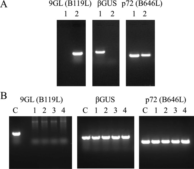
PCR analysis of ASFV-G-Δ9GL virus DNA using specific primers targeting the 9GL (B119L), p72 (B646L), or β-GUS genes. (A) Assessment of purity of the ASFV-G-Δ9GL virus stock by PCR. Lane 1, ASFV-G-Δ9GL; lane 2, ASFV-G. (B) Identification of the presence of parental ASFV-G (lanes 1 to 4) in viruses isolated from animals infected with 104 HAD50 ASFV-G-Δ9GL virus. The control (lane C) consists of a plasmid containing the respective target genes.
Analysis of the ASFV-G-Δ9GL genome sequence relative to parental ASFV-G genome sequence.
To evaluate the accuracy of the genetic modification and the integrity of the genome of the recombinant virus, full genome sequences of ASFV-G-Δ9GL and parental ASFV-G were obtained using NGS on the Ion Torrent PGM and compared. First, a full-length genome comparison between parental ASFV-G and ASFV Georgia 2007/1 (3) was performed. The following differences were observed between these two viruses (nucleotide positions are provided based on ASFV Georgia 2007/1 GenBank accession no. FR682468): (i) two nucleotide insertions, T at position 433 and A at position 441, in a noncoding segment of the genome; (ii) two nucleotide deletions, T at position 1602 and T at position 1603, in the MGF 360-1L gene ORF resulting in a frameshift; (iii) a nucleotide insertion, T at position 1620, in the MGF 360-1L gene ORF resulting in a frameshift; (iv) a nucleotide mutation of A to G at position 97391 resulting in a silent mutation in ORF B438L; (v) a nucleotide mutation of C to G at position 166192 resulting in a residue substitution (Ala to Pro) at residue position 85 in ORF E199L; and (vi) a nucleotide insertion of T at position 183303, a noncoding segment of the genome (Table 1). Second, a full-length genome comparison between ASFV-G-Δ9GL and parental ASFV-G was performed. The DNA sequence of ASFV-G-Δ9GL revealed a deletion of 173 nucleotides in ORF B119L (9GL) relative to parental ASFV-G that corresponds with the introduced modification. The consensus sequence of the ASFV-G-Δ9GL genome showed an insertion of 2,324 nucleotides in ORF B119L corresponding to the p72GUS cassette sequence introduced to generate a 173-nucleotide deletion in the targeted gene. Besides the insertion of the cassette, only one additional difference was observed between ASFV-G-Δ9GL and ASFV-G genomes: a G-to-C point mutation at position 36465 resulting in amino acid substitution E224Q in ORF MGF 505-4R. In summary, ASFV-G-Δ9GL virus did not accumulate any significant mutations during the process of homologous recombination and consequent plaque purification steps.
TABLE 1.
Summary of differences between the full-length genome sequence of ASFV-G-Δ9GL and the parental ASFV-G compared with ASFV Georgia 07/1
| NPNa | Type of modificationb | Result for virus |
|
|---|---|---|---|
| ASFV-G | ASFV-G-Δ9GL | ||
| 433 | T insertion | + | + |
| 411 | A insertion | + | + |
| 1602 | MGF 360-1L TT deletion FS | + | + |
| 1620 | MGF 360-1L T insertion FS | + | + |
| 36465 | MGF 505-4R G to C Glu224Gln | − | + |
| 97391 | B438L A to G SM | + | + |
| 166192 | E199L C to G Ala85Pro | + | + |
| 183303 | T insertion in NCR | + | + |
NPN, nucleotide position number based on the sequence of the ASFV Georgia 2007/1 isolate published by Chapman et al. in 2011 (3).
FS, nucleotide modification causes frameshift in the corresponding ORF; SM, nucleotide modification causes silent mutation; NCR, noncoding region.
Replication of ASFV-G-Δ9GL in primary swine macrophages.
In vitro growth characteristics of ASFV-G-Δ9GL were evaluated in cultures of primary swine macrophages, the primary cell targeted by ASFV during infection in swine, and compared relative to parental ASFV-G in multistep growth curves (Fig. 2). Cell cultures were infected at an MOI of either 0.1 or 0.01, and samples were collected at 2, 24, 48, 72, and 96 h postinfection (hpi). ASFV-G-Δ9GL virus displayed a growth kinetic significantly slower than that of the parental ASFV-G virus. Depending on the time point and MOI utilized to infect macrophages, the recombinant virus exhibited titers 10- to 10,000-fold lower than those of the parental virus. Therefore, and as observed with ASFV Malawi Lil-20/1-Δ9GL, deletion of the 9GL (B119L) gene significantly affects the ability of the virus to replicate in vitro in primary swine macrophage cell cultures.
FIG 2.
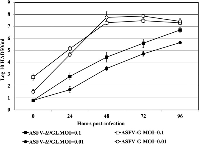
In vitro growth kinetics of the ASFV-G-Δ9GL and parental ASFV-G viruses. Primary swine macrophage cell cultures were infected (MOI of 0.1 or 0.01) with either ASFV-G-Δ9GL or parental ASFV-G virus. Virus yields were estimated at the indicated times postinfection by titration in primary swine macrophage cell cultures. Data represent means and standard deviations from two independent experiments. The sensitivity of virus detection was ≥1.8 HAD50/ml.
Assessment of ASFV-G-Δ9GL virulence in swine.
Deletion of the 9GL (B119L) gene from the genomes of ASFV isolates Malawi Lil-20/1 and Pretoriuskop/96/4 has been shown to drastically reduce virulence in swine (10, 12). In those reports, it was observed that i.m. inoculation of pigs with the recombinant deletion mutant at doses as high as 104 (10, 12) or even 106 HAD50 (10) induced only a transient rise in body temperature.
Here, 80- to 90-pound pigs inoculated i.m. with 104 HAD50 of ASFV-G exhibited increased body temperature (>104°F) by 3 to 4 days postinfection. Pigs presented clinical signs associated with the disease, including anorexia, depression, purple skin discoloration, staggering gait, and diarrhea. Signs of the disease aggravated progressively over time, and animals either died or were euthanized in extremis by day 7 or 8 postinfection (Table 2). Animals inoculated i.m. with 102 or 103 HAD50 of virulent ASFV-G developed a clinical disease comparable in severity to that observed in animals infected with 104 HAD50 of the same virus, with the exception that both clinical signs and the onset of death were slightly delayed by 1 to 3 days. Pigs presented a short period of fever starting by day 6 to 7 postinfection, with animals dying or euthanized in extremis around 8 to 9 days postinfection. The severity of the clinical signs observed in these animals was similar to those inoculated at higher dose (104 HAD50) (Table 2).
TABLE 2.
Swine survival and fever response following infection with different doses of ASFV-G-Δ9GL and parental ASFV-G
| HAD50 of virus | No. of survivors/total | Mean (SD) time to death, days | Mean (SD) characteristic of fever |
||
|---|---|---|---|---|---|
| No. of days to onset | Duration, days | Maximum daily temp, °F | |||
| 102 | |||||
| ASFV-G | 0/5 | 9.4 (1.22) | 7 (0.0) | 1.2 (0. 82) | 106.7 (0.58) |
| ASFV-G-Δ9GL | 10/10a | 103.0 (0.17) | |||
| 103 | |||||
| ASFV-G | 0/5 | 8.8 (1.1) | 5.6 (0.89) | 3.2 (0.43) | 106.2 (0.27) |
| ASFV-G-Δ9GL | 10/10a | 103.4 (0.32) | |||
| 104 | |||||
| ASFV-G | 0/10a | 7.25 (0.7) | 3.5 (0.76) | 3.75 (0.71) | 107 (0.47) |
| ASFV-G-Δ9GL | 0/5 | 8.25 (1.6) | 5.25 (1.91) | 3.25 (0.46) | 106.5 (0.46) |
| ASFV Mal Δ9GL | 4/4 | 9 (0.0) | 1 (0.82) | 104.2 (0.59) | |
Data from two similar experiments are presented together.
Interestingly, animals inoculated i.m. with 104 HAD50 of ASFV-G-Δ9GL developed clinical disease similar to that observed in animals inoculated i.m. with 104 HAD50 of parental ASFV-G, the only difference being a slight delay in the onset of fever. Conversely, pigs inoculated i.m. with 102 or 103 HAD50 of mutant ASFV-G-Δ9GL did not present any signs of clinical disease during the entire observation period (21 days). Therefore, the degree of virulence of ASFV-G-Δ9GL virus administered i.m. into swine appears to depend on the amount of infectious virus used in the experimental inoculation.
A control group of animals (n = 4) were inoculated with 104 HAD50 of Malawi Lil-20/1-Δ9GL (10). As previously described, these animals remained clinically normal throughout the experimental period, showing a transient and rather mild rise in body temperature (Table 2).
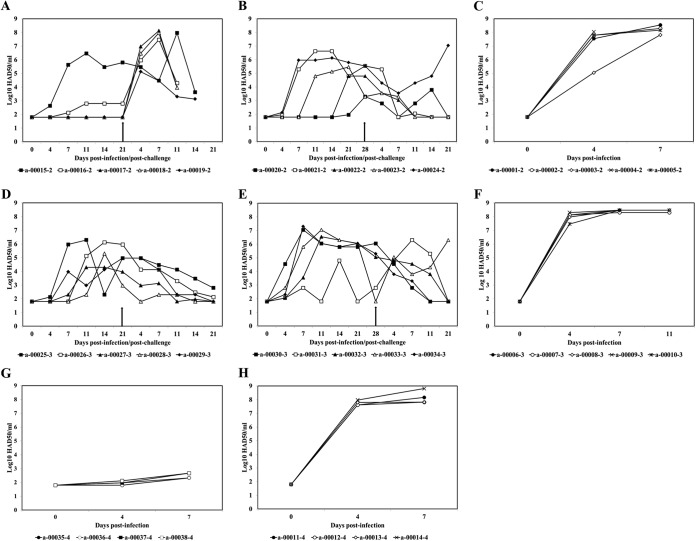
Viremia in experimentally inoculated animals was quantified at different days postinfection by hemadsorption. As expected, animals inoculated with 102, 103, or 104 HAD50 of virulent parental ASFV-G had very high virus titers in blood until the day of their death (Fig. 3C, F, and H). Conversely, animals inoculated with ASFV-G-Δ9GL at any of the utilized doses had relatively low virus titers in blood compared with those of the ASFV-G-inoculated animals (Fig. 3A, B, D, E, and G). Animals inoculated with 104 HAD50 of mutant ASFV-G-Δ9GL presented virus titers in blood 1,000- to 10,000-fold lower than those at the corresponding time point in animals inoculated with similar dose of the ASFV-G virus (Fig. 3G). Therefore, although the severities and kinetics of the presentation of clinical disease were similar, blood titers in both groups were significantly different. Thus, despite a low titer in blood that might indicate limited replication in vivo, ASFV-G-Δ9GL induces a lethal disease in pigs without reaching the viremia levels observed in animals inoculated with parental ASFV-G.
FIG 3.
Virus titers in blood samples (e.g., a-00015-2), obtained from pigs that were infected with either 102 (A and B), 103 (D and E), or 104 (G) HAD50 ASFV-G-Δ9GL and challenged (time of challenge indicated by arrows) at either 21 dpi (A and D) or 28 dpi (B and E). Also shown are viremia titers in pigs infected with either 102 (C), 103 (F), or 104 (H) HAD50 ASFV-G. Values are expressed as log10 HAD50 per milliliter. The sensitivity of virus detection was ≥log10 1.8 HAD50/ml. Numbers with the “a-” prefix are animal identification numbers.
Generally, animals inoculated with either 102 HAD50 or 103 HAD50 of mutant ASFV-G-Δ9GL had relatively low virus titers in blood compared with those of the ASFV-G-inoculated animals (Fig. 3A, B, D, and E). Animals inoculated with 102 HAD50 of ASFV-G-Δ9GL showed very heterogeneous viremia titers. While 5 of the 10 inoculated animals showed undetectable virus titers in blood until 14 to 21 dpi, 2 of the 10 pigs showed intermediate virus titers (ranging from 103 to 104 HAD50/ml), while 3 of 10 animals exhibited high virus titers in blood (ranging from 106 to 107 HAD50/ml). In general, regardless of the observed virus titers, viremia tended to peak around 21 dpi (Fig. 3A and B). Similarly, 6 of 10 animals inoculated with 103 HAD50 of ASFV-G-Δ9GL presented high titers (ranging from 106 to107 HAD50/ml), 2 of 10 presented intermediate titers (approximately 104 HAD50/ml), and 3 of 10 presented low titers (ranging from 102 to 103 HAD50/ml), while the remaining pig presented undetectable titers in blood until the time of challenge (Fig. 3D and E). Despite the observed heterogeneity of viremia titers among animals inoculated with ASFV-G-Δ9GL, it is interesting to notice the absence of clinical signs in infected animals, demonstrating a lack of correlation between the presence and severity of disease with virus titers in blood. It is clear that this lack of correlation between viremia titers and severity of disease is present in animals dying after being inoculated with 104 HAD50 of ASFV-G-Δ9GL as well as in animals inoculated with sublethal doses of ASFV-G-Δ9GL, which although presenting variable virus titers in blood are clinically normal.
Since viremia titers in animals infected with sublethal doses of ASFV-G-Δ9GL are rather heterogeneous, with most of the animals presenting medium to low virus titers in blood compared to animals inoculated with the wild-type virus, it is unlikely that these animals will shed virus. Virus shedding was assessed by virus isolation, using primary cell cultures of swine macrophages, from nasal swab samples obtained from pigs (n = 4) inoculated i.m. with either 102 or 103 HAD50/ml of ASFV-G-Δ9GL (Fig. 4). Virus was not detected in the nasal cavities of inoculated pigs during the monitoring period, although viremia titers in these animals (Fig. 4) resemble those observed in other infected pigs (Fig. 3). Also, virus was not detected in blood or nasal swabs obtained from unninoculated sentinel pigs brought in contact with each group of inoculated animals. Interestingly, these contact pigs did not seroconvert as they were ASFV-specific antibody negative by 28 dpi (data not shown). Therefore, contact pigs show neither viremia nor ASFV-specific antibodies, suggesting that ASFV-G-Δ9GL is not being eliminated by exposed animals.
FIG 4.

Viremia and virus shedding (nasal swabs) observed in animals (n = 4 per group) infected with either 102 or 103 HAD50 ASFV-G-Δ9GL. The average and standard deviation of values from each group along a 28-day observation period are presented. Values are expressed as log10 HAD50 per milliliter. The sensitivity of virus detection was ≥log10 1.8 HAD50/ml.
To rule out that the disease observed in the animals inoculated i.m. with 104 HAD50 of ASFV-G-Δ9GL was caused due to contamination of the inoculum with parental ASFV-G (previously shown to be undetectable by PCR in Fig. 1A), viruses isolated from blood at 7 dpi were tested by PCR using primers that target the p72 (B646L), 9GL (B119L), and β-GUS genes. All four ASFV-G-Δ9GL viruses isolated from blood of inoculated animals tested negative for parental ASFV-G. The 9GL (B119L) gene was not detected in these viruses, whereas amplification of the p72 (B646L) and β-GUS genes was recorded in all instances (Fig. 1B). Furthermore, sequencing was conducted on blood-isolated viruses to assess the integrity of the p72GUS cassette inserted by homologous recombination into ASFV-G. Obtained sequences revealed that p72GUS and both flanking regions were not modified in these viruses (data not shown). Since these data indicated the absence of contamination of the inoculum with parental ASFV-G, it is concluded that ASFV-G-Δ9GL virus inoculated at high doses (104 HAD50) is able to induce a clinical disease indistinguishable from that induced by the parental virus.
The ASFV 9GL (B119L) gene is highly conserved.
Sequence analysis of the 9GL (B119L) genes from several ASFV isolates obtained from various temporal and geographic origins, including those from tick and pig sources, reveals a high degree of conservancy. Isolates compared include Malawi Lil-20/1 (1983), Crocodile/96/1 (1996), Crocodile/96/3 (1996), Pretoriuskop/96/5 (1996), Pretoriuskop/96/4 (1996), Fairfield/96/1, and Wildebeeslaagte/96/1 from ticks, domestic Georgia 2007/1 (2007), Killean 3, European-70 (1970), European-75 (1975), Kimakia (1964), Victoria Falls, La Granja (1963), Lisbon60 (1960), Spencer (1951), Tengani (1962), Zaire (1967), and Haiti 811 (1980) from domestic pigs, Uganda (1961) from a warthog, and Lee (1955) from a bush pig. Among these isolates, the amino acid identity for 9GL (B119L) ranges between 93% and 100%. In the particular case of the Malawi Lil-20/1 or Pretoriuskop/96/4 isolates, identity with the Georgia 2007 isolate is 93%. Clearly, the 9GL (B119L) gene is highly conserved, suggesting a common and conserved function for the gene across ASFV isolates. Therefore, evidence suggests that differences in virulence between Δ9GL Malawi Lil-20/1 and ASFV-G mutant viruses may be a multigenic effect involving viral genes other than the 9GL (B119L) gene. Amino acid identities of translational products of predicted open reading frames (ORFs) of ASFV-G and Malawi Lil-20/1 genomes were compared using CLC Genomics Workbench (CLC Bio) and the Basic Local Alignment Search Tool (BLAST) (18). A total of 189 predicted ORFs were used for this analysis. It was observed that 102/189 of the predicted proteins encoded by these ORFs retain a high percentage of identity that is over 90%; 29/189 have identities ranging between 80% and 90%, 16/189 are 70% to 80% identical, 7/189 predicted proteins have identities that range between 60% and 70%, and 35/189 seem to be dissimilar, with percentages of identity of less than 60%. Altogether the observed genotypic difference between Malawi Lil-20/1 and ASFV-G should account for the phenotypic differences observed between the derived Δ9GL mutants. In fact, this analysis supports a previous phylogenic analysis of known ASFV isolates in which Georgia 2007/1 and Malawi Lil-20/1 were found to be distantly related (3).
Animals inoculated with sublethal doses of ASFV-G-Δ9GL virus are protected against challenge with virulent parental virus.
In order to assess induction of protection of the mutant virus against challenge with the virulent parental virus, animals inoculated i.m. with ASFV-G-Δ9GL (Table 3) were challenged with the parental virus ASFV-G. Groups 1 and 2, which received 102 HAD50 and 103 HAD50 of ASFV-G-Δ9GL, respectively, were challenged i.m. with 103 HAD50 of ASFV-G at 21 dpi. Groups 3 and 4, which received 102 HAD50 and 103 HAD50 of ASFV-G-Δ9GL, respectively, were challenged i.m. with 103 HAD50 of ASFV-G at 28 dpi. After challenge, animals were monitored daily for clinical signs and changes in body temperature. Five additional naive animals in a control group were challenged i.m. with 103 HAD50 of ASFV-G. In this control group, the onset of ASF-related signs was observed by 5 days postchallenge (dpc), evolving to a more severe disease in the following days, with all animals dying or being euthanized by 8 dpc.
TABLE 3.
Swine survival and fever response in animals infected with ASFV-G-Δ9GL after challenge with parental ASFV-G
| Treatment by day challenged | No. of survivors/total | Mean (SD) time to death, days | Mean (SD) characteristic of fever |
||
|---|---|---|---|---|---|
| No. of days to onset | Duration, days | Maximum daily temp, °F | |||
| 21 dpi | |||||
| 102 HAD50 (group 1) | 2/5 | 8.33 (0.58)a | 6.4 (0.55)a | 2.6 (0.55)a | 106.2 (0.87)a |
| 103 HAD50 (group 2) | 5/5 | 9.0 (0)b | 3 (0)b | 105.8 (0)b | |
| 28 dpi | |||||
| 102 HAD50 (group 3) | 5/5 | 14 (0)b | 7 (0)b | 106.2 (0)b | |
| 103 HAD50 (group 4) | 5/5 | 103.0 (0.17) | |||
| Control | 0/5 | 8.2 (1.1) | 5.20 (1.31) | 3.0 (0.70) | 106.5 (0.46) |
Values calculated considering only animals dying from the disease. The two surviving animals, although presenting ASF-related clinical signs, did not present body temperatures that were ≥104°F.
Values calculated considering only the sick animals.
Animals in group 1 (102 HAD50 ASFV-G-Δ9GL/challenge at 21 days) started showing clinical signs of the disease by 6 dpc. Progress toward a more severe clinical stage of the disease was observed in 3 pigs that died by 8 dpc, whereas the remaining two animals of the group displayed a milder form of the disease without a rise in body temperature. These animals survived the challenge with virulent ASFV-G during the entire observation period (21 days) (Table 3 and Fig. 5). All animals in group 3 (102 HAD50 ASFV-G-Δ9GL/challenge at 28 days) survived the infection with the parental virulent virus. In this group, four pigs remained clinically normal during the observational period, while the remaining pig presented a late onset of body temperature increase (14 dpc) that lasted until the end of the observational period (Table 3 and Fig. 5).
FIG 5.
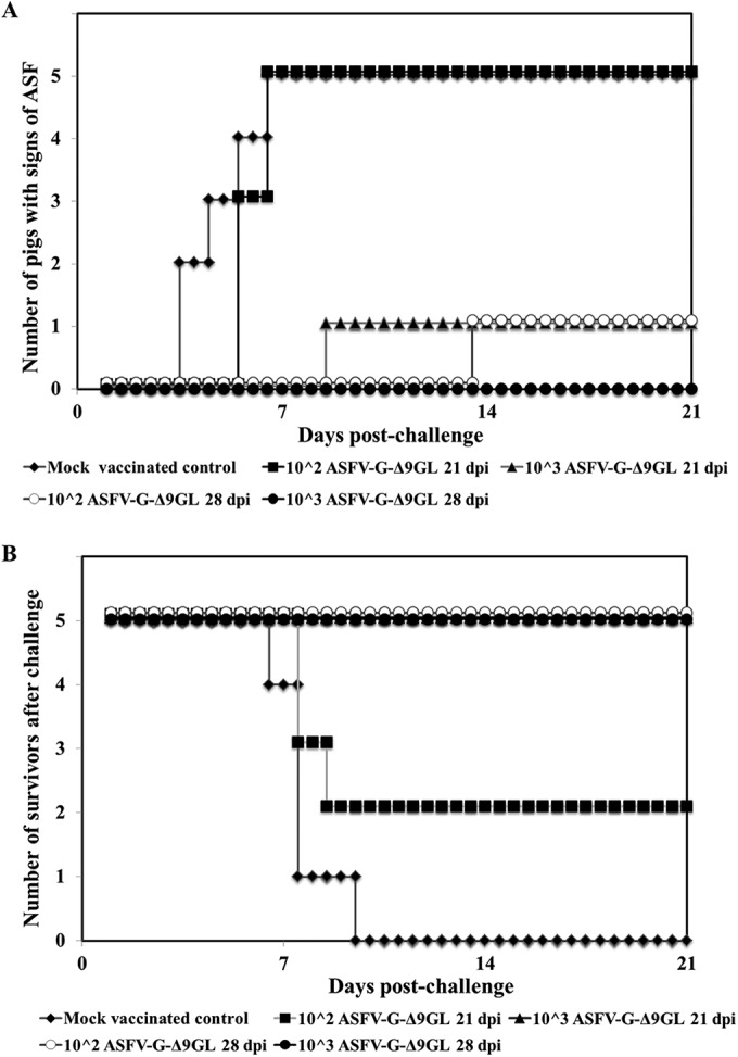
Morbidity (A) and mortality (B) observed in animals infected with either 102 or 103 HAD50 of ASFV-G-Δ9GL and challenged at either 21 or 28 dpi with the parental virulent ASFV-G virus.
In group 2 (103 HAD50 ASFV-G-Δ9GL/challenge at 21 days), four pigs remained clinically normal during the observational period, while the remaining pig presented a transient increase in body temperature for 3 days starting by day 9 dpc without additional signs of ASF (Table 3 and Fig. 5). In a similar experiment performed previously under the same conditions described here, complete protection against ASF was observed in 4 of 5 animals (data not shown). Pigs in group 4 (103 HAD50 ASFV-G-Δ9GL/challenge at 28 days) survived challenge, remaining clinically normal throughout the observational period (Table 3 and Fig. 5). This experiment was repeated under exactly the same conditions in which 5 of 5 animals were completely protected against ASFV-G (data not shown). In summary, inoculation of pigs with sublethal doses of ASFV-G-Δ9GL effectively induced protection against clinical disease and death induced by challenging pigs with parental virulent ASFV-G. This phenomenon was even more pronounced when higher doses of ASFV-G-Δ9GL were used in the inoculation of pigs, and it was not fully observed before 28 dpi.
Viremias observed after challenge could be caused by the primary infection with ASFV-G-Δ9GL or represent the replicative activity of ASFV-G in challenged pigs. The presence of ASFV-G or ASFV-G-Δ9GL in blood samples taken after challenge was determined by conventional PCR (Table 4). In group 1 (102 HAD50 ASFV-G-Δ9GL/challenge at 21 days), a drastically increased viremia was observed in four of five pigs by 4 dpc (Fig. 3A). Three of these four animals developed severe clinical disease and died or were euthanized in extremis. One of these four animals became sick transitorily and survived challenge. The virus detected in the blood of these 4 animals at 4 dpc was parental ASFV-G, but it was not detected in the remaining surviving animal of the group. Animals in group 3 (102 HAD50 ASFV-G-Δ9GL/challenge at 28 days) presented postchallenge viremia titers similar to the virus titers observed at 0 dpc (Fig. 3B). ASFV-G was not detected in the blood of these animals at 4 dpc. A similar situation was observed with animals in group 2 (103 HAD50 ASFV-G-Δ9GL/challenge at 21 days) (Fig. 3D). Also animals in group 4 (103 HAD50 ASFV-G-Δ9GL/challenge at 28 days) showed the same general patterns of viremia after challenge observed in groups 2 and 3. In group 4, only one animal showed high virus titers in blood after challenge, and ASFV-G was the detected virus, while only ASFV-G-Δ9GL was detected in blood from the four remaining pigs in the group (Fig. 3D).
TABLE 4.
Virus detected in blood, clinical signs, and outcome of disease in pigs exposed to ASFV-G-Δ9GL and challenged with parental ASFV-G
| ASFV-G-Δ9GL dose/day of challenge for blood sample shown | Virus detected at 7 dpca |
Disease presentation and outcome |
||
|---|---|---|---|---|
| ASFV-G-Δ9GL | ASFV-G | Clinical signs | Survival | |
| 102 HAD50/21 dpi | ||||
| a-00015-2b | + | − | Yes | Yes |
| a-00016-2 | + | + | Yes | No |
| a-00017-2 | + | + | Yes | No |
| a-00018-2 | + | + | Yes | No |
| a-00019-2 | + | + | Yes | Yes |
| 102 HAD50/28 dpi | ||||
| a-00020-2 | + | − | No | Yes |
| a-00021-2 | + | − | No | Yes |
| a-00022-2 | + | − | No | Yes |
| a-00023-2 | + | − | No | Yes |
| a-00024-2 | + | − | Yes | Yes |
| 103 HAD50/21 dpi | ||||
| a-00025-3 | + | − | No | Yes |
| a-00026-3 | + | − | No | Yes |
| a-00027-3 | + | − | No | Yes |
| a-00028-3 | + | − | No | Yes |
| a-00029-3 | + | − | No | Yes |
| 103 HAD50/28 dpi | ||||
| a-00030-3 | + | − | No | Yes |
| a-00031-3 | + | + | No | Yes |
| a-00032-3 | + | − | No | Yes |
| a-00033-3 | + | − | No | Yes |
| a-00034-3 | + | − | No | Yes |
Virus detection by PCR: +, positive; −, negative.
Numbers with the “a-” prefix are animal identification numbers.
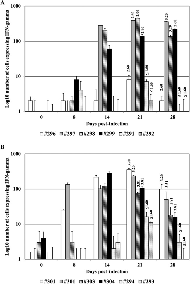
The immune response to ASFV-G-Δ9GL infection was evaluated in pigs at different time points until day 28 postinfection. Three groups of pigs were i.m. inoculated (n = 4) with either 102 or 103 HAD50 of ASFV-G-Δ9GL or mock infected, and their immune responses were evaluated by assessing the presence of ASFV-specific antibodies and the presence of ASFV-specific IFN-γ-producing cells in blood. Pigs in the groups inoculated with ASFV-G-Δ9GL developed a robust antibody response against ASFV (detected at 21 and 28 dpi) with no significant differences in the antibody titers between the two sampling time points (Fig. 6). Similarly, ASFV-specific IFN-γ-producing cells were detected in ASFV-G-Δ9GL-infected animals (with the sole exception of animal 296, who remained nonresponsive). IFN-γ-producing cells were detectable by day 14 postinfection, generally peaking by day 21 postinfection and decreasing toward day 28 postinfection. Control animals did not show ASFV-specific antibody or IFN-γ responses (Fig. 6). At 28 dpi, animals in all groups were i.m. challenged with 103 HAD50 of ASFV-G. The ASFV-G-Δ9GL-exposed groups each included an uninoculated contact pig. As expected, all animals in the control group as well as the contact pigs in the ASFV-G-Δ9GL-exposed groups became sick and died or were euthanized in extremis by days 8 to 9 postchallenge. Pigs in the 103-HAD50 ASFV-G-Δ9GL group survived challenge, although showing transient periods of fever (1 to 2 days). Similarly, 3/4 pigs in the 102 HAD50 ASFV-G-Δ9GL group survived challenge, showing transient periods of fever, with the exception of animal 296, who developed severe ASF (euthanized on day 10 postchallenge). Noticeable is the fact that this animal was the only one among all ASFV-G-Δ9GL-infected swine that did not develop ASFV-specific antibody or IFN-γ responses.
FIG 6.
Assessment of ASFV-specific antibodies and IFN-γ-producing cell responses in pigs infected with either 102 (animals 296 to 299) or 103 (animals 301 to 304) HAD50 ASFV-G-Δ9GL or mock infected (animals 291 to 294). Bars represent the IFN-γ-producing cells in each of the animals at different times postinfection (expressed as number of cells producing IFN-γ per 5 × 10⁵ cells). Anti-ASFV antibody titers are shown on the top of the bars (titers measured at 21 or 28 dpi) and are expressed as the log10 value of the inverse of the highest serum dilution still recognizing ASFV-infected cells (as described in Materials and Methods).
DISCUSSION
No vaccines are available to prevent ASFV infection. Only live attenuated virus strains have been useful in protecting pigs against challenge with homologous virulent isolates. These attenuated viruses have been regularly produced by sequential passages in cell cultures and, more recently, by genetic manipulation. Attenuated viruses obtained by genetic manipulation involve the deletion of specific genes by a process of homologous recombination. Independent deletion of four different genes from ASFV has been shown to attenuate virulent viruses. Independent deletions of the NL (DP71L) (11) or the UK (DP69R) (19) genes from ASFV E75, deletion of the TK (A240L) gene (9) from ASFV adapted to Vero cells, Malawi Lil-20/1, and Haiti, and deletion of the 9GL (B119L) gene also from Malawi Lil-20/1 (10) and Pretoriuskop/96/4 (12) isolates rendered recombinant deletion mutant viruses with significantly reduced virulence in swine. In all of these cases, animals inoculated with each of these genetically modified viruses survived the infection and became protected against ASFV when challenged with the corresponding virulent parental virus (homologous challenge) (9–12, 19). Those findings suggest that development of attenuated ASFV recombinant viruses by genetic manipulations of target genes is an effective approach for vaccine development.
The NL (DP71L) gene product exists in two different forms: a long form (184 amino acids) and a short form (70 to 72 amino acids), depending on the ASFV isolate (11). Although deletion of this gene in the ASFV E70 isolate (short form) rendered an attenuated virus, the deletion of the NL (DP71L) gene from ASFV Malawi Lil-20/1 (long form) or Pretoriuskop/96/4 (short form) did not result in attenuation of the virus (20). A deletion of the TK (A240L) gene, a highly conserved gene among all ASFV isolates that is involved in DNA synthesis, has been introduced into the genome of the pathogenic Vero cell-adapted Malawi Lil-20/1 and Haiti H811 viruses. The Malawi Lil-20/1 mutant virus was less virulent in vivo than a revertant virus (wild-type-like virus), but it was not completely attenuated in swine (9). The UK (DP69R) gene is located in the right variable region of certain ASFV isolates. Deletion of this gene from the ASFV E70 isolate rendered a virus exhibiting reduced virulence (19). Although the UK (DP69R) gene is conserved, it is not present in every ASFV isolate (e.g., Malawi Lil-20/1), limiting its use as a candidate target gene for producing attenuated viruses.
The 9GL (B119L) gene is highly conserved among the ASFV isolates sequenced thus far, including those from both tick and pig sources. The fact that deletion of the gene from virulent Malawi Lil-20/1 (10) or Pretoriuskop/96/4 (12) effectively reduced virulence in swine and induced protection makes 9GL (B119L) a candidate target gene for modification to produce an attenuated virus that can confer effective protection against ASFV. Interestingly, here we observed that deletion of 9GL (B119L) from the ASFV-G isolate does not have the same effect in terms of attenuation reported for Malawi Lil-20/1 or Pretoriuskop/96/4. Only when ASFV-G-Δ9GL was administered at a low dose to swine was it possible to observe a significant reduction in virus virulence. Data presented here indicate that the 9GL (B119L) gene is not absolutely required for ASFV-G virulence, suggesting that other virulence factors may be involved in the process. As observed with deletions of NL (DP71L) in the E70, Malawi Lil-20/1, and Pretoriuskop/96/4 isolates that lead to different phenotypes (11, 20), deletions of 9GL (B119L) have produced similar outcomes, suggesting that virulence of ASFV is the result of a multigene effect.
The NL proteins encoded by E70 (short form) and Malawi Lil-20/1 (long form) differ significantly, and that may explain the phenotypic differences observed in swine inoculated with the respective deletion mutant viruses. However, protein identity matrices indicate that the 9GL protein is highly similar among ASFV isolates, where ASFV-G, Malawi Lil-20/1, and Pretoriuskop/96/4 share over 93% amino acid identity, making it unlikely that ASFV attenuation relies solely on protein divergence. Since the observed phenotypes are most likely mediated by the effect of multiple genes (9–12, 19), the evidence accumulated so far makes it difficult to speculate what is indeed the spectrum of genes mediating virulence in the ASFV Georgia 2007 isolate. It is possible that the number or function of additional virulence-associated genes among different ASFV strains may alter the intrinsic effect of the 9GL (B119L) gene on the general balance of the virulence in a particular virus strain. Therefore, it remains to be determined why the deletion of 9GL (B119L), a gene that has been associated with virus virulence in Malawi Lil-20/1 and Pretoriuskop/96/4 isolates, does not drastically alter virulence of ASFV-G. Sublethal doses of ASFV-G-Δ9GL were effective at inducing protection against challenge with the virulent parental isolate.
The protection induced by ASFV-G-Δ9GL delivered either at 102 or 103 HAD50/ml was more effective when animals were challenged at 28 dpi, suggesting the need for some degree of maturation of the host immune mechanism(s) mediating protection against ASFV. The observed ASFV-specific antibody levels in ASFV-G-Δ9GL-exposed pigs were not significantly different at 21 or 28 dpi, whereas circulating ASFV-dependent IFN-γ-producing cells appear to peak at 21 dpi and decrease toward 28 dpi. Thus, there is no evident association between the measured immune parameters at 28 dpi and the observed protection against ASFV-G challenge. Challenging at the same time naive sentinel pigs commingling with ASFV-G-Δ9GL-exposed pigs resulted in less effective protection against ASFV-G. Clearly under these conditions, cohabitation of ASFV-G-Δ9GL-inoculated pigs with contact naive animals that developed ASF while shedding large amounts of ASFV-G resulted in highly stringent challenge. This was evidenced by the fact that some of the ASFV-G-Δ9GL-inoculated pigs developed transient fever or even ASF (i.e., pig 296). Interestingly, this particular pig failed to develop an IFN-γ response against ASFV. This observation is in agreement with previous reports that indirectly support the role of the T-cell response in the protection against ASFV (21–23). The immune mechanisms mediating protection against ASF are still not well understood. The results presented here show no substantial differences in the antibody or T-cell responses observed in pigs at 21 or 28 dpi (with the exception of the T-cell response in pig 296). It is possible that under the experimental conditions tested here, still unidentified immune mechanisms mediating protection against ASFV in ASFV-G-Δ9GL-infected animals evolve or mature after a certain period of time. In fact, we have observed a similar progressive acquisition of immunity to challenge with parental homologous virus until 28 dpi in animals infected with ASFV-Pret4-Δ9GL (unpublished data).
Our findings hinder the possibility of using the deletion of the 9GL (B119L) gene as the sole target for developing a live attenuated vaccine candidate against the ASFV-G isolate. The results shown here, demonstrating that sublethal doses of ASFV-G-Δ9GL did induce protection against ASF, open the possibility for using the ASFV-G-Δ9GL genome as platform for incorporating additional genetic modifications that could lead to a safer and useful vaccine candidate against ASF.
In summary, we present evidence of the differential attenuation effect of the deletion of the ASFV 9GL gene in the ASFV Georgia isolate. We have shown complete protection of domestic pigs against challenge with the highly virulent, epidemiologically relevant, ASFV Georgia isolate.
ACKNOWLEDGMENTS
We thank the Plum Island Animal Disease Center Animal Care Unit staff for excellent technical assistance. We wish to particularly thank Melanie V. Prarat for editing the manuscript.
This project was funded through an interagency agreement with the Science and Technology Directorate of the U.S. Department of Homeland Security under award no. HSHQDC-11-X-00077 and HSHQPM-12-X-00005. We thank ARS/USDA—University of Connecticut SCA 58-1940-1-190 for partially supporting this work.
REFERENCES
- 1.Costard S, Wieland B, de Glanville W, Jori F, Rowlands R, Vosloo W, Roger F, Pfeiffer DU, Dixon LK. 2009. African swine fever: how can global spread be prevented? Philos Trans R Soc Lond B Biol Sci 364:2683–2696. doi: 10.1098/rstb.2009.0098. [DOI] [PMC free article] [PubMed] [Google Scholar]
- 2.Tulman ER, Delhon GA, Ku BK, Rock DL. 2009. African swine fever virus. Curr Top Microbiol Immunol 328:43–87. [DOI] [PubMed] [Google Scholar]
- 3.Chapman DA, Darby AC, Da Silva M, Upton C, Radford AD, Dixon LK. 2011. Genomic analysis of highly virulent Georgia 2007/1 isolate of African swine fever virus. Emerg Infect Dis 17:599–605. doi: 10.3201/eid1704.101283. [DOI] [PMC free article] [PubMed] [Google Scholar]
- 4.Coggins L. 1974. African swine fever virus. Pathogenesis Prog Med Virol 18:48–63. [PubMed] [Google Scholar]
- 5.Kihm U, Ackerman M, Mueller H, Pool R. 1987. Approaches to vaccination, p 127–144. In Becker Y. (ed), African swine fever. Martinus Nijhoff Publishing, Boston, MA. [Google Scholar]
- 6.Mebus CA. 1988. African swine fever. Adv Virus Res 35:251–269. [DOI] [PubMed] [Google Scholar]
- 7.Hamdy FM, Dardiri AH. 1984. Clinical and immunologic responses of pigs to African swine fever virus isolated from the Western Hemisphere. Am J Vet Res 45:711–714. [PubMed] [Google Scholar]
- 8.Ruiz-Gonzalvo F, Carnero ME, Bruyel V. 1981. Immunological responses of pigs to partially attenuated ASF and their resistance to virulent homologous and heterologous viruses, p 206–216. In Wilkinson PJ. (ed), FAO/CEC Expert Consultation in ASF Research, Rome, Italy. [Google Scholar]
- 9.Moore DM, Zsak L, Neilan JG, Lu Z, Rock DL. 1998. The African swine fever virus thymidine kinase gene is required for efficient replication in swine macrophages and for virulence in swine. J Virol 72:10310–10315. [DOI] [PMC free article] [PubMed] [Google Scholar]
- 10.Lewis T, Zsak L, Burrage TG, Lu Z, Kutish GF, Neilan JG, Rock DL. 2000. An African swine fever virus ERV1-ALR homologue, 9GL, affects virion maturation and viral growth in macrophages and viral virulence in swine. J Virol 74:1275–1285. doi: 10.1128/JVI.74.3.1275-1285.2000. [DOI] [PMC free article] [PubMed] [Google Scholar]
- 11.Zsak L, Lu Z, Kutish GF, Neilan JG, Rock DL. 1996. An African swine fever virus virulence-associated gene NL-S with similarity to the herpes simplex virus ICP34.5 gene. J Virol 70:8865–8871. [DOI] [PMC free article] [PubMed] [Google Scholar]
- 12.Neilan JG, Zsak L, Lu Z, Burrage TG, Kutish GF, Rock DL. 2004. Neutralizing antibodies to African swine fever virus proteins p30, p54, and p72 are not sufficient for antibody-mediated protection. Virology 319:337–342. doi: 10.1016/j.virol.2003.11.011. [DOI] [PubMed] [Google Scholar]
- 13.Reed LJ, Muench HA. 1938. A simple method of estimating fifty per cent endpoints. Am J Hyg 27:493–497. [Google Scholar]
- 14.Neilan JG, Lu Z, Kutish GF, Zsak L, Burrage TG, Borca MV, Carrillo C, Rock DL. 1997. A BIR motif containing gene of African swine fever virus, 4CL, is nonessential for growth in vitro and viral virulence. Virology 230:252–264. doi: 10.1006/viro.1997.8481. [DOI] [PubMed] [Google Scholar]
- 15.Sanger F, Nicklen S, Coulson AR. 1977. DNA sequencing with chain-terminating inhibitors. Proc Natl Acad Sci U S A 74:5463–5467. doi: 10.1073/pnas.74.12.5463. [DOI] [PMC free article] [PubMed] [Google Scholar]
- 16.Huang X, Madan A. 1999. CAP3: A DNA sequence assembly program. Genome Res 9:868–877. doi: 10.1101/gr.9.9.868. [DOI] [PMC free article] [PubMed] [Google Scholar]
- 17.Krug PW, Holinka LG, O'Donnell V, Reese B, Sanford B, Fernandez-Sainz I, Gladue DP, Arzt J, Rodriguez L, Risatti GR, Borca MV. 2015. The progressive adaptation of a Georgian isolate of African swine fever virus to Vero cells leads to a gradual attenuation of virulence in swine corresponding with major modifications of the viral genome. J Virol 89:2324–2332. doi: 10.1128/JVI.03250-14. [DOI] [PMC free article] [PubMed] [Google Scholar]
- 18.Altschul SF, Gish W, Miller W, Myers EW, Lipman DJ. 1990. Basic local alignment search tool. J Mol Biol 215:403–410. doi: 10.1016/S0022-2836(05)80360-2. [DOI] [PubMed] [Google Scholar]
- 19.Zsak L, Caler E, Lu Z, Kutish GF, Neilan JG, Rock DL. 1998. A nonessential African swine fever virus gene UK is a significant virulence determinant in domestic swine. J Virol 72:1028–1035. [DOI] [PMC free article] [PubMed] [Google Scholar]
- 20.Afonso CL, Zsak L, Carrillo C, Borca MV, Rock DL. 1998. African swine fever virus NL gene is not required for virus virulence. J Gen Virol 79:2543–2547. [DOI] [PubMed] [Google Scholar]
- 21.Argilaguet JM, Perez-Martin E, Nofrarias M, Gallardo C, Accensi F, Lacasta A, Mora M, Ballester M, Galindo-Cardiel I, Lopez-Soria S, Escribano JM, Reche PA, Rodriguez F. 2012. DNA vaccination partially protects against African swine fever virus lethal challenge in the absence of antibodies. PLoS One 7:e40942. doi: 10.1371/journal.pone.0040942. [DOI] [PMC free article] [PubMed] [Google Scholar]
- 22.King K, Chapman D, Argilaguet JM, Fishbourne E, Hutet E, Cariolet R, Hutchings G, Oura CA, Netherton CL, Moffat K, Taylor G, Le Potier MF, Dixon LK, Takamatsu HH. 2011. Protection of European domestic pigs from virulent African isolates of African swine fever virus by experimental immunisation. Vaccine 29:4593–4600. doi: 10.1016/j.vaccine.2011.04.052. [DOI] [PMC free article] [PubMed] [Google Scholar]
- 23.Oura CA, Denyer MS, Takamatsu H, Parkhouse RM. 2005. In vivo depletion of CD8+ T lymphocytes abrogates protective immunity to African swine fever virus. J Gen Virol 86:2445–2450. doi: 10.1099/vir.0.81038-0. [DOI] [PubMed] [Google Scholar]


