Abstract
Mucosal immunity plays a crucial role in controlling human respiratory tract infections. This study characterizes the naturally acquired mucosal antibody levels to three Streptococcus pneumoniae (Spn) protein antigens, pneumococcal histidine triad protein D (PhtD), pneumococcal choline binding protein A (PcpA), and pneumolysin (Ply), and assesses the association of the mucosal antibody levels with occurrence of acute otitis media (AOM) caused by Spn. Both nasophargyeal (NP) IgG and IgA levels to all three proteins slightly decreased in children from 6 to 9 months of age and then gradually increased through 24 months of age. Spn NP colonization was associated with higher mucosal antibody levels to all three proteins. However, children with Spn AOM had 5-8 fold lower IgG and 3-6 fold lower IgA levels to the three proteins than children without AOM but asymptomatically colonized with Spn. Antigen-specific antibody levels in the middle ear fluid (MEF) were correlated with antibody levels in the NP. Children with AOM caused by Spn had lower antibody levels in both the MEF and NP than children with AOM caused by other pathogens. These results indicate that higher naturally acquired mucosal antibody levels to PhtD, PcpA and Ply are associated with reduced AOM caused by Spn.
Keywords: Streptococcus pneumoniae, mucosal antibody, pneumococcal protein vaccine, acute otitis media, pneumococcal histidine triad protein D, pneumococcal choline binding protein A, pneumolysin D1
Introduction
Streptococcus pneumoniae (Spn) is responsible for a large spectrum of infectious diseases in children and adults such as sepsis, pneumonia, meningitis, sinusitis and acute otitis media (AOM).1-3 To date, 94 distinct serotypes have been documented based on capsular composition.3,4 Current licensed 23-valent pneumococcal polysaccharide vaccine (PPV-23) and 7-, 10- and 13-valent pneumococcal conjugate vaccines (PCV-7, -10, -13) significantly reduce invasive pneumococcal diseases1,5, but coverage is limited to the vaccine serotypes. After introduction of PCV-7, and more recently PCV-13, there has been a decrease in carriage caused by vaccine serotypes but an emergence of non-vaccine replacement serotypes.1,3,6-8 Furthermore, current polysaccharide-based vaccines are less effective in non-invasive pneumoniae and acute otitis media.9,10 In addition, PPV-23 is currently recommended in the elderly and high-risk adults. It is poorly immunogenic in children, especially under 2 years of age.11 Due to these shortcomings, protein-based vaccine candidates have been sought to replace or complement current polysaccharide-based vaccines.9,12,13
A number of pneumococcal protein antigens have been studied as vaccine candidates against pneumococcal infection, and multiple proteins have shown sufficient promise to enter human clinical trials.12,13 Monovalent protein vaccine candidates of PhtD (pneumococcal histidine triad protein D),14 PcpA (pneumococcal choline binding protein A),15 PspA (pneumococcal surface protein A)12 and dPly (detoxified pneumolysin),16 bivalent protein vaccine candidates PhtD/PcpA,15 PspA/PsaA,12 PhtA/PhtB,12 and PhtD/Ply alone or along with PCV-7,12 trivalent protein vaccine candidates PhtD/PcpA/PlyD1,12 PhtD/Ply with protein D of Haemophilus influenzae13 were or are in human clinical trials. Our laboratory has been studying three pneumococcal proteins PhtD, PcpA and PlyD1 (genetic detoxified pneumolysin), which are the components of a candidate vaccine in a trial sponsored by Sanofi Pasteur. PhtD is a divalent cation regulated surface protein, shown to elicit protection against nasopharyngeal colonization, and is highly conservation across pneumococcal serotypes.17 PcpA is another divalent cation regulated surface protein that plays a major role in pneumococcal adherence15. Both PhtD and PcpA have been shown to be involved in adherence of Spn to human NP epithelial cells in vitro.18,19 Ply is a cholesterol-dependent secreted cytolysin which is a key virulence factor contributing to bacterial pathogenesis at both early and late stages of infection.16 In mice studies, immunization with PhtD, PcpA and PlyD1 have been shown to elicit protection.20-24 In our previous studies, we have detected specific antibodies to PhtD, PcpA, and PlyD1 antibodies in children from natural exposure to Spn in sera after nasopharyngeal (NP) colonization and AOM.25 However, upon further analysis, no correlation between serum antibody titers to these proteins and protection of occurrence of Spn AOM could be identified (unpublished data). We therefore hypothesize that mucosal immunity plays a critical role in control of pneumococcal mucosal diseases such as AOM, sinusitis, and non-bacteremic pneumonia.
Although Spn NP colonization is a necessary pre-requisite for infections to develop, carriage is mostly asymptomatic.10 However, when the condition of the host is altered, such as by an upper respiratory viral infection, Spn may cause AOM.26 Unfortunately, the human mucosal immune response against pneumococci10 and to pneumococcal proteins after natural Spn exposure and AOM is poorly understood. In the present study we characterized the induced mucosal antibody levels in the NP to PhtD, PcpA and PlyD1, and assessed the association of theses antibody responses with the occurrence of natural Spn AOM infections in children 6 - 24 months of age. In addition, in a previous study, we found MEF antibody in humans originates predominantly from sera and NP secretions.27 Here we assessed the correlation of antibody levels in NP secretions with middle ear fluid (MEF).
Materials and Methods
Study design
This study derives from a 5-year (2006-2011) prospective longitudinal evaluation of immunity to Spn and NTHi NP colonization and AOM in young children ages 6 to 24 months, supported by the U.S. National Institute of Deafness and Communication Disorders. Healthy children without previous episodes of AOM were enrolled at 6 months of age from a middle class, suburban sociodemographic pediatric practice in Rochester, NY (Legacy Pediatrics). NP samples were obtained every 3 to 6 months prospectively from healthy children at 6-24 months of age. When AOM occurred tympanocentesis was performed to collect MEF and confirm the diagnosis of AOM, as previously described.28 At the time of an AOM diagnosis NP and MEF samples were concurrently obtained. All children in this study who developed an AOM had common clinical symptoms of viral upper respiratory infection (URI) such as cough, sore throat, runny nose, nasal congestion, headache, low grade fever and sneezing. All of the children received standard vaccinations including the PCV-7 or PCV-13 pneumococcal conjugate vaccine (Prevnar, Pfizer Pharmaceuticals, Collegeville, PA) at the appropriate age. The study was approved by the Institutional Review Board (IRB) of the University of Rochester and Rochester General Hospital, and written informed consent was obtained from parents or guardians of all child subjects.
Sample collection
NP swab samples were obtained by inserting a cotton-tipped wire swab deeply into both nares. NP wash samples were obtained by instilling 1 ml of sterile phosphate buffered saline and aspirating from both nares for antibody measurement. MEF samples for antibody measurement varied in quantity of material obtained from 50 to 250 μl and the entire sample was added to one ml of PBS (pH 7.4). The NP wash samples and MEF samples were centrifuged at 3000 rpm (1100g) at 4°C for 10 minutes and the supernatants were stored at -80°C until use. NP swab samples and MEF samples were for microbiological culture, and NP wash samples and MEF samples were for antibody measurements.
Microbiology
Three potential bacterial pathogens, Spn, Haemophilus influenzae, and Moraxella catarrhalis, were isolated and identified according to tests of the eighth edition of the Manual of Clinical Microbiology.29
Quantitative ELISA to detect pneumococcal antigen-specific IgG and IgA antibodies
PhtD, PcpA and PlyD1-specific antibody IgG and IgA concentrations in the NP and MEF were determined by quantitative ELISA as previously described30 with modification. Briefly, 384-well high binding microplates (Greiner Bio-One, USA) were coated with 20 ng of individual purified recombinant proteins in 20 μl of coating buffer (bicarbonate, pH 9.6) at 4°C overnight, then blocked with 60 μl of PBS containing 4.0 % skim milk at 37°C for 1h. The samples were 2-fold serially diluted in 20 μl PBS containing 4.0 % skim milk at an initial dilution of 1:5, and incubated at room temperature for 1 hour followed by an incubation with HP-conjugated anti-human IgG or IgA (Bethyl Laboratories, Inc, Montgomery, TX). The reaction products were developed with TMB Microwell Peroxidase Substrate System (KPL, Gaithersburg, MD), stopped by 20 μl of 1.0 molar phosphoric acid, and read by an automated ELISA reader using a 450-nm filter. A standard curve was generated using a four-parameter logistic-log function and a reference human serum containing known specific antibody concentrations to corresponding individual antigens. Antigen-specific IgG and IgA levels against the three pneumococcal antigens in the reference serum were quantified using Human IgA and IgG ELISA Quantitation Sets (Bethyl Laboratories, Inc) as described previously.30 The assay lower limits of detection were 2.4 ng/ml for anti-PhtD IgG, 0.18 ng/ml for anti-PhtD IgA; 2.2 ng/ml for anti-PcpA IgG, 0.28 ng/ml for anti-PcpA IgA; 1.0 ng/ml for anti-PlyD1 IgG, and 0.22 ng/ml for anti-PlyD1 IgA. An internal control was included in each plate for each antigen and the inter-assay coefficient of variation was ≤ 20% for all antigens and secondary antibody combinations. For the purpose of statistical analysis, undetectable samples were arbitrarily assigned a value equivalent to half the lower limit of detection of corresponding specific antibodies. ELISAs were fully validated according to ICH Guidance and performed in a GLP laboratory.
To correct for differential dilution effects that occurred during the NP wash and MEF sample collection, total IgG and IgA were determined using Human IgG and IgA ELISA Quantitation Kits (Bethyl Laboratories, Montgomery, TX) according to manufacturer's protocol. Mucosal antigen-specific IgG and IgA in NP and MEF samples were then corrected according to the total IgG or IgA in the sample. Results were expressed as a ratio of specific IgG to total IgG or specific IgA to total IgA in the same sample (ng/μg) as described previously.31,32 Samples with a total IgG or IgA < 0.05 μg/ml were excluded because in preliminary studies we determined that such samples were from children with a difficult or failed sampling process, and had undetectable antigen-specific antibodies. There were 5% of MEF and 20% of NP samples were excluded in this study according to this criterion.
Statistics
Statistical analysis was performed with R Project version 2.13.2 (www.r-project.org/). Antibody levels were expressed as geometric means (GM) with 95% confidence intervals (CI) of ratios of specific to total IgG or IgA. The associations between antibody levels and AOM occurrences and ages were analyzed using Generalized Estimating Equations (GEE) to fit a repeated measures logistic regression models. The AOM odds ratios between the 95th and 5th percentile antibody level were estimated using GEE models with AR1 subject level correlation and quadratic terms. The paired NP and MEF samples that were collected from the same AOM visits of the same subject were used to analyze the correlation between antibody levels in the NP and MEF. Because antibodies were found to correlate significantly with age, to control for a spurious latent correlation, antibody levels were adjusted for age using a GEE model, assuming a within-subject auto-regressive correlation. Antibody levels were subject to Box-Cox power transformations, and age was log-transformed33. Confidence intervals and levels of significance for (age-adjusted) antibody correlations were estimated using a bootstrap procedure with subject-level resampling. Antibody levels between age-matched groups (colonization, non-colonization, Spn AOM, non-Spn AOM groups) were compared using the non-parametric two-tailed Mann-Whitney test using GraphPad Prism 6.0. P<0.05 was considered to indicate statistical significance.
Results
Study cohort
This analysis involved a total of 424 NP and 152 MEF samples collected during 234 health and 208 AOM visits from 176 children between the ages of 6 and 24 months. 133 (76%) children had both health and AOM visits and 43 (24%) children had only AOM visits. The characteristics of the children are shown in Table 1. Since our group has previously shown age-related differences in serum antibody response to the studied antigens,25,34 the NP and MEF samples for this study were age-matched.
Table 1. Characteristics of children (n = 176).
| Number (%) | |
|---|---|
| Female | 80 (45%) |
| Male | 96 (55%) |
|
| |
| Breast feed | 55 (31%) |
| Formula | 67 (38%) |
| Both | 54 (31%) |
|
| |
| Smoke exposure | 13 (7%) |
| Non- smoke exposure | 163 (93%) |
|
| |
| Daycare | 70 (40%) |
| Non- Daycare | 106 (60%) |
|
| |
| PCV-7 up-to-date | 176 (100%) |
Natural acquisition of mucosal antibody in the NP over time
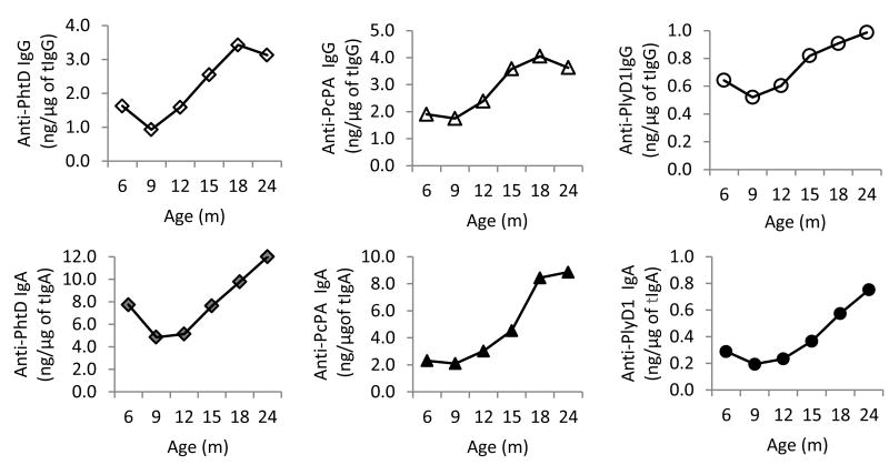
First, we evaluated the natural acquisition of NP mucosal antibody in 233 NP samples from 55 children who had at least 4 sequential health visits during the study period. Figure 1 shows GM of antibody levels to PhtD, PcpA and PlyD1 in the NP of children at 6, 9, 12, 15, 18 and 24 months of age. Both IgG and IgA antibody to all three Spn proteins decreased from 6 to 9 months and then increased steadily over time with a peak at 18 to 24 months of age.
Figure 1. Natural acquisition of NP mucosal antibody over time.
NP samples were collected from children who had at least 4 regular perspective visits to determine rates of pneumococcal specific to total IgG and IgA. Antibody levels were expressed as GM of the ratio of specific to total antibody, and association between age and antibody levels was analyzed using repeated measures logistic regression. N= 47, 45, 38, 35, 33, 34 for 6, 9, 12, 15, 18 and 24 months of age, respectively. A, anti-PhtD IgG; B, Anti-PcpA IgG; C, Anti-PlyD1 IgG,; D, anti-PhtD IgA; E , Anti-PcpA IgA; F, Anti-PlyD1 IgA,; tIgG, tIgA, total amount of IgG, IgA.
Mucosal antibody levels in the NP of Spn colonized versus non-colonized children
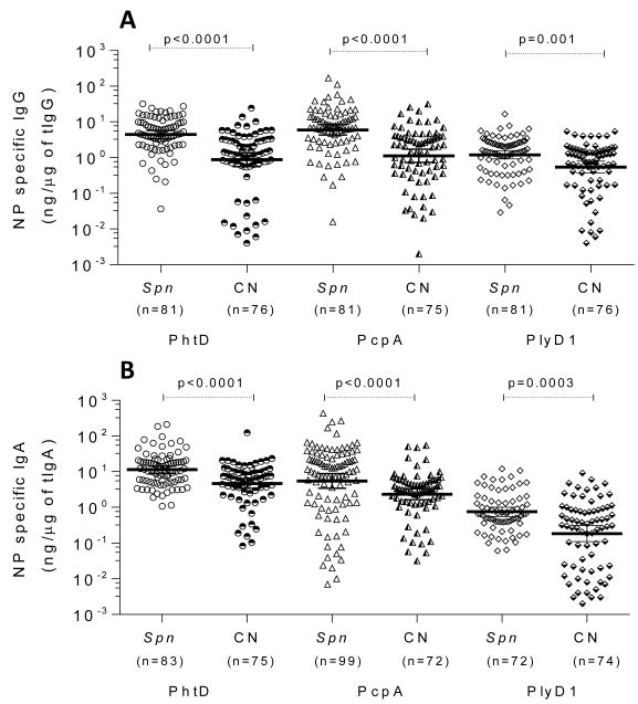
Next, we sought to determine differences in detectable antibodies in nasal secretions in Spn colonized versus non-colonized children. It is well-established that Spn colonization in children occurs in the first 6 months of life.35 Therefore, we anticipated that detectable mucosal antibodies would be present at the first sampling of our study cohort at 6 months of age. Our hypothesis was that detectable Spn colonization would result in higher mucosal IgG and IgA antibody levels to the pneumococcal proteins. We compared IgG and IgA levels to PhtD, PcpA and PlyD1 in NP samples from children who had Spn detected in their NP secretions at the time of sampling versus those who were Spn culture-negative. Children with Spn had significantly higher GM of specific IgG and IgA antibody in the NP than Spn culture-negative children (Figure 2). There was a 5–fold higher GM of IgG anti-PhtD (4.39 vs 0.87 ng/μg, p < 0.0001), a 5-fold higher GM of IgG anti-PcpA (5.93 vs 1.11 ng/μg, p < 0.0001), and a 2–fold higher GM of IgG anti-PlyD1 (1.20 vs 0.54 ng/μg, p = 0.001). The NP IgA levels to PhtD, PcpA and PlyD1 were higher by 2-fold (11.38 vs 4.67 ng/μg p <0.0001), 2-fold (5.49 vs 2.33 ng/μg, p < 0.0001), and 4-fold (0.75 vs 0.19 ng/μg, p = 0.0003) in children colonized with Spn versus non-colonized, respectively.
Figure 2. Colonized-children by Spn had higher mucosal GM of antibody levels than non-colonized children.
NP samples were collected from age-matched healthy children with or without Spn colonization. The rates of pneumococcal specific to total IgG and total IgA were determined by ELISA, and compared using nonparametric Mann-Whitney Test between culture positive and negative for Spn. A, anti-PhtD, anti-PcpA, and anti-PlyD1 IgG; B, anti-PhtD, anti-PcpA, and anti-PlyD1 IgA; NP, nasopharyngeal; CN: culture negative; Spn: S. pneumoniae; tIgG,tIgA, total amount of IgG, IgA. Lines represent GM with 95% CI.
Higher mucosal GM of antibody levels in the NP are associated with reduced AOM caused by Spn
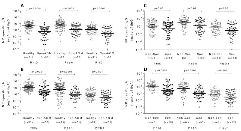
We next investigated whether there is an association of mucosal antibody levels with development of Spn AOM. We compared antibody levels to the pneumococcal proteins in the NP between children with Spn AOM and healthy children asymptomatically colonized with Spn. We found that children who developed Spn AOM had significantly lower mucosal IgG and IgA levels in their NP to all three studied Spn proteins compared to Spn colonized children who did not progress to AOM (Figure 3A-B). The NP GM of antibody levels of children with Spn AOM were 8-fold lower in anti-PhtD IgG (0.56 vs 4.39 ng/μg, p < 0.0001), 5-fold lower in anti-PcpA IgG (1.18 vs 5.93 ng/μg, p < 0.0001), 4-fold lower in anti-PlyD1 IgG (0.29 vs 1.19 ng/μg, p < 0.0001) , 4-fold lower in anti-PhtD IgA (2.87 vs 11.38 ng/μg, p < 0.0001), 6-fold lower in anti-PcpA IgA (0.97 vs 5.49 ng/μg, p < 0.0001), and 3-fold lower in anti-PlyD1 IgA (0.28 vs 0.75 ng/μg, p = 0.001) compared to those of healthy children. GM of antibody levels in NP were negatively associated with occurrence of Spn AOM in anti-PhtD IgG (OR = 0.04 , 95% CI 0.02 - 0.09, P < 0.0001), anti-PcpA IgG (OR = 0.06, 95% CI 0.03 - 0.12, P < 0.01), anti-PlyD1 IgG (OR = 0.24, 95% CI 0.15 - 0.37, P = 0.0001), anti-PhtD IgA (OR = 0.03, 95% CI 0.01 - 0.06, P < 0.0001), anti-PcpA IgG (OR = 0.02, 95% CI 0.00 - 0.07, P < 0.0001), anti-PlyD1 IgG (OR = 0.13 95 % CI, 0.07 - 0.24, P = 0.0008).
Figure 3. Higher mucosal antibody levels in the NP are associated with reduced AOM caused by Spn.
NP samples were collected from age-matched healthy Spn-colonized asymptomatic children and children with AOM. The levels of pneumococcal specific to total IgG and IgA (ng/μg) were compared between children with Spn AOM and children with asymptomatic Spn colonization (panels A and B), and between children with Spn AOM and children with non-Spn AOM (panels C and D) susing Mann-Whitney Test. A,C, anti-PhtD, anti-PcpA, and anti-PlyD1 IgG; B,D, anti-PhtD, anti-PcpA, and anti-PlyD1 IgA; Spn: S. pneumoniae; tIgG,tIgA, total amount of IgG, IgA. Lines represent GM with 95% CI.
A similar association was found in the NP GM of antibody levels between children with AOM caused by Spn and children with AOM caused by other pathogens. At onset of AOM, when co-colonization in the NP of Spn with other potential pathogens occurs, the children with Spn AOM have slightly lower GM of IgG but significantly lower GM of IgA to all three pneumococcal proteins than children with non-Spn AOM (Figure 3C-D). Compared with children with non-Spn AOM, children with Spn-AOM had 2-fold lower GM of anti-PhtD IgG (0.57 vs 1.10 ng/μg, p=0.08), 2fold lower GM of anti-PcpA IgG (1.18 vs 2.82 ng/μg, p = 0.02), and 2-fold lower GM of anti-PlyD1 IgG (0.29 vs 0.63 ng/μg, p = 0.06). The GM of IgA in the NP of children with Spn AOM were 4-fold lower in anti-PhtD (2.88 vs 12.25 ng/μg, p < 0.0001), 5-fold lower in anti-PcpA (0.97 vs 5.21 ng/μg, p < 0.0001) and 3-fold lower in anti-PlyD1 (0.28 vs 0.80 ng/μg, p = 0.001).
Children with Spn AOM have lower GM of antibody levels in the MEF than children with non-Spn AOM
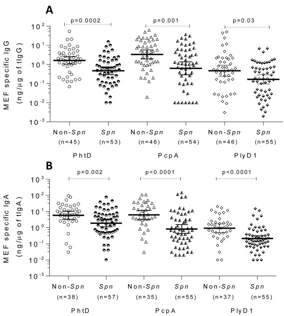
We also examined the MEF antibody levels to the three studied Spn proteins in children with AOM. At the onset of AOM, children with Spn AOM, compared to children who experienced AOM caused by other otopathogens, had 2-fold lower GM of anti-PhtD IgG (0.47 vs 1.64 ng/μg, p = 0.0002), 5-fold lower GM of anti-PcpA IgG (0.63 vs 3.37 ng/μg, p = 0.001) , and 3-fold lower GM of anti-PlyD1 IgG (0.17 vs 0.47 ng/μg, p = 0.03) (Figure 4A). The MEF IgA GM in Spn AOM children were 3-fold lower in anti-PhtD (1.87 vs 5.81 ng/μg, p = 0.002), 8-fold lower in anti-PcpA (0.82 vs 6.17 ng/μg, p < 0.0001) and 4-fold lower in anti-PlyD1 (0.2 vs 0.9 ng/μg, p < 0.0001) (Figure 4B).
Figure 4. Higher mucosal antibody levels in the MEF are associated with reduced AOM caused by Spn.
NP samples were collected from age-matched healthy children. The levels of pneumococcal specific to total IgG and IgA (ng/μg) were compared using Mann-Whitney Test. A, anti-PhtD, anti-PcpA, and anti-PlyD1 IgG; B, anti-PhtD, anti-PcpA, and anti-PlyD1 IgA; Spn: S. pneumoniae; tIgG, tIgA, total amount of IgG, IgA. Lines represent GM with 95% CI.
Correlation between antibody levels in the NP and antibody levels in MEF at AOM
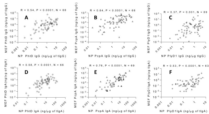
Antibody in NP secretions refluxes from the NP through the Eustachian tube to the middle ear during viral URIs. We have previously shown that MEF antibody originates predominantly from sera and NP secretions.27 Therefore, we selected available paired NP and MEF samples simultaneously collected from same children to analyze the correlation between antibody levels in the NP and MEF. Because antibodies were found to correlate significantly with age, to control potential spurious latent correlation, antibody levels were adjusted for age. Both IgG and IgA levels to all three Spn antigens in the NP were significantly and positively correlated with those in MEF of children at onset of AOM (all p values < 0.01) (Figure 5).
Figure 5. Correlations of antibody levels in the NP to MEF.
Paired NP and MEF samples simultaneously collected from same visit of same children, and correlation of IgG and IgA levels in NP to MEF was analyzed using Spearman Coefficient. A, anti-PhtD IgG, B, anti-PcpA IgG; C; anti-PlyD1 IgG; D, anti-PhtD IgA, E, anti-PcpA IgA; F; anti-PlyD1 IgA.
Discussion
Mucosal immunity is thought to play a crucial role in control of respiratory tract infections.36 Here we address gaps in knowledge of the immune response mounted by children who experience Spn NP colonization and AOM. We characterized the natural acquisition of mucosal antibody to Spn candidate vaccine proteins in young children who experience Spn colonization and consequent AOM. IgG and IgA antibody levels to PhtD, PcpA and PlyD1 in nasal secretions increased over time, suggesting that sequential natural exposure to Spn results in boosting antibody in mucosal NP secretions. We found that Spn colonization of the NP was associated with higher mucosal antibody responses to the vaccine candidate antigens studied versus non-Spn colonized NP samples. The results are consistent with previous observations regarding serum antibody responses to the same antigens.25,30 Most importantly, we found that children who developed Spn AOM had lower levels of IgG and IgA in nasal secretions at the onset of AOM compared to the antibody levels of children who were NP colonized with Spn but did not progress to AOM infection. Furthermore, when children developed Spn AOM they had lower IgG and IgA levels in their MEF to all three studied proteins compared to children who experienced AOM caused by other otopathogens. These results suggest an association between higher NP antibody levels to Spn proteins and a significantly reduced risk of Spn AOM.
Although numerous studies have shown that serum antibodies rise following pneumococcal carriage,25,30,37-39 very limited information is available regarding mucosal antibody responses to respiratory bacterial pathogens in humans, especially infants and children. In large part this is because reproducible collection of nasal secretions is challenging due to variability in acquiring mucus from the NP. Saliva is another source for study of mucosal immunity and its collection is easier than NP samples. Zhang Q et al (2006) found that children at 2-12 years of age who were culture-positive for Spn in their NP had higher IgG but not IgA in serum and saliva to pneumococcal choline binding protein A (CbpA) and Ply but not pneumococcal surface adhesin A (PsaA) or pneumococcal surface protein A (PspA). However, NP mucosal antibody assays are more reproducible and reliable than saliva antibody levels because saliva antibody levels are significantly influenced by many variables such as saliva flow.40,41 There are no previous reports on mucosal antibody to PhtD, PcpA and Ply in nasal secretions of young children by natural Spn NP colonization. The results of this study, along with our prior findings regarding serum antibody responses to Spn NP colonization in the same cohorts of subjects,25,30 clearly show that Spn colonization is an immunizing event for both systemic- and mucosal-acquired immune responses. In addition, PhtD, PcpA and Ply are highly immunogenic following natural exposure to Spn.
The elicitation of serum antibody following NP colonization by potential respiratory pathogens has been well documented in previous studies. Holmlund E et al (2006) 37 reported an increase in serum antibody concentrations to PsaA and Ply in infants who were colonized with Spn. Simell B et al (2009)38 showed that children with prior positive NP cultures for Spn had significantly higher serum anti-CbpA and anti-PhtD IgG titers. Prevaes SM et al (2012) 39 reported that colonization with Spn induced serum IgG against 14 pneumococcal proteins. Verkaik NJ (2010) 42 found that children at 6-24 months of age colonized by Staphylococcus aureus had higher serum IgG and IgA levels to a number of staphylococcal proteins than non-colonized children. Our research group previously reported that colonization with either Spn or H. influenzae elicited serum IgG and IgA responses to homologous bacterial species.25,30,34
The most important observation in this report is the identification of an association between higher mucosal antibody levels in NP and MEF to PhtD, PcpA and PlyD1 and reduced risk of AOM. This implies the NP mucosal antibody levels to PhtD, PcpA and Ply may play an important role in preventing Spn AOM in young children. Both IgG and IgA antibody levels in MEF are significantly associated with antibody levels detected in nasal secretions. Also of great relevance to vaccine design, we found that antibodies to these 3 vaccine candidate proteins did not require eradication of Spn from the NP to positively impact the occurrence of Spn AOM. We hypothesize that protection from AOM occurred by reduction of the effective Spn inoculum (bacterial load) in the NP below a pathogenic threshold level. We are initiating a study to investigate this notion.
Our study has limitations. The colonization in our study was defined based on bacterial cultures at a moment in time when samples were collected. We did not determine NP bacterial or viral loads which have been proven to be critical variants influencing host immune responses.43 The antibody levels in this study were free antibody. The results might be influenced by bacterial load, bacterial agglutination with antibody, and soluble antigens (e.g. pneumolysin) released from Spn. The NP is the ecological niche for a variety of commensal microbiota as well as potential respiratory disease causing bacteria and viruses.44,45 Cross-reactive antigens that are expressed by other non-pneumococcal species (e.g. S. mitis) may influence the detected antibody titers. Capsule is regarded as a critical virulence factor for pathogenesis of pneumococcal infections. We did not measure mucosal anti-capsular titers, and thus do not exclude the possibility that Spn AOM resulted from lack of capsular antibody to the Spn serotypes. In addition, 5% of MEF and 20% of NP samples that had undetectable or very low total antibody levels were excluded in this study. Since these samples distributed randomly to each age, colonization, and AOM group, and represented a small fraction of the total samples, the exclusion of those samples had limited influence on the conclusions of this study.
In summary, this is the first report to characterize the natural acquisition of mucosal NP and MEF antibody responses to Spn candidate vaccine proteins PhtD, PcpA and PlyD1 in young children who experience Spn colonization and AOM. We found that colonization by Spn is positively associated with mucosal antibody levels to PhtD, PcpA and PlyD1 proteins. Most importantly we identified an association of higher mucosal antibody levels in the NP to PhtD, PcpA and PlyD1 with reduced AOM caused by Spn. The results imply that NP mucosal antibody against PhtD, PcpA and Ply are potential markers of anti-pneumococcal immunity and these three proteins may elicit protection against Spn mucosal infections, especially AOM.
Acknowledgments
This study was supported by an investigator-initiated grant from Sanofi pasteur and NIH NIDCD RO1 08671. We thank the nurses and staff of Legacy Pediatrics and the collaborating pediatricians from Sunrise Pediatrics, Westfall Pediatrics, Lewis Pediatrics and Long Pond Pediatrics and the parents who consented and the children who participated in this long and challenging study. We also thank Dr. Anthony Almudevar (Department of Biostatistics and Computational Biology, University of Rochester Medical Center) for data satatsitical analysis, Jill Mangiafesto, Czup, Katerina, Virginia Judson and Emily Newman, for assistant with ELISA, and Dr. Robert Zagursky for reviewing, editing and commenting the manuscript.
References
- 1.Lynch JP, 3rd, Zhanel GG. Streptococcus pneumoniae: epidemiology and risk factors, evolution of antimicrobial resistance, and impact of vaccines. Curr Opin Pulm Med. 2010;16:217–225. doi: 10.1097/MCP.0b013e3283385653. [DOI] [PubMed] [Google Scholar]
- 2.Pavia M, Bianco A, Nobile CG, Marinelli P, Angelillo IF. Efficacy of pneumococcal vaccination in children younger than 24 months: a meta-analysis. Pediatrics. 2009;123:e1103–1110. doi: 10.1542/peds.2008-3422. [DOI] [PubMed] [Google Scholar]
- 3.van der Linden M, Reinert RR, Kern WV, Imohl M. Epidemiology of serotype 19A isolates from invasive pneumococcal disease in German children. BMC infectious diseases. 2013;13:70. doi: 10.1186/1471-2334-13-70. [DOI] [PMC free article] [PubMed] [Google Scholar]
- 4.Calix JJ, et al. Biochemical, genetic, and serological characterization of two capsule subtypes among Streptococcus pneumoniae Serotype 20 strains: discovery of a new pneumococcal serotype. J Biol Chem. 2012;287:27885–27894. doi: 10.1074/jbc.M112.380451. [DOI] [PMC free article] [PubMed] [Google Scholar]
- 5.Ruckinger S, van der Linden M, Reinert RR, von Kries R. Efficacy of 7-valent pneumococcal conjugate vaccination in Germany: An analysis using the indirect cohort method. Vaccine. 2010;28:5012–5016. doi: 10.1016/j.vaccine.2010.05.021. [DOI] [PubMed] [Google Scholar]
- 6.Besser TE, McGuire TC, Gay CC, Pritchett LC. Transfer of functional immunoglobulin G (IgG) antibody into the gastrointestinal tract accounts for IgG clearance in calves. Journal of virology. 1988;62:2234–2237. doi: 10.1128/jvi.62.7.2234-2237.1988. [DOI] [PMC free article] [PubMed] [Google Scholar]
- 7.Pichichero ME, Casey JR. Emergence of a multiresistant serotype 19A pneumococcal strain not included in the 7-valent conjugate vaccine as an otopathogen in children. Jama. 2007;298:1772–1778. doi: 10.1001/jama.298.15.1772. [DOI] [PubMed] [Google Scholar]
- 8.Kaplan SL, et al. Serotype 19A Is the most common serotype causing invasive pneumococcal infections in children. Pediatrics. 2010;125:429–436. doi: 10.1542/peds.2008-1702. [DOI] [PubMed] [Google Scholar]
- 9.Moffitt KL, Malley R. Next generation pneumococcal vaccines. Current opinion in immunology. 2011;23:407–413. doi: 10.1016/j.coi.2011.04.002. [DOI] [PMC free article] [PubMed] [Google Scholar]
- 10.Jambo KC, Sepako E, Heyderman RS, Gordon SB. Potential role for mucosally active vaccines against pneumococcal pneumonia. Trends in microbiology. 2010;18:81–89. doi: 10.1016/j.tim.2009.12.001. [DOI] [PMC free article] [PubMed] [Google Scholar]
- 11.Vila-Corcoles A, et al. Effectiveness of the 23-valent polysaccharide pneumococcal vaccine against invasive pneumococcal disease in people 60 years or older. BMC infectious diseases. 2010;10:73. doi: 10.1186/1471-2334-10-73. [DOI] [PMC free article] [PubMed] [Google Scholar]
- 12.Darrieux M, Goulart C, Briles D, Leite LC. Current status and perspectives on protein-based pneumococcal vaccines. Critical reviews in microbiology. 2013:1–11. doi: 10.3109/1040841X.2013.813902. [DOI] [PubMed] [Google Scholar]
- 13.Feldman C, Anderson R. Review: Current and new generation pneumococcal vaccines. The Journal of infection. 2014;69:309–325. doi: 10.1016/j.jinf.2014.06.006. [DOI] [PubMed] [Google Scholar]
- 14.Seiberling M, et al. Safety and immunogenicity of a pneumococcal histidine triad protein D vaccine candidate in adults. Vaccine. 2012;30:7455–7460. doi: 10.1016/j.vaccine.2012.10.080. [DOI] [PubMed] [Google Scholar]
- 15.Bologa M, et al. Safety and immunogenicity of pneumococcal protein vaccine candidates: monovalent choline-binding protein A (PcpA) vaccine and bivalent PcpA-pneumococcal histidine triad protein D vaccine. Vaccine. 2012;30:7461–7468. doi: 10.1016/j.vaccine.2012.10.076. [DOI] [PubMed] [Google Scholar]
- 16.Kamtchoua T, et al. Safety and immunogenicity of the pneumococcal pneumolysin derivative PlyD1 in a single-antigen protein vaccine candidate in adults. Vaccine. 2013;31:327–333. doi: 10.1016/j.vaccine.2012.11.005. [DOI] [PubMed] [Google Scholar]
- 17.Bersch B, et al. New Insights into Histidine Triad Proteins: Solution Structure of a Streptococcus pneumoniae PhtD Domain and Zinc Transfer to AdcAII. PloS one. 2013;8:e81168. doi: 10.1371/journal.pone.0081168. [DOI] [PMC free article] [PubMed] [Google Scholar]
- 18.Khan MN, Sharma SK, Filkins LM, Pichichero ME. PcpA of Streptococcus pneumoniae mediates adherence to nasopharyngeal and lung epithelial cells and elicits functional antibodies in humans. Microbes and infection / Institut Pasteur. 2012;14:1102–1110. doi: 10.1016/j.micinf.2012.06.007. [DOI] [PMC free article] [PubMed] [Google Scholar]
- 19.Barocchi MA, Censini S, Rappuoli R. Vaccines in the era of genomics: the pneumococcal challenge. Vaccine. 2007;25:2963–2973. doi: 10.1016/j.vaccine.2007.01.065. [DOI] [PubMed] [Google Scholar]
- 20.Plumptre CD, Ogunniyi AD, Paton JC. Vaccination against Streptococcus pneumoniae Using Truncated Derivatives of Polyhistidine Triad Protein D. PloS one. 2013;8:e78916. doi: 10.1371/journal.pone.0078916. [DOI] [PMC free article] [PubMed] [Google Scholar]
- 21.Verhoeven D, Perry S, Pichichero ME. Contributions to protection from Streptococcus pneumoniae infection using the monovalent recombinant protein vaccine candidates PcpA, PhtD, and PlyD1 in an infant murine model during challenge. Clinical and vaccine immunology: CVI. 2014;21:1037–1045. doi: 10.1128/CVI.00052-14. [DOI] [PMC free article] [PubMed] [Google Scholar]
- 22.Verhoeven D, Xu Q, Pichichero ME. Vaccination with a Streptococcus pneumoniae trivalent recombinant PcpA, PhtD and PlyD1 protein vaccine candidate protects against lethal pneumonia in an infant murine model. Vaccine. 2014;32:3205–3210. doi: 10.1016/j.vaccine.2014.04.004. [DOI] [PubMed] [Google Scholar]
- 23.Glover DT, Hollingshead SK, Briles DE. Streptococcus pneumoniae surface protein PcpA elicits protection against lung infection and fatal sepsis. Infection and immunity. 2008;76:2767–2776. doi: 10.1128/IAI.01126-07. [DOI] [PMC free article] [PubMed] [Google Scholar]
- 24.Mann B, et al. Broadly protective protein-based pneumococcal vaccine composed of pneumolysin toxoid-CbpA peptide recombinant fusion protein. The Journal of infectious diseases. 2014;209:1116–1125. doi: 10.1093/infdis/jit502. [DOI] [PMC free article] [PubMed] [Google Scholar]
- 25.Pichichero ME, et al. Antibody response to Streptococcus pneumoniae proteins PhtD, LytB, PcpA, PhtE and Ply after nasopharyngeal colonization and acute otitis media in children. Hum Vaccin Immunother. 2012;8:799–805. doi: 10.4161/hv.19820. [DOI] [PMC free article] [PubMed] [Google Scholar]
- 26.Heikkinen T, Chonmaitree T. Importance of respiratory viruses in acute otitis media. Clinical microbiology reviews. 2003;16:230–241. doi: 10.1128/CMR.16.2.230-241.2003. [DOI] [PMC free article] [PubMed] [Google Scholar]
- 27.Kaur R, Kim T, Casey JR, Pichichero ME. Antibody in middle ear fluid of children originates predominantly from sera and nasopharyngeal secretions. Clinical and vaccine immunology : CVI. 2012;19:1593–1596. doi: 10.1128/CVI.05443-11. [DOI] [PMC free article] [PubMed] [Google Scholar]
- 28.Pichichero ME, Wright T. The use of tympanocentesis in the diagnosis and management of acute otitis media. Curr Infect Dis Rep. 2006;8:189–195. doi: 10.1007/s11908-006-0058-9. [DOI] [PubMed] [Google Scholar]
- 29.Murray PR, B E, Pfaller MA, Jorgensen JH, Yolken RH, editors. Manual of clinical microbiology. 8th. 2003. [Google Scholar]
- 30.Xu Q, Pichichero ME. Co-colonization by Haemophilus influenzae with Streptococcus pneumoniae enhances pneumococcal-specific antibody response in young children. Vaccine. 2014;32:706–711. doi: 10.1016/j.vaccine.2013.11.096. [DOI] [PMC free article] [PubMed] [Google Scholar]
- 31.Hammitt LL, et al. Kinetics of viral shedding and immune responses in adults following administration of cold-adapted influenza vaccine. Vaccine. 2009;27:7359–7366. doi: 10.1016/j.vaccine.2009.09.041. [DOI] [PubMed] [Google Scholar]
- 32.Durrer P, et al. Mucosal antibody response induced with a nasal virosome-based influenza vaccine. Vaccine. 2003;21:4328–4334. doi: 10.1016/s0264-410x(03)00457-2. [DOI] [PubMed] [Google Scholar]
- 33.Venables WN, Ripley BD. Modern Applied Statistics with S. Springer; New York, NY: 2010. [Google Scholar]
- 34.Pichichero ME, et al. Antibody response to Haemophilus influenzae outer membrane protein D, P6, and OMP26 after nasopharyngeal colonization and acute otitis media in children. Vaccine. 2010;28:7184–7192. doi: 10.1016/j.vaccine.2010.08.063. [DOI] [PMC free article] [PubMed] [Google Scholar]
- 35.Syrjanen RK, Kilpi TM, Kaijalainen TH, Herva EE, Takala AK. Nasopharyngeal carriage of Streptococcus pneumoniae in Finnish children younger than 2 years old. The Journal of infectious diseases. 2001;184:451–459. doi: 10.1086/322048. [DOI] [PubMed] [Google Scholar]
- 36.Sato S, Kiyono H. The mucosal immune system of the respiratory tract. Current opinion in virology. 2012;2:225–232. doi: 10.1016/j.coviro.2012.03.009. [DOI] [PubMed] [Google Scholar]
- 37.Holmlund E, Quiambao B, Ollgren J, Nohynek H, Kayhty H. Development of natural antibodies to pneumococcal surface protein A, pneumococcal surface adhesin A and pneumolysin in Filipino pregnant women and their infants in relation to pneumococcal carriage. Vaccine. 2006;24:57–65. doi: 10.1016/j.vaccine.2005.07.055. [DOI] [PubMed] [Google Scholar]
- 38.Simell B, et al. Pneumococcal carriage and acute otitis media induce serum antibodies to pneumococcal surface proteins CbpA and PhtD in children. Vaccine. 2009;27:4615–4621. doi: 10.1016/j.vaccine.2009.05.071. [DOI] [PubMed] [Google Scholar]
- 39.Prevaes SM, et al. Nasopharyngeal colonization elicits antibody responses to staphylococcal and pneumococcal proteins that are not associated with a reduced risk of subsequent carriage. Infection and immunity. 2012;80:2186–2193. doi: 10.1128/IAI.00037-12. [DOI] [PMC free article] [PubMed] [Google Scholar]
- 40.Bellussi L, Cambi J, Passali D. Functional maturation of nasal mucosa: role of secretory immunoglobulin A (SIgA) Multidisciplinary respiratory medicine. 2013;8:46. doi: 10.1186/2049-6958-8-46. [DOI] [PMC free article] [PubMed] [Google Scholar]
- 41.Brandtzaeg P. Do salivary antibodies reliably reflect both mucosal and systemic immunity? Annals of the New York Academy of Sciences. 2007;1098:288–311. doi: 10.1196/annals.1384.012. [DOI] [PubMed] [Google Scholar]
- 42.Verkaik NJ, et al. Induction of antibodies by Staphylococcus aureus nasal colonization in young children. Clinical microbiology and infection : the official publication of the European Society of Clinical Microbiology and Infectious Diseases. 2010;16:1312–1317. doi: 10.1111/j.1469-0691.2009.03073.x. [DOI] [PubMed] [Google Scholar]
- 43.Franz A, et al. Correlation of viral load of respiratory pathogens and co-infections with disease severity in children hospitalized for lower respiratory tract infection. Journal of clinical virology : the official publication of the Pan American Society for Clinical Virology. 2010;48:239–245. doi: 10.1016/j.jcv.2010.05.007. [DOI] [PMC free article] [PubMed] [Google Scholar]
- 44.Bosch AA, Biesbroek G, Trzcinski K, Sanders EA, Bogaert D. Viral and bacterial interactions in the upper respiratory tract. PLoS pathogens. 2013;9:e1003057. doi: 10.1371/journal.ppat.1003057. [DOI] [PMC free article] [PubMed] [Google Scholar]
- 45.Pettigrew MM, et al. Viral-bacterial interactions and risk of acute otitis media complicating upper respiratory tract infection. Journal of clinical microbiology. 2011;49:3750–3755. doi: 10.1128/JCM.01186-11. [DOI] [PMC free article] [PubMed] [Google Scholar]


