Abstract
Background and Objectives:
The purpose of this study was to analyze the surgical technique, postoperative complications, and possible recurrence after laparoscopic ventral hernia repair (LVHR) in comparison with open ventral hernia repair (OVHR), based on the international literature.
Database:
A Medline search of the current English literature was performed using the terms laparoscopic ventral hernia repair and incisional hernia repair.
Conclusions:
LVHR is a safe alternative to the open method, with the main advantages being minimal postoperative pain, shorter recovery, and decreased wound and mesh infections. Incidental enterotomy can be avoided by using a meticulous technique and sharp dissection to avoid thermal injury.
Keywords: Hernia, Incisional hernia, Laparoscopy, Ventral hernia
INTRODUCTION
Ventral and incisional hernia repair is one of the most common operations performed in everyday clinical practice. Incisional hernia is a common long-term complication of abdominal surgery and is estimated to occur in 11–20% of laparotomy incisions.1,2 Almost 50% of incisional hernias develop within the first 2 years after the primary surgery, and 74% develop after 3 years.3,4 The recurrence rate of incisional hernia after primary suture repair is more than 50%5 and has been reduced to 10–23% after the introduction of prosthetic materials (meshes) in hernia repair.6
However, open hernia repair can be a major operation with considerable morbidity caused by infectious complications. An increasing interest in laparoscopic surgery and the availability of new materials have encouraged the adoption of laparoscopic techniques in ventral hernia repair. Leblanc and Booth7 described the first laparoscopic ventral hernia repair (LVHR) in 1991. It is based on the same physical and surgical principles as the open underlay procedure described by Stoppa,8 Rives et al,9 and Wantz.10 LVHR is now being used with increasing frequency, even for the management of complex incisional hernias. Most reports on this topic have supported minimal postoperative morbidity, a shorter convalescence period, and an acceptable recurrence rate.11–13
Up to date, more than 270 studies on LVHR have been published, although most of them are case series with no control groups. Studies of large samples have been conducted recently, because of increasing experience.14,15 Herein, we analyze the existing literature on LVHR, focusing in the optimal surgical technique, complications, and long-term results of the procedure.
OPEN REPAIR TECHNIQUES
Primary open ventral hernia repair (OVHR) is based on suture approximation of aponeurosis on each side of the hernia defect. However, recurrence rates after this procedure range from 41 to 52% in the long term.6 The introduction of prosthetic meshes in hernia repair has reduced recurrence rates. Indeed, Luijendijk et al6 demonstrated a significant reduction in the recurrence rate for first-time incisional hernia repairs, from 43% (after suture repair) to 24% (after mesh repair). However, the mesh repairs require wide dissection of soft tissue, which contributes to an increased incidence of wound-related complications (more than 12%).16
Among the open repairs, the onlay technique was the most widely used one. In this method, a polypropylene mesh is sutured onto the anterior rectus sheath.17 The procedure is easy to perform, but it has a considerable morbidity rate and recurrence rate and therefore is not used often at present (8–27%17–20).
In the inlay technique, the mesh is sutured to the margin of the aponeurosis. This technique has been used to cover large aponeurotic gaps, and it carries extremely high recurrence rates.21,22 In the extraperitoneal underlay technique, the mesh is placed retromuscularly and preperitoneally, as described by Stoppa in 1989.8 This technique requires limited soft tissue dissection; therefore, it carries low morbidity and recurrence rates.22,23
The intraperitoneal underlay technique was introduced by McCarthy and Twiest24 in 1981. First, they used polypropylene mesh, which was sutured to the peritoneal edge of the hernia sac. However, polypropylene caused the formation of adhesions to the adjacent bowel loops and was replaced by the expanded polytetrafluoroethylene (ePTFE) mesh or bilayer PTFE and polypropylene mesh.25 Millikan et al25 applied this technique by using full-thickness transfascial sutures in 102 patients, with a 0% recurrence rate in a median follow-up of 28 months. This approach has been regarded as the gold standard for ventral hernia repair by the American Hernia Society.26 By placing the mesh posterior to the abdominal wall, the underlay technique applies Pascal's law, which states that any pressure exerted on an enclosed fluid is transmitted undiminished throughout the fluid and acts equally in all directions. Therefore as the intraabdominal pressure increases, equal amounts of force are exerted across the mesh. On the contrary, in the onlay technique, any increase in intra-abdominal pressure exerts lifting forces against the mesh. Consequently, the underlay repair seems mechanically advantageous.27
The relatively high morbidity and recurrence rates of the open repair techniques prompted the development of the laparoscopic approach in an effort to improve the clinical outcome. The laparoscopic repair follows the principles of the open underlay repair and has the same advantages.
LAPAROSCOPIC REPAIR
Since the introduction of the laparoscopic approach in ventral hernia repair by LeBlanc and Booth in 1993,7 the laparoscopic technique has been used worldwide, as it offers earlier recovery, decreased hospital stay, and low recurrence rates.28 Moreover, it is well accepted that the primary advantage of the laparoscopic approach is that wound infections are less frequent compared with the open approach.27
Contraindications
The laparoscopic approach is not indicated in emergency situations, especially in cases with hemodynamic instability or incarcerated hernia, with or without gangrenous bowel. Also severe coagulopathy is an absolute contraindication for laparoscopy. An open approach also may be indicated for patients with a hostile abdomen. For instance, enterocutaneous fistulae, history of an open abdomen, severe abdominal injuries, or previous extensive operations may be associated with diffuse adhesions and may render the laparoscopic approach very tedious or impossible. In addition, patients with previous mesh repairs may have dense adhesions. In those cases, the decision to proceed with the laparoscopic approach should be based on the surgeon's expertise and the probability that safe access can be obtained.27
Preoperative Preparation
Smoking and obesity are known risk factors for the development of infectious complications and recurrence. Therefore cessation of smoking for at least 2 weeks before surgery is desirable. Obese patients should try to lose weight on a dietary program for 2 months before surgery. For morbidly obese individuals, a weight loss procedure is often necessary in the first stage, together with temporary closure of the hernia defect, to be completed with a definitive hernia repair 12 to 18 months later, when the patient has lost substantial weight and is less prone to infectious complications.27,29
Abdominal imaging with computed tomography (CT) is necessary in patients with large or recurrent hernias, as well as those with strangulated hernias. For complex hernias or those along the abdominal border, knowing the proximity of the edges of the hernia defect to the bony landmarks is useful in preoperative planning for mesh fixation.27
Technique
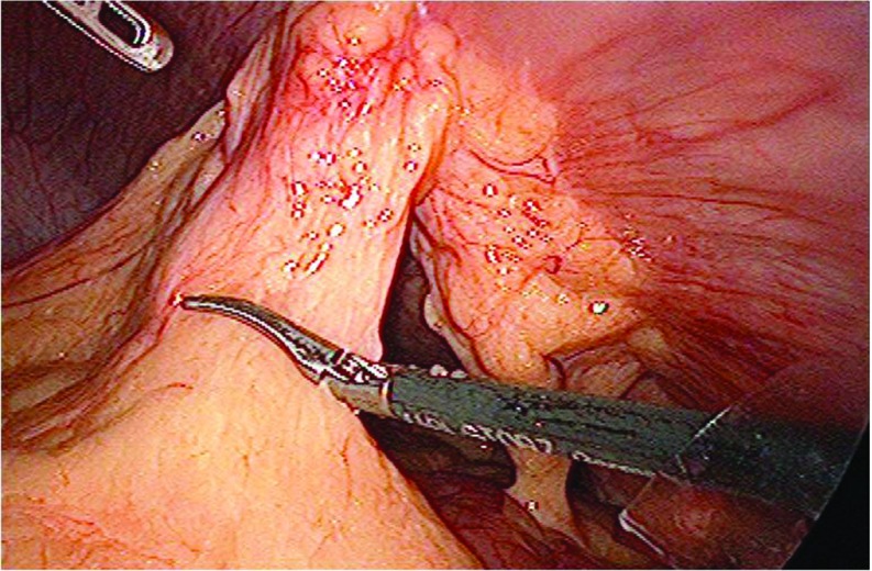
The procedure starts with entry into the peritoneal cavity with a Veress needle, an open Hasson method, or an optical trocar. Three trocars are used, one 10-mm and two 5-mm trocars, which are placed as laterally as possible on the abdominal wall, so they are at an adequate distance from the hernia orifice. The position of the first trocar should be several centimeters from scars from previous surgeries and as far from the hernia as possible, but should still provide adequate instrument reach.27 Most operations can be completed with 3 trocars. The next step of the operation is the most tedious one: adhesiolysis. The adhesions in the abdomen are lysed with electrocautery or an ultrasonic scalpel. The abdominal contents of the hernia sac are reduced into the peritoneal cavity (Figure 1). No cauterization should be done that may injure the bowel wall. Perforation of the intestine is the most serious injury associated with LVHR.30 In this case, there should be a low threshold to conversion to an open procedure. If an open approach is used, the hernia should be repaired primarily or by implanting a biological mesh.31 Otherwise the injury can be repaired laparoscopically, the adhesiolysis can be completed, and the hernia repair can be completed after 1 week.32 Retrospectively, in most published series in which iatrogenic enterotomy occurred, the hernia repair was completed with a laparoscopically placed mesh, and only 43% were converted to an open procedure. A recognized enterotomy was associated with a mortality rate of 1.7%, whereas an unrecognized enterotomy had a mortality rate of 7.7%.31,33
Figure 1.
The adhesions in the abdomen are lysed by electrocautery or with an ultrasonic scalpel. The contents of the hernia sac (omentum or bowel loops) are reduced into the peritoneal cavity.
After adhesiolysis, the sac contents are gently reduced into the peritoneal cavity with atraumatic graspers, while the hernia sac is left in situ. However, it may be necessary to excise a portion of the sac if the bowel is closely adherent. If the hernia content cannot be reduced, conversion to an open procedure is necessary.27
Primary Fascial Closure
At this point, some authors suggest primary fascial closure. This technique has been developed in an effort to reduce postoperative bulging and formation of seroma after laparoscopic ventral hernia repair. Given LaPlace's law, a central nonfunctional portion of the abdominal wall acts like a “sail in the wind” and is prone to bulging. Primary fascial closure restores normal anatomy by reapproximating the abdominal wall under physiologic tension, which may restore its function.34,35
The techniques for closure include intracorporeal closure, extracorporeal closure, or a mixed technique. The most commonly used technique is extracorporeal suturing, according to which small skin incisions are made after which a suture passer is used to close the defect. By eliminating the dead space, this technique decreases the incidence of seromas and wound complications. Moreover, it allows wider lateral mesh overlap that reduces the possibility of recurrence.35 However, to date, there have been no randomized studies supporting closure versus nonclosure of the hernia defect in LVHR.
Mesh Repair
The peritoneal surface is cleared extensively, by lysing adhesions well away from the defect. For hernias located in the upper midline, the falciform ligament should be dissected from the abdominal wall by using an energy source. The pneumoperitoneum is then reduced to 5 to 8 mm Hg, so that the abdominal wall is minimally stretched revealing the true size of the hernia defect. The periphery of the hernia defect is evaluated by direct vision and palpation and is marked on the abdominal wall skin with a marker. The craniocaudal and lateral measurements are taken, to define the size of the prosthetic mesh. Because most meshes are associated with significant postoperative shrinkage, most surgeons suggest a 5-cm overlap.27 It is very important to identify all hernia defects and include all of them within the measured distances. The size of mesh that most closely approaches this measurement is selected for the repair. Four main types of mesh have been used: polypropylene (Prolene; Ethicon, Somerville, New Jersey), ePTFE (dual mesh; Gore-Tex; Gore Medical, Flagstaff, Arizona), composite polypropylene+PTFE (Composix; Bard Davol, Warwick, Rhode Island), or composite polypropylene+collagen (Parietene; Sofradim, Trevoux, France). Polypropylene prosthesis has been abandoned in the laparoscopic approach, because it may create adhesions with bowel loops. It has been replaced by Proceed (Ethicon), which is composed of polypropylene covered with oxidized regenerated cellulose (ORC).36 A newer mesh composed of polypropylene covered by a layer of polyglecaprone-25 on both sides (Physiomesh; Ethicon) has been added recently to the surgical practice (Table 1).37
Table 1.
Meshes Used Intraperitoneally for the Repair of Ventral and Incisional Hernias
| Group/Mesh | Material | Company |
|---|---|---|
| PTFE | ||
| Dulex | ePTFE | Bard Davol, Inc., Warwick, RI |
| Mycromesh | ePTFE | W. L. Gore, Newark, DE |
| Dual Mesh | ePTFE | W. L. Gore |
| Composite mesh with absorbable coated barrier | ||
| Proceed | PP with ORC layer | Ethicon, Somerville, NJ |
| Parietene | PP with collagen coated | Covidien, Mansfield, MA |
| Parietex Composite | Polyester with collagen coated | Covidien |
| Symbotex | Polyester with collagen film | Covidien |
| Permacol | Porcine dermal collagen implant | Covidien |
| Physiomesh | PP with polyglecaprone 25 | Ethicon |
| C-Qur | PP with omega 3 fatty acid coating | Atrium Medical, Hudson, NH |
| Sepramesh IP Composite | PP with hydrogel safety coating | Bard Davol, Inc. |
| Composite mesh with permanent coated barrier | ||
| Composix | PP/ePTFE | Bard Davol, Inc. |
| Ventrio | PP/ePTFE | Bard Davol, Inc. |
| Intramesh T1 | PP/ePTFE | Cousin Biotech, Wervicq-Sud, France |
| Intramesh W3 | Polyester mesh with silicone layer | Cousin Biotech |
ePTFE, expanded polytetrafluoroethylene; PP, polypropylene; ORC, oxidized regenerated cellulose.
The clinical experience with all these types of mesh varies from country to country and from center to center. Each particular material may have unique advantages and disadvantages. Gore-Tex (ePTFE) mesh has been used worldwide; Composix was more popular in the early years. The collagen- and cellulose-based meshes have been widely used for the past decade with satisfactory results. Biological meshes are mainly used to reconstruct the abdominal wall in an infected field, but they are of limited use in LVHR.38
After the selection of the appropriate sized prosthesis, 4 sutures are placed on the axial edges of the mesh. Permanent sutures are most widely used. The suture sites are numbered with a marker to ensure correct orientation of the mesh in the abdominal cavity. The mesh is rolled tightly and is inserted in the peritoneal cavity through the 10–11-mm trocar. It is unrolled inside the abdomen and spread under the defect. Assisted by a suture passer, the 4 transfascial sutures are used to fix the mesh to the interior of the abdominal wall,39 avoiding postoperative migration of the mesh. The mesh is further secured with 5-mm titanium tacks, applied with a Protack device (Covidien, Mansfield, Massachusetts); with absorbable tacks, applied with the Absorbatack device (Covidien); with the SorbaFix (Bard Davol); or by using titanium clips (EMS; Ethicon).40,41 A recently introduced fixation device, the Secure Strap (Ethicon) uses absorbable straps to fix the mesh, with promising results.42
The tacks are placed circumferentially at the margins of the mesh at 1-cm intervals, to prevent the bowel from becoming incarcerated between the mesh and the abdominal wall (single-crown technique). A second row of tacks is recommended at approximately 2-cm intervals and 2 cm from the edge, to provide a more robust mesh fixation to the peritoneal surface (double-crown technique).27,39 Novel positioning devices, such as the AccuMesh positioning system (Covidien) or the Echo PS positioning system (Bard Davol) have been developed, which, with their expandable frame and precise articulation control, facilitate greatly the positioning of mesh over the abdominal wall.
Most surgeons support the use of transfascial sutures as a measure of preventing mesh migration after surgery. Further they support the placement of additional sutures, every 5 cm around the perimeter of the mesh, as this method offers a repair with long-term durability.27 Other authors, however, believe that the use of sutures may increase surgery time, the incidence of pain, and the risk of infection, without offering any substantial advantage in preventing recurrence.43
Recurrent Hernia
Before surgery, it is useful to have CT imaging to help guide the approach. The old mesh can be left in place if it is well incorporated. If the mesh is bulky or has a curled edge, it may be excised partially. If the mesh is palpable externally and bothersome to the patient, the surgeon may have to use an open approach to excise the mesh. If a portion of the mesh is densely adherent to the bowel a small piece of it should be excised and left attached to the bowel, to prevent deserosalization or opening of the bowel wall, most often with an open approach.27
Complications
Hemorrhage.
Intraoperative bleeding may occur initially during the insertion of trocars, usually from branches of the inferior epigastric vessels. If the bleeding is persistent, cauterization or suture placement may be necessary.
During adhesiolysis, bleeding may occur from the cut omentum or adhesive bands. The source of bleeding can be controlled by electrocautery or ultrasonic shears or by suturing or clipping if the bleeding site is on the bowel or mesentery.
During mesh fixation, the inferior epigastric vessels should be visualized to avoid any injury. If these vessels are injured, they should be ligated by placing a simple transfascial suture or a figure-of-eight transfascial suture.27
Incidental Enterotomy.
Iatrogenic enterotomy is a serious complication during LVHR with an incidence from 0 to 14%.31 The poorest surgical outcome is observed in patients in whom enterotomy is recognized in the postoperative period (mortality 40%, morbidity 100%).33 Dense bowel adhesions, recurrent hernias, and use of external energy devices for adhesiolysis contribute to the risk of this serious complication. According to Leblanc et al,31 a recognized enterotomy is repaired by conversion to an open method in 43% of cases. After conversion to open to repair the enterotomy, the bowel is returned to the abdominal cavity, and the hernia repair can be accomplished laparoscopically after an interval of 1 week. The enterotomy is recognized after surgery in approximately 18% of cases and is best managed by re-exploration in an open or laparoscopic procedure.31,33 The injured segment of the intestine should be resected with or without creating a stoma, and the prosthetic biomaterial should be removed.30 Primary suture repair of the hernia follows and the definite mesh repair is postponed for at least 4 to 6 weeks. Most surgeons believe it is acceptable to use biological meshes in a contaminated field, but this practice has not been verified yet.
Seroma.
Seroma can be detected by ultrasonography in up to 100% of patients after LVHR. Formation of a seroma most often occurs at postoperative day 7 and it is resolved usually by day 90.30
Seromas are usually asymptomatic; however, 30–35% of patients experience symptoms, such as pain, pressure, and erythema. Risk factors for development of seroma are nonreducible hernia, multiple incisions, recurrent hernia, and suture placement through the hernia sac during the repair. So far, no specific mesh type has been found to be associated with seroma. For prevention, cauterizing the hernia sac may afford a lower risk of seroma. In addition, compression dressing for 1 week after surgery reduces the occurrence of seroma.30
As for treatment, expectant management is reasonable, since most seromas resolve spontaneously. Aspiration is justified in large symptomatic seromas, but there is always a risk of mesh infection, especially if it is repeated several times.
Postoperative Bulging.
Abdominal bulging is a specific problem associated with the laparoscopic repair of large incisional hernias and is observed in 1.6–17.4% of patients.30 Bulging may be treated expectantly, if it is asymptomatic. Symptomatic bulging after LVHR, although not a recurrence, is an undesirable outcome and may necessitate a second repair.
Orenstein et al44 modified their laparoscopic approach to routine closure of the hernia defect (the “shoelacing technique”). In their experience, this modification eliminated postoperative seroma and reduced bulging.
Chronic Pain.
The LVHR procedure may lead to residual pain in almost 26% of patients. Nonmidline LVHR is more often associated with chronic pain. The evidence on whether the type of suture, tack, glue, or mesh used alters the incidence of chronic pain is not conclusive. Transfascial sutures with tacks do not result in higher pain scores than tacks only. Absorbable fixation tacks are associated with few cases of chronic pain at 1 year after surgery. Suture fixation at 2- to 3-cm intervals results in a higher number of patients with pain at 6 months after surgery, compared with tacks-only fixation.30 However, a randomized trial by Wassernaar et al45 comparing the 3 fixation techniques most commonly used (ie, absorbable sutures with tacks; tacks only, in a double-crown configuration; and nonabsorbable sutures with tacks) failed to show a significant difference in chronic postoperative pain associated with the 3 techniques.
Prolonged intractable pain is usually due to nerve entrapment by a suture or tack. Injection of a local anesthetic at the suture sites or intercostal nerve block is a helpful method in the treatment of chronic pain.46 In persistent cases, removal of a suture or tack will usually resolve the pain.47 In intractable cases, mesh removal can be considered for the treatment of chronic pain.30
Infectious Complications.
The incidence of infectious complications in LVHR is lower than in the open approach (range from 16–18% to 2–3%).43 Indeed, laparoscopic operations have a lower incidence of surgical site infection (SSI) than do open operations, because the length of the incision is shorter, reducing the risk that bacteria will enter the subcutaneous tissue. Castro et al40 in a recent meta-analysis found that LVHR was associated with an infection rate of 4.4% versus a rate of 23.53% in open repair. Infectious complications are significantly associated with larger hernias, previous herniorrhaphy, longer operating times, and extended hospital stays.30
Patient characteristics that increase the risk of SSI include smoking, old age, steroid use, obesity, diabetes, malnutrition, and remote site infection. Before surgery, any known risk factors for SSI should be treated if feasible. To reduce the risk of perioperative infection, the operative time and the hospital stay should be as short as possible.30
Recurrence.
The mechanisms of recurrence in decreasing order of frequency are: infection, lateral detachment of the mesh, inadequate mesh fixation, inadequate size of mesh, inadequate overlap, missed hernias, increased intra-abdominal pressure, and trauma. Recurrence may be a 2-step process, beginning first with an intraoperative shift of the mesh, followed by contraction, which may accentuate the shift.30
The recurrence rate reported in the literature after LVHR is not greater than 7%, which represents a lower margin of recurrence rates than that recorded after open repair.41,43 However, a recent multicenter controlled trial from the Netherlands presented a higher recurrence rate after LVHR (≤18%).48 In this trial, hernia size correlated positively with recurrence rate.
Generally, there is no difference in recurrence rates in connection with the type of prosthesis used. Moreover, Muysoms et al49 showed that recurrence rate is not associated with fixation method (ie, transfascial sutures and tacks compared to tacks only), in agreement with results in an earlier study.50 Wassenaar et al51 in a case–control study with 505 patients compared the impact of fixation on the outcome of surgical technique and found a recurrence rate of 1.85% with a mean 31.3-month follow-up. Recurrence rate did not correlate with fixation technique, whether transfascial sutures combined with 1 row of spiral tacks or tacks placed in a double-crown pattern was used. In addition, a recent systematic review by Reynvoet et al52 failed to show any advantage in recurrence rate in association with the fixation method used (ie, the use of tacks and sutures, tacks only, or sutures only). However, the general opinion of most surgeons is that a dual method of fixation with tacks and sutures is necessary for a more robust repair.27
A mesh overlap of at least 5 cm and fixation of the lower margin of the mesh to the Cooper's ligament confers increased durability and reduces the possibility of recurrence in patients with suprapubic hernia. Insufficient coverage of the incision scar is a risk factor for recurrence after LVHR. Therefore, the entire incision and not just the hernia must be covered with mesh.30
Advantages
LVHR achieves adequate closure of the hernia defect using intraperitoneal mesh fixation with minimal soft tissue dissection. The technique has all the advantages of the laparoscopic approach, such as less postoperative pain, earlier recovery, and shorter hospital stay and convalescence period than the OVHR,53 although these results were not supported by another study.48 Moreover, the patients feel more comfortable and tolerate oral intake earlier than after the open procedure. For patients undergoing LVHR, there is also a significant cosmetic advantage. However, for patients undergoing laparoscopic repair of an incisional hernia, the cosmetic benefit is limited.
The laparoscopic and open approaches do not differ substantially in operating room time.30,48 However, there are several studies, such as the one by Itani et al,54 that report that LVHR involves a significantly shorter hospital stay than OVHR. The laparoscopic approach has a major technical advantage, in that it enables complete visibility of the internal abdominal wall, so that the mesh fixation is more accurate. In addition, LVHR offers the possibility of uncovering occult hernias that were not detected during the preoperative workup.55
Regarding complications, the laparoscopic approach has a significantly lower risk for wound infections, but it carries a higher risk of bowel injury than the open approach.30 In addition, the open approach carries a higher risk of respiratory complications, renal insufficiency, deep venous thrombosis, and sepsis than the laparoscopic approach, although the mortality rate is similar after the use of either approach.56
The laparoscopic procedure carries higher operative cost than does open repair. However, it offers better cost effectiveness as it is associated with a shorter hospital stay, reduced morbidity, significantly lower mortality, and fewer intensive care unit (ICU) admissions and 30-day readmissions, and thus it significantly reduces overall hospital costs.30,57
Laparoscopic repair offers a quality of life and patient satisfaction comparable or better than that afforded by open repair. Hope et al58 studied prospectively the quality of life after surgery of a group of patients who underwent LVHR or OVHR. Postoperative quality of life scores on the Carolinas Comfort Scale were significantly improved in the laparoscopic group compared to the open group. In an earlier report, Eriksen et al59 found that LVHR is associated with considerable postoperative pain and fatigue in the first postoperative month, which is mainly associated with the use of tacks, and they proposed the use of fibrin glue for mesh fixation. However, fibrin glue has not been widely used in LVHR. Moreover, a previously mentioned study by Wassenaar et al45 showed that the fixation method (ie, the use of absorbable sutures with tacks, nonabsorbable sutures with tacks, or tacks alone) usually does not have a significant impact on quality of life after LVHR. In addition, in most reports, patient satisfaction is reported as higher after LVHR than after OVHR.30
ROBOT-ASSISTED VENTRAL HERNIA REPAIR
During the past decade, several surgeons with expertise in robotic surgery have used robotic systems in ventral hernia repair. Ballantyne et al60 reported in 2003 the use of the da Vinci robotic system (Intuitive Surgical) in 2 cases of ventral hernia. These operations were accomplished within a virtual operative field, and they carried the advantages of the robotic system. The use of angulation in the instruments enabled the surgeons to reach adhesions to the anterior abdominal wall and to dissect in various directions around the adhesions.
Allison et al61 recently reported the use of robot-assisted ventral hernia repair in a series of 13 patients. They successfully performed intracorporeal suturing of the fascial defect and mesh fixation with circumferential fascial fixation. They noted the advantage of the da Vinci robot, which has 6 degrees of freedom provided by the EndoWrist instruments (Intuitive Surgical, Sunnyvale, California), which enable intraabdominal articulations and true 3-dimensional imaging. The authors noted that there was less abdominal wall trauma and postoperative pain at the working trocar ports, because the fulcrum was not entirely at the abdominal wall but at the articulating joint of the EndoWrist instruments.61 For mesh fixation, they used continuous circumferential suturing without the need for tacks.
The use of robotics in ventral hernia repair is still very limited because of the excessive expense associated with the technique. Further research is needed to show the superiority of the technique in recurrence rate or postoperative pain, so as to justify the use of such expensive procedures.62
INDICATIONS FOR OVHR
Despite the extensive use of LVHR in daily surgical practice, the open approach is preferred in the following cases:
Umbilical hernias in many centers are best managed with the use of polypropylene mesh placed in the onlay position or Ventralex-type mesh (Bard Davol) placed intraperitoneally in patients under local anesthesia in a day-surgery setting.63,64
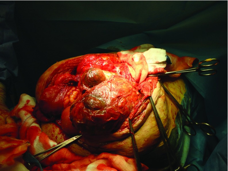
For very large, nonreducible hernias and strangulated hernias, especially when there is gangrenous bowel, open repair is indicated.27 The presence of intestinal ischemia and necrosis, with or without bowel perforation, renders the surgical field septic, and therefore the use of mesh is contraindicated, at least in the first operation (Figure 2). The abdominal cavity is closed by means of fascia approximation with primary suturing, and a definitive repair with mesh can follow after a 2-month period.
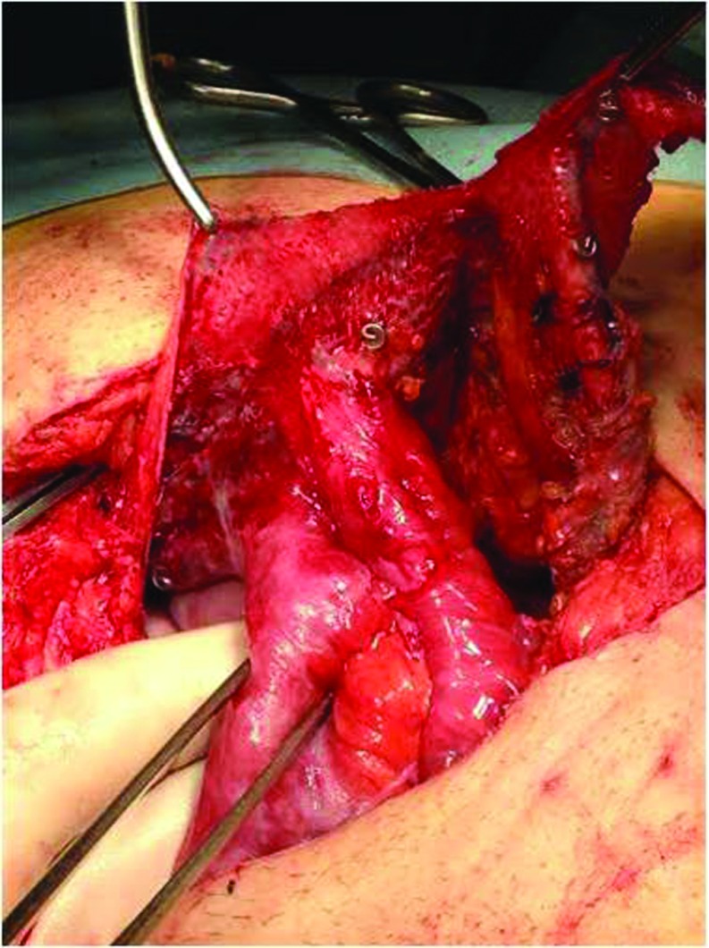
After a failed mesh repair or multiple failed repairs, when intestinal loops are attached to the mesh, an open exploration is recommended. If a piece of mesh is densely adherent to the bowel, a small piece may be excised and left attached to the bowel to prevent deserosalization or an enterotomy (Figure 3).27
Cases of previous repair with a mesh infection usually necessitate abdominal exploration, washout of the contaminated wound, drainage of fluid collections, and often, mesh removal. After an interval period of 2 to 3 months, a second open repair is necessary, with placement of the biological mesh.65
Open surgery is necessary in cases with severe contraindications for laparoscopy in the patient's status, such as cardiorespiratory insufficiency, abnormalities of hemostasis, or ascites.
Figure 2.
For very large nonreducible or strangulated hernias, especially with a gangrenous bowel, open repair is indicated.
Figure 3.
In cases in which a piece of mesh from a prior repair is densely adherent to the bowel, a small piece of mesh may be excised and left attached to the bowel to prevent deserosalization or an opening of the bowel wall.
The Component Separation Technique for Large Complex Hernias
An LVHR repair of a large hernia defect (>8 cm) does not achieve satisfactory muscular strength, and an open repair with open/laparoscopic component separation technique (CST) may be a more functional repair for the patient. CST is a natural method of fascia–fascia closure without the complication of an artificial implant caused by creation of a linea alba, which provides a midline anchor.66 This repair allows for advancement of the rectus abdominis muscle up to 10 cm per side, facilitating closure of large gaps of the abdominal wall. A recently introduced endoscopic technique with the use of a balloon dissector in the space between the external oblique and internal oblique muscles and laparoscopic division of the external oblique aponeurosis along the midclavicular line may reduce substantially the abdominal wall wound morbidity associated with the CST. However, this technique achieves only 86% of the abdominal wall advancement that is obtained with the open technique.67,68
The posterior CST has also been introduced as a modification of the Rives-Stoppa technique, allowing for significant posterior rectus fascia advancement, wide lateral dissection, and a large space for mesh sublay. Transversus abdominis muscle release (TAR) is a novel approach to posterior CST for the repair of complex abdominal hernias, as well as the repair of recurrent hernias after use of the anterior CST. According to this technique the posterior rectus sheath and the underline transversus abdominis muscle are incised and the lateral space is developed. The dissection proceeds to the arcuate line of Douglas toward the space of Retzius. The posterior rectus sheaths are reapproximated in the midline, and the mesh is placed in the retromuscular space and fixed to the abdominal wall with sutures.69,70 This technique is an important addition to the armamentarium of surgeons undertaking abdominal wall reconstructions and represents the gold standard of open repair according to the American Hernia Society.
CONCLUSIONS
LVHR is a safe and excellent alternative to OVHR for the management of abdominal wall hernias. The technique offers the advantages of the laparoscopic approach (i.e., a short hospital stay and brief convalescence). The approach carries a higher risk of bowel injury during surgery, but it has a significantly lower risk of SSI. Laparoscopic repair offers a quality of life and patient satisfaction comparable to or better than that of open repair, and the recurrence rate is equivalent. Even for the repair of large complex hernias, for which the CST is regarded as the gold standard, the endoscopic CST can help to reduce the abdominal wall complications associated with the open technique. For all these reasons, LVHR is used with increasing frequency in everyday surgical practice.
Contributor Information
Evangelos P. Misiakos, Third Department of Surgery, University of Athens School of Medicine, Attikon University Hospital, Rimini 1, Chaidari, Athens, Greece..
Paul Patapis, Third Department of Surgery, University of Athens School of Medicine, Attikon University Hospital, Rimini 1, Chaidari, Athens, Greece..
Nick Zavras, Third Department of Surgery, University of Athens School of Medicine, Attikon University Hospital, Rimini 1, Chaidari, Athens, Greece..
Panagiotis Tzanetis, Third Department of Surgery, University of Athens School of Medicine, Attikon University Hospital, Rimini 1, Chaidari, Athens, Greece..
Anastasios Machairas, Third Department of Surgery, University of Athens School of Medicine, Attikon University Hospital, Rimini 1, Chaidari, Athens, Greece..
References:
- 1. Bloemen A, van Dooren P, Huizinga BF, et al. Randomized clinical trial comparing polypropylene or polydioxanone for midline abdominal wall closure. Br J Surg. 2011;98:633–639. [DOI] [PubMed] [Google Scholar]
- 2. Van't Riet M, Steyerberg EW, Nellensteyn J, et al. Meta-analysis of techniques for closure of midline abdominal incisions. Br J Surg. 2002;89:1350–1356. [DOI] [PubMed] [Google Scholar]
- 3. Pollock AV, Evans M. Early prediction of late incisional hernias. Br J Surg. 1989;76:953–954. [DOI] [PubMed] [Google Scholar]
- 4. Anthony T, Bergen PC, Kim LT, et al. Factors affecting recurrence following incisional herniorrhaphy. World J Surg. 2000;24:95–100; discussion 101. [DOI] [PubMed] [Google Scholar]
- 5. Shell DH, de la Torre J, Andrades T, Vasconez LO. Open repair of ventral hernia incisions. Surg Clin North Am. 2008;88:61–83. [DOI] [PubMed] [Google Scholar]
- 6. Luijendijk R, Hop W, Van den Tol MP, et al. A comparison of suture repair with mesh repair for incisional hernia. N Eng J Med. 2000;343:392–398. [DOI] [PubMed] [Google Scholar]
- 7. Leblanc KA, Booth WV. Laparoscopic repair of incisional abdominal hernias using polytetrafluoroethylene: preliminary findings. Surg Laparosc Endosc. 1993;3:39–41. [PubMed] [Google Scholar]
- 8. Stoppa RE. The treatment of complicated groin and incisional hernias. World J Surg. 1989;13:545–554. [DOI] [PubMed] [Google Scholar]
- 9. Rives J, Pire JC, Flament JB, et al. Treatment of large eventrations: new therapeutic indications apropos of 322 cases. Chirurgie. 1985;111:215–225. [PubMed] [Google Scholar]
- 10. Wantz GE. Incisional hernioplasty with Mersilene. Surg Gynecol Obstetr. 1991;172:129–137. [PubMed] [Google Scholar]
- 11. Franklin ME, Dorman JP, Glass JL, Balli JE, Gonzalez JJ. Laparoscopic ventral and incisional hernia repair. Surg Laparosc Endosc. 1998;8:294–299. [PubMed] [Google Scholar]
- 12. Heniford BT, Park A, Ramshaw BJ, Voeller G. Laparoscopic ventral and incisional hernia repair in 407 patients. J Am Coll Surg. 2000;190:645–650. [DOI] [PubMed] [Google Scholar]
- 13. LeBlanc KA, Booth WV, Whitaker JM, Bellanger DE. Laparoscopic incisional and ventral herniorrhaphy: our initial 100 patients. Hernia. 2001;5:41–45. [DOI] [PubMed] [Google Scholar]
- 14. Heniford BT, Park A, Ramshaw BJ, Voeller G. Laparoscopic repair of ventral hernias: nine years' experience with 850 consecutive hernias. Ann Surg. 2003;238:391–400. [DOI] [PMC free article] [PubMed] [Google Scholar]
- 15. Olmi S, Erba L, Magnone S, Bertolini A, Croce E. Prospective clinical study of laparoscopic treatment of incisional and ventral hernia using a composite mesh: indications, complications and results. Hernia. 2006;10:243–247. [DOI] [PubMed] [Google Scholar]
- 16. Heniford BT, Park A, Ramshaw BJ, Voeller G. Laparoscopic ventral and incisional hernia repair in 407 patients. J Am Coll Surg. 2000;190:645–650. [DOI] [PubMed] [Google Scholar]
- 17. Misra MC, Bansal VK, Kulkarni MP, Pawar DK. Comparison of laparoscopic and open repair of incisional and primary ventral hernia: results of a prospective randomized study. Surg Endosc. 2006;20:1839–1845. [DOI] [PubMed] [Google Scholar]
- 18. LeBlanc KA, Booth WV, Whitaker JM, Bellanger DE. Laparoscopic incisional and ventral herniorrhaphy in 100 patients. Am J Surg. 2000;180:193–197. [DOI] [PubMed] [Google Scholar]
- 19. LeBlanc KA, Whitaker JM, Bellanger DE, Rhynes VK. Laparoscopic incisional and ventral hernioplasty: lessons learned from 200 patients. Hernia. 2003;7:118–124. [DOI] [PubMed] [Google Scholar]
- 20. Machairas A, Misiakos EP, Liakakos T, Karatzas G. Incisional hernioplasty with extraperitoneal onlay polyester mesh. Am Surg. 2004;70:726–729. [PubMed] [Google Scholar]
- 21. Perrone JM, Soper NJ, Eagon JC, et al. Perioperative outcomes and complications of laparoscopic ventral hernia repair. Surgery. 2005;138:708–715; discussion 715–716. [DOI] [PubMed] [Google Scholar]
- 22. Cobb WS, Kercher KW, Heniford BT. Laparoscopic repair of incisional hernias, ix. Surg Clin North Am. 2005;85:91–103. [DOI] [PubMed] [Google Scholar]
- 23. Cassar K, Munro A. Surgical treatment of incisional hernia. Br J Surg. 2002;89:534–545. [DOI] [PubMed] [Google Scholar]
- 24. McCarthy JD, Twiest MW. Intraperitoneal polypropylene mesh support of incisional herniorrhaphy. Am J Surg. 1981;142:707–711. [DOI] [PubMed] [Google Scholar]
- 25. Millikan KW, Baptista M, Amin B, Deziel DJ, Doolas A. Intraperitoneal underlay ventral hernia repair utilizing bilayer expanded polytetrafluoroethylene and polypropylene mesh. Am Surg. 2003;69:287–291. [PubMed] [Google Scholar]
- 26. Jin J, Rosen MJ. Laparoscopic versus open ventral hernia repair. Surg Clin North Am. 2008;88:1083–1100. [DOI] [PubMed] [Google Scholar]
- 27. Alexander AM, Scott DJ. Laparoscopic ventral hernia repair. Surg Clin North Am. 2013;93:1091–1110. [DOI] [PubMed] [Google Scholar]
- 28. Zhang V, Zhou H, Chai Y, Cao C, Jin K, Hu Z. Laparoscopic versus open incisional and ventral hernia repair: a systematic review and meta-analysis. World J Surg. 2014;38:2233–2240. [DOI] [PubMed] [Google Scholar]
- 29. Newcomb WL, Polhil JL, Chen AY, et al. Staged hernia repair preceded by gastric bypass for the treatment of morbidly obese patients with complex ventral hernias. Hernia. 2008;12:465–469. [DOI] [PubMed] [Google Scholar]
- 30. Bittner R, Bingener-Casey J, Dietz U, et al. Guidelines for laparoscopic treatment of ventral and incisional abdominal wall hernias (International Endohernia Society [IEHS]), Part 2. Surg Endosc. 2014;28:353–379. [DOI] [PMC free article] [PubMed] [Google Scholar]
- 31. LeBlanc KA, Elieson MJ, Corder JM., III Enterotomy and mortality rates of laparoscopic incisional and ventral hernia repair: a review of the literature. JSLS. 2007;11:408–414. [PMC free article] [PubMed] [Google Scholar]
- 32. LeBlanc KA. Laparoscopic incisional and ventral hernia repair: complications: how to avoid and handle. Hernia. 2004;8:323–331. [DOI] [PubMed] [Google Scholar]
- 33. Sharma A, Khullar R, Soni V, et al. Iatrogenic enterotomy in laparoscopic ventral/incisional hernia repair: a single center experience of 2,346 patients over 17 years. Hernia. 2013;17:581–587. [DOI] [PubMed] [Google Scholar]
- 34. Kurmann A, Visth E, Candinas D, et al. Long term follow-up of open and laparoscopic repair of large incisional hernias. World J Surg. 2011;35:297–301. [DOI] [PubMed] [Google Scholar]
- 35. Nguyen DH, Ngugen MT, Askenasy EP, Kao LS, Liang MK. Primary fascial closure with laparoscopic ventral hernia repair: systematic review. World J Surg. 2014;38:3097–3104. [DOI] [PubMed] [Google Scholar]
- 36. Bhanot P, Franklin BR, Patel KM. ProceedTM mesh for laparoscopic ventral hernia repair. JSLS. 2013;17:565–569. [DOI] [PMC free article] [PubMed] [Google Scholar]
- 37. Tollens T, Maxime E, Anthony B, Aelvoet C. Retrospective study on the use of a composite mesh (Physiomesh) in laparoscopic ventral hernia repair. Surg Technol Int. 2012;22:141–145. [PubMed] [Google Scholar]
- 38. Mariette C, Wind P, Micelli Lupinacci R, et al. Practice patterns in Comlex ventral hernia repair and place of biological grafts: a national survey among French digestive academic surgeons. J Visc Surg. 2014;151:9–16. [DOI] [PubMed] [Google Scholar]
- 39. Misiakos EP, Machairas A, Patapis P, Liakakos T. Laparoscopic ventral hernia repair: pros and cons compared with open hernia repair. JSLS. 2008;12:117–125. [PMC free article] [PubMed] [Google Scholar]
- 40. Castro PMV, Rabelato JT, Monteiro GGR, Guerra GC, Mazzurana M, Alvarez A. Laparoscopy versus laparotomy in the repair of ventral hernias: systematic review and meta-analysis. Arq Gastroenterol. 2014;51:205–211. [DOI] [PubMed] [Google Scholar]
- 41. Moreno-Egea A, Castillo JA, Girela BE, Aguayo-Albasini JL. Long-term results of laparoscopic repair of incisional hernias using an intraperitoneal composite mesh. Surg Endosc. 2010;24:359–365. [DOI] [PubMed] [Google Scholar]
- 42. Deeken CR, Matthews BD. Ventralight and Sorbafix versus Physiomesh and Securestrap in a porcine model. JSLS. 2013;17:549–559. [DOI] [PMC free article] [PubMed] [Google Scholar]
- 43. Cuccurullo D, Piccoli M, Agresta F, et al. Laparoscopic ventral incisional hernia repair: evidence-based guidelines of the first Italian Consensus Conference. Hernia. 2013;17:557–566. [DOI] [PubMed] [Google Scholar]
- 44. Orenstein SB, Dumeer JL, Monteagudo J, Poi MJ, Novitsky YW. Outcomes of laparoscopic ventral hernia repair with routine defect closure using “shoelacing” technique. Surg Endosc. 2011;25:1452–1457. [DOI] [PubMed] [Google Scholar]
- 45. Wassenaar E, Schoenmaeckers E, Raymakers J, van der Palen J, Rakic S. Mesh-fixation method and pain and quality of life after laparoscopic ventral or incisional hernia repair; a randomized trial of three fixation techniques. Surg Endosc. 2010;24:1296–1302. [DOI] [PMC free article] [PubMed] [Google Scholar]
- 46. Carbonell AM, Harold KL, Mahmutovic AJ, et al. Local injection for the treatment of suture site pain after laparoscopic ventral hernia repair. Am Surg. 2003;69:688–692. [PubMed] [Google Scholar]
- 47. Wassenaar EB, Raymakers JT, Rakic S. Removal of transabdominal sutures for chronic pain after laparoscopic ventral and incisional hernia repair. Surg Laparosc Endosc Percutan Tech. 2007;17:514–516. [DOI] [PubMed] [Google Scholar]
- 48. Eker HH, Hansson BME, Buunen M, et al. Laparoscopic vs open incisional hernia repair: a randomized clinical trial FREE. JAMA Surg. 2013;148:259–263. [DOI] [PubMed] [Google Scholar]
- 49. Muysoms F, Vander Mijnsbrugge G, Pletinckx P, et al. Randomized clinical trial of mesh fixation with “double crown” versus “sutures and tackers” in laparoscopic ventral hernia repair. Hernia. 2013;17:603–612. [DOI] [PubMed] [Google Scholar]
- 50. Heniford T, Park A, Ramshaw BJ, Voeller G. Laparoscopic repair of ventral hernias nine years' experience with 850 consecutive hernias. Ann Surg. 2003;238:391–400. [DOI] [PMC free article] [PubMed] [Google Scholar]
- 51. Wassenaar EB, Schoenmaeckers EJ, Raymakers JT, Rakic S. Recurrences after laparoscopic repair of ventral and incisional hernia: lessons learned from 505 repairs. Surg Endosc. 2009;23:825–832. [DOI] [PubMed] [Google Scholar]
- 52. Reynvoet E, Deschepper E, Rogiers X, Troisi R, Berrevoet F. Laparoscopic ventral hernia repair: is there an optimal mesh fixation technique? A systematic review. Langebecks Arch Surg. 2014;399:55–63. [DOI] [PubMed] [Google Scholar]
- 53. Poelman M, Apers J, van den Brand H, et al. The INCh-trial: a multicentre randomized controlled trial comparing the efficacy of conventional open surgery and laparoscopic surgery for incisional hernia repair. BMC Surg. 2013;13:18. [DOI] [PMC free article] [PubMed] [Google Scholar]
- 54. Itani KM, Hu K, Kim LT, et al. Veterans Affairs Ventral Incisional Hernia Investigators. Comparison of laparoscopic and open repair with mesh for the treatment of ventral incisional hernia: a randomised study. Arch Surg. 2010;145:322–328. [DOI] [PubMed] [Google Scholar]
- 55. Cocozza E, Berselli M, Latham L, et al. Laparoscopic repair of ventral hernia in a laparoscopic experienced surgical center: low recurrence rate, morbidity, and chronic pain are achievable. Surg Laparosc Endosc Perutan Tech. 2014;24:168–172. [DOI] [PubMed] [Google Scholar]
- 56. Aher CV, Kubasiak JC, Daly SC, et al. The utilization of laparoscopy in ventral hernia repair: an update of outcomes analysis using ACS-NSQIP data. Surg Endosc. 2015;29:1099–1104. [DOI] [PubMed] [Google Scholar]
- 57. Beldi G, Ipaktchi R, Wagner M, Gloor B, Candinas D. Laparoscopic ventral hernia repair is safe and cost effective. Surg Endosc. 2006;20:92–95. [DOI] [PubMed] [Google Scholar]
- 58. Hope WW, Lincourt AE, Newcomb WL, Schmelzer TM, Kercher KW, Heniford BT. Comparing quality-of-life outcomes in symptomatic patients undergoing laparoscopic or open ventral hernia repair. J Laparoendosc Adv Surg Tech. 2008;18:567–571. [DOI] [PubMed] [Google Scholar]
- 59. Eriksen JR, Poornoroozy P, Jørgensen LN, Jacobsen B, Friis-Andersen HU, Rosenberg J. Pain, quality of life and recovery after laparoscopic ventral hernia repair. Hernia. 2009;13:13–21. [DOI] [PubMed] [Google Scholar]
- 60. Ballantyne GH, Hourmont K, Wasielewski A. Telerobotic laparoscopic repair of incisional ventral hernias using intracorporeal prosthetic mesh. JSLS. 2003;7:7–14. [PMC free article] [PubMed] [Google Scholar]
- 61. Allison N, Tieu K, Snyder B, Pigazzi A, Wilson E. Technical feasibility of robot-assisted ventral hernia repair. World J Surg. 2012;36:447–452. [DOI] [PubMed] [Google Scholar]
- 62. Beldi G. Technical feasibility of a robotic-assisted ventral hernia repair. World J Surg. 2012;36:453–454. [DOI] [PubMed] [Google Scholar]
- 63. Kulacoglu H, Yazicioglu D, Ozyaylali I. Prosthetic repair of umbilical hernias in adults with local anesthesia in a day-case setting: a comprehensive report from a specialized hernia center. Hernia. 2012;16:163–170. [DOI] [PubMed] [Google Scholar]
- 64. Tollens T, Den Hondt M, Devroe K, Terry C, Speybroeck S, Aelvoet C. Retrospective analysis of umbilical, epigastric, and small incisional hernia repair using the Ventralex™ hernia patch. Hernia. 2011;15:531–540. [DOI] [PubMed] [Google Scholar]
- 65. Mariette C, Wind P, Micelli Lupinacci R, et al. Practice patterns in complex ventral hernia repair and place of biological grafts: a national survey among French digestive academic surgeons. Visc Surg. 2014;151:9–16. [DOI] [PubMed] [Google Scholar]
- 66. Shih PK. Difficult abdominal wall closure: component separation versus partition technique. Hernia. 2015;19:301–305. [DOI] [PubMed] [Google Scholar]
- 67. Giurgius M, Bendure L, Davenport DL, Roth JS. The endoscopic component separation technique for hernia repair results in reduced morbidity compared to the open component separation technique. Hernia. 2012;16:47–51. [DOI] [PubMed] [Google Scholar]
- 68. Milburn ML, Shah PK, Friedman EB. Laparoscopically assisted components separation technique for ventral incisional hernia repair. Hernia. 2007;11:157–161. [DOI] [PubMed] [Google Scholar]
- 69. Novitsky YW, Elliott HL, Orenstein SB, Rosen MJ. Transversus abdominis muscle release: a novel approach to posterior component separation during complex abdominal wall reconstruction. Am J Surg. 2012;204:709–716. [DOI] [PubMed] [Google Scholar]
- 70. Petro CC, Como JJ, Yee S, et al. Posterior component separation and transverses abdominis muscle release for complex incisional hernia repair inpatients with a history of an open abdomen. J Trauma Acute Care Surg. 2015;78:422–429. [DOI] [PubMed] [Google Scholar]


