Abstract
Some neoplasms are associated with granulomatous inflammation. Granuloma formation in tumor tissue is caused by the cytokines derived from either the main tumor or other cells surrounding the tumor. In other instances, granulomatous inflammation is observed in the lymph nodes draining a tumor. This has been recognized as a sarcoid-like reaction. Herein, we report of a 75-year-old man with pulmonary squamous cell carcinoma (SCC), where granulomatous inflammation was observed extensively at the primary site. The carcinoma seemed to partly regress. In the regressing area, tumor cell debris was surrounded by granuloma. In contrast, no granuloma was identified in the dissected regional lymph nodes. To the best of our knowledge, such a case of SCC had not been described thus far. More case studies are required to determine whether tumor-related granuloma is the main cause of regression or whether it is just a secondary phenomenon caused by the attack and destruction of the tumor by lymphocytes.
Keywords: Granulomatous inflammation, granuloma, squamous cell carcinoma, lung, sarcoid-like
Introduction
Granulomatous inflammation is often encountered during infection by mycobacteria, fungi, or parasites and in non-infectious etiologies, such as intrusion by foreign bodies. In addition, granuloma formation is a diagnostic symptom of some diseases such as sarcoidosis and Crohn’s disease.
Certain neoplasms are also associated with granulomatous inflammation, including seminoma of the testis [1] and its counterparts in various locations [2], renal cell carcinoma [3,4], and nasopharyngeal carcinoma [5]. Granuloma formation in tumor tissue is largely caused by the cytokines derived from either the main tumor or non-cancerous cells near the tumor [6]. In some instances, granulomatous inflammation is observed in the lymph nodes draining a tumor where it is known as a sarcoid-like reaction [7]. It has been suggested that T-cell mediated immunological reaction to antigens expressed by the tumor cells and soluble antigens shed by the tumor might result in granulomatous inflammation [8].
Herein, we report of a 75-year-old man with pulmonary squamous cell carcinoma (SCC), in which granulomatous inflammation was observed extensively at the primary site. The carcinoma seemed to partly regress due to granulomatous inflammation. To the best of our knowledge, such a phenomenon has not been previously reported in pulmonary SCC. In contrast, no granuloma was identified in the dissected regional lymph nodes.
Clinical summary
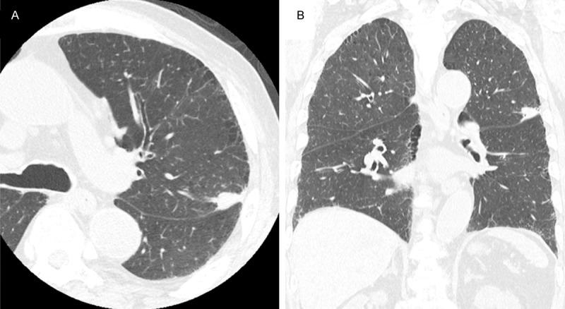
A 75-year-old man presented with an irregular mass detected on computed tomography performed during his regular cerebral infarction follow-up. The mass was located in the left upper lobe and showed extensive contact with the interlobar pleura; it measured 18×12×10 mm (Figure 1A, 1B). He is currently smoking 5 cigarettes per day. Physical examination and laboratory tests revealed no abnormalities. Being unable to rule out the possibility of a primary lung carcinoma, surgery was performed. Intraoperative pathological examination identified a few carcinoma cells. Therefore, left upper lobectomy and regional lymph nodes dissections were conducted. His postoperative course was uneventful.
Figure 1.

Computed tomography findings. A. Axial; B. Coronal: The mass is located in the left upper lobe and shows extensive contact with the interlobar pleura; the mass measured 18×12×10 mm.
Pathological findings
The surgically resected specimen revealed a mass with anthracotic discoloration. The entire mass was fixed using formalin and then embedded in paraffin blocks.
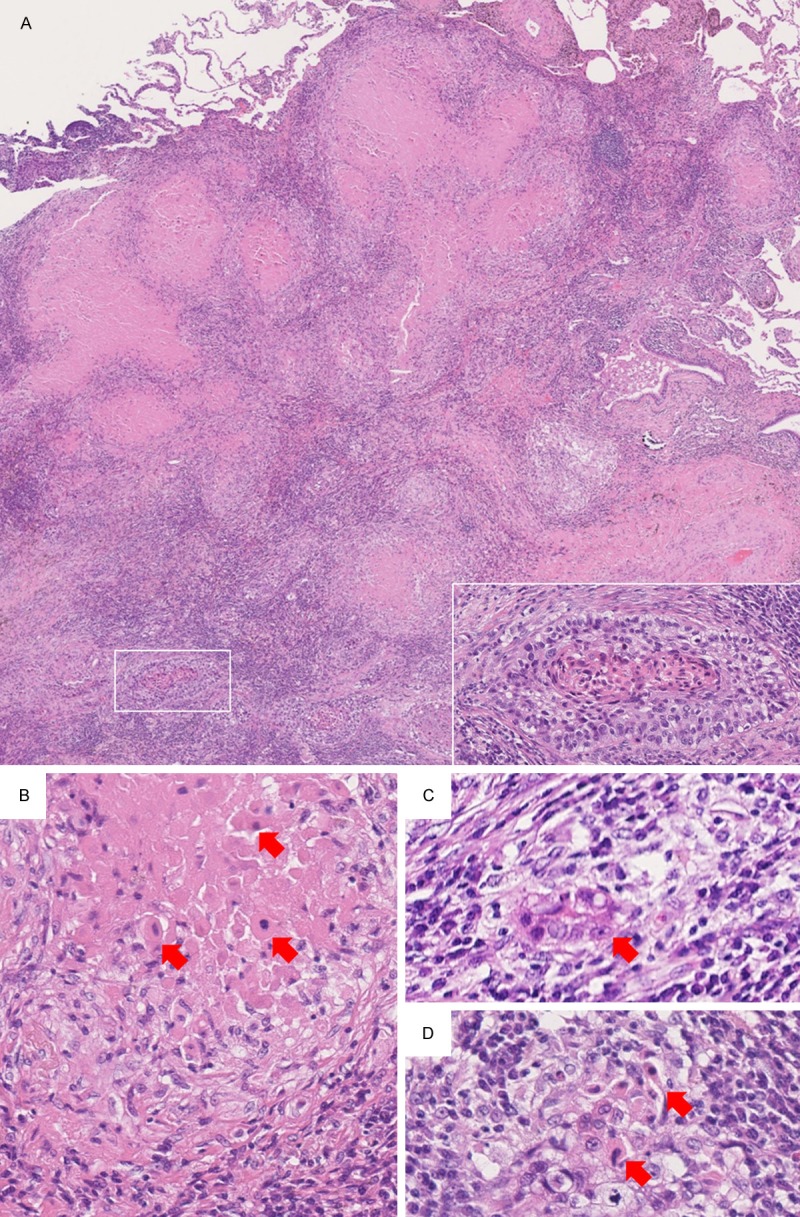
According to the histopathology results, approximately 70% of the mass was composed of granulomatous inflammation accompanied by the formation of prominent epithelioid cell granulomas with a central necrotic substance. In the remaining 30% of the mass, viable well-to-moderately differentiated SCC was present (Figure 2A). The necrotic substance inside the granulomas consisted of necrotic tumor cells, with some retaining their shape and having pyknotic nuclei (Figure 2B). Some of the tumor nests were surrounded by macrophages and/or epithelioid histiocytes (Figure 2C); several such tumor nests were degenerated, and the remaining necrotic tumor cells retained cellular shape and pyknotic nuclei (Figure 2D). Special stains, such as Ziehl-Neelsen and Grocott stains, did not reveal pathogenic microorganisms inside the granulomas. The surgical margin was free of the tumor. No granulomas were identified in the dissected regional lymph nodes.
Figure 2.

Microscopic findings. A. Approximately 70% of the mass is composed of granulomatous inflammation accompanied by formation of prominent epithelioid cell granulomas with a central necrotic substance. In the remaining 30% of the mass, viable well-to-moderately differentiated squamous cell carcinoma is present (×20). Inset: Higher magnification of the boxed area shows a viable tumor nest with keratinization (×200). B. The necrotic substance inside the granulomas were consists of necrotic tumor cells that retained cellular shape and exhibited pyknotic nuclei (arrows) (×400). C. Tumor nest surrounded by macrophages and/or epithelioid histiocytes (arrow) (×400). D. Tumor nest with degeneration that showed remaining necrotic tumor cells with retained cellular shape and pyknotic nuclei (arrows) (×400).
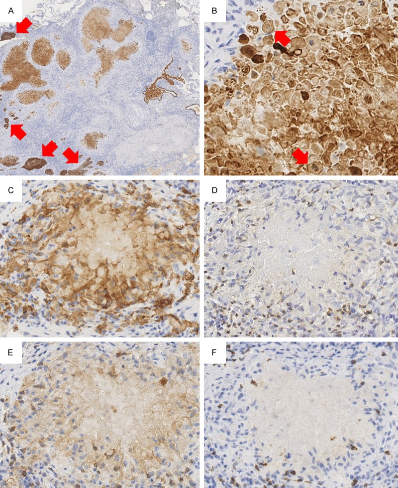
Using immunohistochemistry, viable tumor nests were strongly stained for CK5/6 (D5/16 B4, 1:100; Dako, Glostrup, Denmark). The necrotic substance was moderately stained for CK5/6 (Figure 3A). The necrotic substance inside granulomas revealed CK5/6-positive necrotic tumor cells that retained cellular shape and pyknotic nuclei (Figure 3B). Granulomas were composed of CD68-positive (PG-M1, 1:00; Dako) macrophages and/or epithelioid histiocytes (Figure 3C). T lymphocytes expressing CD3 (F7.2.38, 1:100; Dako) were abundant inside and outside the granulomas (Figure 3D). Among T lymphocytes, the proportion of CD4-expressing (4B12, 1:100; Dako) lymphocytes was slightly higher than that of CD8-expressing (C8/144B, 1:100; Dako) lymphocytes both inside and outside the granulomas (Figure 3E, 3F).
Figure 3.

Immunohistochemistry findings. (A) Viable tumor nests showing strong staining for CK5/6 (arrows). Necrotic substance showing moderate staining for CK5/6 (×20). (B) Necrotic substance inside a granuloma showing CK5/6-positive necrotic tumor cells that retained cellular shape and pyknotic nuclei (arrows) (×400). (C) Granulomas are composed of CD68-positive macrophages and/or epithelioid histiocytes (×400). (D) T lymphocytes expressing CD3 are abundant inside and outside the granulomas (×400). (E, F) Among T lymphocytes, CD4-expressing lymphocytes (E) are slightly more numerous than CD8-expressing lymphocytes (F) (×400).
Discussion
Granulomatous tissue could be caused by infections, autoimmune diseases, or tumors. Although the causes of granulomas are varied, they result in similar morphological features that are indistinguishable from one another. This similarity could be due to closely related pathogenesis involving the same molecular mechanisms. Close examination of the cellular subcomponents of infection-related and tumor-related granulomatous inflammations showed an accumulation of gamma/delta T-cells [9]. These gamma/delta T-cells not only help keep infections with intracellular microorganisms under control [10], but also participate in autologous tumor killing activity [11]. The molecular similarities of infection-related and tumor-related granuloma formation could be attributed to tumor necrosis factor-alpha, interferon-gamma, and granulocyte-macrophage colony stimulating factor produced by gamma/delta T-cells. These similarities could also explain their indistinguishable histological appearance [9-11].
Tumor-related tissue reactions leading to granuloma formation have been described for a long time [12,13]. Such granuloma formation may take place in lymph nodes draining the primary tumor site, in tumoral tissue, and even in areas unrelated to the primary tumor site [13]. While granulomas occur in approximately 4% of carcinomas, this reaction is considered to be exceedingly rare in sarcomas [13].
It was proposed that granulomatous reactions could prevent tumor expansion [14]. Although in our case and other cases such as seminoma [1], germinoma [2], renal cell carcinoma [4], and nasopharyngeal carcinoma [5], granuloma formation occurred within the tumor, there exists a renal cell carcinoma case in which granuloma formation was observed in the peritumoral stroma alone [3]. In addition, many granulomas do not form within the tumor but rather in the regional lymph nodes by a sarcoid-like reaction [15].
As for searching cellular subpopulation of tumor-related granuloma in pathological practice, T-cell markers, such as CD3, CD4, CD8, in addition to monocyte/macrophage marker, CD68, were often used; specific gamma/delta T-cell markers were not commonly available. For tumor-related granulomas, it was found that CD4-positive T-cells tended to be present in the internal area of the granuloma rather than its surroundings, which predominantly displayed CD8-positive T-cells. This is the same distribution pattern found in sarcoidosis [16]. In our case, CD4-positive T-cells were slightly more numerous than CD8-positive T-cells both inside and outside the granulomas. Differences between the internal and surrounding areas of the granulomas were not apparent. This may be because almost all the granulomas in our case had a tumor element inside of them. Tumor-related granulomas do not usually contain tumor elements in them.
The tumor-related granuloma described in this study seemed to be involved in partial regression of SCC of the lung. It is generally recognized that natural killer (NK) cells and cytotoxic T-cells (a subset of CD8-positive T-cells) play an important role in tumor regression through direct contact with tumor cells [17]. In addition, CD4-positive T-cell tumor infiltration is important for melanoma regression, a well-known phenomenon, as it can directly break up tumor nests [18]. Therefore, other case studies are required to determine whether formation of tumor-related granuloma is an initiating phenomenon of tumor regression, or whether it is only involved in eliminating cellular debris after NK cells and T-cells destroyed the tumor.
In conclusion, this is an extremely rare case of pulmonary SCC with partial regression accompanied by granulomatous inflammation. Inside the tumor-related granulomas, necrotic tumor cells were frequently observed. More cases are required to determine whether formation of tumor-related granulomas is a primary cause of tumor regression or just a secondary phenomenon following the attack and destruction of the tumor by lymphocytes.
Disclosure of conflict of interest
None.
References
- 1.Ulbright TM. Germ cell neoplasms of the testis. Am J Surg Pathol. 1993;17:1075–1091. doi: 10.1097/00000478-199311000-00001. [DOI] [PubMed] [Google Scholar]
- 2.Mueller W, Schneider GH, Hoffmann KT, Zschenderlein R, von Deimling A. Granulomatous tissue response in germinoma, a diagnostic pitfall in endoscopic biopsy. Neuropathology. 2007;27:127–132. doi: 10.1111/j.1440-1789.2006.00749.x. [DOI] [PubMed] [Google Scholar]
- 3.Ouellet S, Albadine R, Sabbagh R. Renal cell carcinoma associated with peritumoral sarcoid-like reaction without intratumoral granuloma. Diagn Pathol. 2012;7:28. doi: 10.1186/1746-1596-7-28. [DOI] [PMC free article] [PubMed] [Google Scholar]
- 4.Hes O, Hora M, Vanecek T, Sima R, Sulc M, Havlicek F, Beranova M, Michal M. Conventional renal cell carcinoma with granulomatous reaction: a report of three cases. Virchows Arch. 2003;443:220–221. doi: 10.1007/s00428-003-0860-9. [DOI] [PubMed] [Google Scholar]
- 5.Chen CL, Su IJ, Hsu MM, Hsu HC. Granulomatous nasopharyngeal carcinoma: with emphasis on difficulty in diagnosis and favorable outcome. J Formos Med Assoc. 1991;90:353–356. [PubMed] [Google Scholar]
- 6.Haralambieva E, Rosati S, van Noesel C, Boers E, van Marwijk Kooy M, Schuuring E, Kluin P. Florid granulomatous reaction in Epstein-Barr virus-positive nonendemic Burkitt lymphomas: report of four cases. Am J Surg Pathol. 2004;28:379–383. doi: 10.1097/00000478-200403000-00011. [DOI] [PubMed] [Google Scholar]
- 7.Gregorie HB Jr, Othersen HB Jr, Moore MP Jr. The significance of sarcoid-like lesions in association with malignant neoplasms. Am J Surg. 1962;104:577–586. doi: 10.1016/0002-9610(62)90399-9. [DOI] [PubMed] [Google Scholar]
- 8.Kobayashi K, Kaneda K, Kasama T. Immunopathogenesis of delayed-type hypersensitivity. Microsc Res Tech. 2001;53:241–245. doi: 10.1002/jemt.1090. [DOI] [PubMed] [Google Scholar]
- 9.Wangoo A, Johnson L, Gough J, Ackbar R, Inglut S, Hicks D, Spencer Y, Hewinson G, Vordermeier M. Advanced granulomatous lesions in Mycobacterium bovis-infected cattle are associated with increased expression of type I procollagen, gammadelta (WC1+) T cells and CD68+ cells. J Comp Pathol. 2005;133:223–234. doi: 10.1016/j.jcpa.2005.05.001. [DOI] [PubMed] [Google Scholar]
- 10.Ehlers S. Tumor necrosis factor and its blockade in granulomatous infections: differential modes of action of infliximab and etanercept? Clin Infect Dis. 2005;41(Suppl 3):S199–203. doi: 10.1086/429998. [DOI] [PubMed] [Google Scholar]
- 11.Zhao X, Wei YQ, Kariya Y, Teshigawara K, Uchida A. Accumulation of gamma/delta T cells in human dysgerminoma and seminoma: roles in autologous tumor killing and granuloma formation. Immunol Invest. 1995;24:607–618. doi: 10.3109/08820139509066861. [DOI] [PubMed] [Google Scholar]
- 12.Gorton G, Linell F. Malignant tumours and sarcoid reactions in regional lymph nodes. Acta Radiol. 1957;47:381–392. doi: 10.3109/00016925709170911. [DOI] [PubMed] [Google Scholar]
- 13.Brincker H. Sarcoid reactions in malignant tumours. Cancer Treat Rev. 1986;13:147–156. doi: 10.1016/0305-7372(86)90002-2. [DOI] [PubMed] [Google Scholar]
- 14.Pavic M, Debourdeau P, Vacelet V, Rousset H. [Sarcoidosis and sarcoid reactions in cancer] . Rev Med Interne. 2008;29:39–45. doi: 10.1016/j.revmed.2007.09.041. [DOI] [PubMed] [Google Scholar]
- 15.Kennedy MP, Jimenez CA, Mhatre AD, Morice RC, Eapen GA. Clinical implications of granulomatous inflammation detected by endobronchial ultrasound transbronchial needle aspiration in patients with suspected cancer recurrence in the mediastinum. J Cardiothorac Surg. 2008;3:8. doi: 10.1186/1749-8090-3-8. [DOI] [PMC free article] [PubMed] [Google Scholar]
- 16.Hojo H, Suzuki S, Kikuta A, Ito M, Abe M. Sarcoid reaction in primary neuroblastoma: case report. Pediatr Dev Pathol. 2000;3:584–590. doi: 10.1007/s100240010113. [DOI] [PubMed] [Google Scholar]
- 17.Ricci SB, Cerchiari U. Spontaneous regression of malignant tumors: Importance of the immune system and other factors (Review) Oncol Lett. 2010;1:941–945. doi: 10.3892/ol.2010.176. [DOI] [PMC free article] [PubMed] [Google Scholar]
- 18.Printz C. Spontaneous regression of melanoma may offer insight into cancer immunology. J Natl Cancer Inst. 2001;93:1047–1048. doi: 10.1093/jnci/93.14.1047. [DOI] [PubMed] [Google Scholar]


