Abstract
A novel neutron irradiation facility at the Radiological Research Accelerator Facility (RARAF) has been developed to mimic the neutron radiation from an Improvised Nuclear Device (IND) at relevant distances (e.g. 1.5 km) from the epicenter. The neutron spectrum of this IND-like neutron irradiator was designed according to estimations of the Hiroshima neutron spectrum at 1.5 km. It is significantly different from a standard reactor fission spectrum, because the spectrum changes as the neutrons are transported through air, and it is dominated by neutron energies from 100 keV up to 9 MeV. To verify such wide energy range neutron spectrum, detailed here is the development of a combined spectroscopy system. Both a liquid scintillator detector and a gas proportional counter were used for the recoil spectra measurements, with the individual response functions estimated from a series of Monte Carlo simulations. These normalized individual response functions were formed into a single response matrix for the unfolding process. Several accelerator-based quasi-monoenergetic neutron source spectra were measured and unfolded to test this spectroscopy system. These reference neutrons were produced from two reactions: T(p,n)3He and D(d,n)3He, generating neutron energies in the range between 0.2 and 8 MeV. The unfolded quasi-monoenergetic neutron spectra indicated that the detection system can provide good neutron spectroscopy results in this energy range. A broad-energy neutron spectrum from the 9Be(d,n) reaction using a 5 MeV deuteron beam, measured at 60 degrees to the incident beam was measured and unfolded with the evaluated response matrix. The unfolded broad neutron spectrum is comparable with published time-of-flight results. Finally, the pair of detectors were used to measure the neutron spectrum generated at the RARAF IND-like neutron facility and a comparison is made to the neutron spectrum of Hiroshima.
Keywords: Neutron spectroscopy, radiation, accelerator, liquid scintillator, proportional counter, unfolding
1. Introduction
A new beam line at Columbia University’s Radiological Research Accelerator Facility (RARAF) has been built to generate neutrons mimicking an IND (Improvised Nuclear Device) exposure [1]. This accelerator driven radiation source produces neutron beams which simulate realistic potential IND scenarios [2] (e.g. a gun-type Hiroshima device) for irradiation experiments using mice and human blood. The neutron spectrum is generated through bombarding a thick beryllium (Be) target with single and molecular hydrogen and deuterium ions. Through the 9Be(p,n) (low neutron energy) and 9Be(d,n) (high neutron energy) reactions, the full energy spectrum of a Hiroshima-like IND from 0.1 MeV to 9 MeV can be recreated. In order to characterize the spectrum of this broad energy neutron radiator, we have developed and validated a dual detector spectroscopy system.
The neutron spectrum are acquired indirectly through a proton recoil pulse height spectrum, therefore, an algorithm is necessary to unfold the raw pulse height data into the actual neutron energy spectra. To cover a wide range of energies, the recoil spectra were measured using an EJ-301 liquid scintillator detector [3] and a spherical hydrogen gas-filled proportional counter [4]. The measured recoil spectra were unfolded using a standard technique [5, 6]. This neutron spectroscopy approach for evaluating neutron spectra [3, 4, 7] is well established; it utilizes the relationship between the neutron energy spectrum and the recoil proton spectrum which allowed us to identify and quantify a broad-energy neutron field.
Two tests of the unfolding were made: first using quasi-monoenergetic neutrons for single energy analysis and second, using a broad-energy neutron spectrum from the 9Be(d,n) reaction with 5 MeV deuteron beam. The unfolded result was comparable with the result from a time-of-flight spectroscopy measurement made at Ohio University using the method.
Using the developed response matrices verified through the quasi-monenergetic measurements and the deuteron beam test, the full neutron spectrum of the RARAF IND-like neutron facility [1] was measured. The combined spectra from the two detectors are comparable to the neutron spectrum from the Hiroshima bomb at 1.5 km [8].
2. Detector design
2.1. Liquid scintillator detector
The Liquid scintillator detector is a 1.8 inch diameter by 1.8 inch high cylindrical aluminum chamber filled with EJ-301 liquid scintillator (ELJEN Technology, Sweetwater, TX) and was chosen for neutron spectrometry in the energy range 1 MeV to 8 MeV. Liquid scintillator has good pulse shape characteristics for neutron-gamma discrimination since the rise time of the light pulses from the scintillator are approximately 130 nsec when it is excited by neutrons and approximately 10 nsec when it is excited by gamma rays. The wavelength of emission of EJ-301 is about 425 nm.
The liquid scintillator detector was assembled at RARAF, Columbia University. The detector liquid was bubbled with Argon gas to remove oxygen, which may quench the slower neutron-induced signal and reduce the light output. A small amount of inert gas remained above the liquid in the sealed chamber.
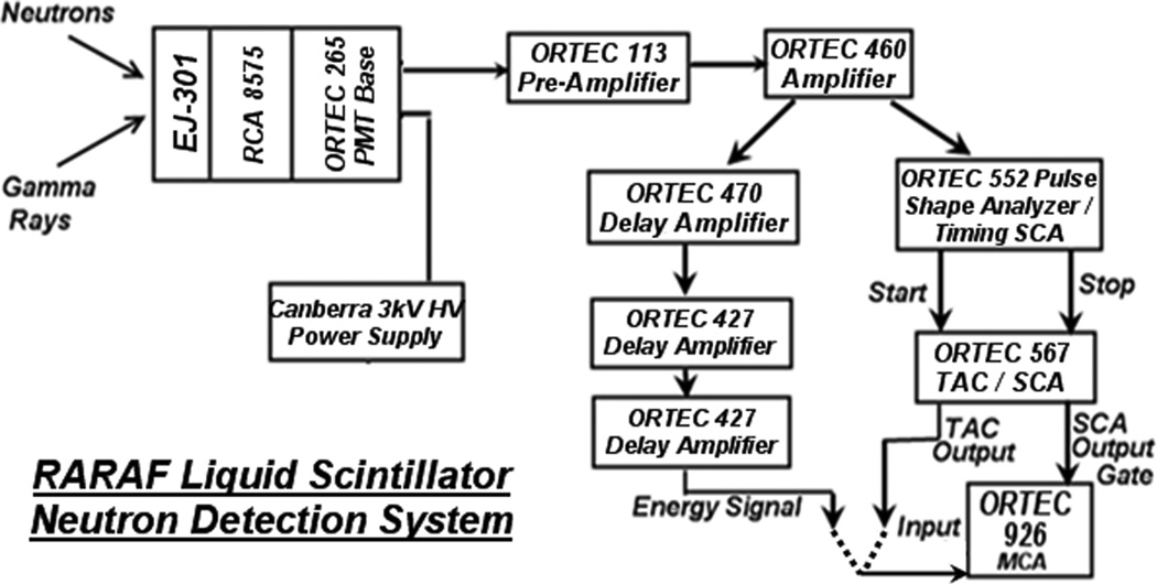
The liquid scintillator chamber was attached to an RCA 8575 photomultiplier, which produces about 1.7 photoelectrons/keV (corresponding to the light generated from the energy deposited in the scintillator) with a pulse rise time of less than 100 nsec. A Pyrex glass window and Dow Corning Q2-3067 optical coupling grease were used to provide optical transmission of the emission light. The measurement data were filtered and recorded by an ORTEC (AMETEK, Inc., Berwyn, PA) 926 Multi-channel Analyzer (MCA). The photomultiplier was powered by a 3 kV, 0.75 A CANBERRA high voltage power supply (CANBERRA Industries Inc., Meriden, CT). The diagram of the data acquisition (DAQ) system including pulse shape discrimination (PSD) is shown in Fig. 1.
Figure 1.
The light pulse height spectra from neutron production were evaluated using different radiation sources. A 10 μCi 22Na source was used for the recoil energy calibration. The operating voltage and the amplifier gains were adjusted with this source to produce a reasonable pulse height range. The MCA was calibrated using the Compton edge (1.06 MeV) of the 22Na source. Because both gamma rays and neutrons produce scintillation light signals, our data analysis used a rise-time discrimination method to separate the pulses from the two types of radiation. This is facilitated by the fact that the two types of radiation produce differently shaped pulses. Pulses from gamma rays have very short rise times while pulses from neutrons have relatively longer rise times.
2.2. Hydrogen-filled proportional counter
A hydrogen gas-filled proportional counter (LND Inc, Oceanside, NY) was chosen for neutron spectroscopy in energy range between 200 keV and 1 MeV. This gas counter is spherical with a diameter of ~2 cm. The shell of this detector is made of stainless steel and the detector can be filled with gas up to 10 atm pressure. In our case, the chamber was filled with about 3 atm of ultra-pure hydrogen (99.99%). A thin wire runs across the diameter of the sphere as the anode. The high voltage is supplied to the anode wire through an SHV connector and the signal is transferred to a low-noise preamplifier through the same connector and extracted by a high pass filter in the preamplifier. A low-intensity 241Am alpha particle source is deposited on the center of the wire for detector self-calibration. In our case (3 atm Hydrogen, 2cm diameter chamber), the internal alpha absorption peak is at about 1 MeV (light output). An operating voltage of up to 3.7 kV can be applied to the detector; in our measurements we used 1.9 kV provided by a Burle stand-alone HV power supply (PHOTONIS USA PENNSYLVANIA, INC., Lancaster, PA). An ORTEC 142PC preamp (AMETEK) and an ORTEC 572 amplifier (AMETEK) were used. The shaping constant was set to 8 μs to give enough time for pulse integration.
3. Quasi-monoenergetic neutron spectra measurements
3.1. Liquid scintillator detector
The EJ-301 liquid scintillator was tested with quasi-monoenergetic neutrons from two types of neutron production reactions to cover the neutron energy range. The detection efficiency for this type of detector is very low below 1 MeV, therefore it is only used for the higher energy end of the spectrum. For neutrons from 1 MeV up to 3.5 MeV, a tritium target was impinged on by a proton beam, resulting in neutrons via the T(p,n)3He reaction. For higher energy ranges up to 8 MeV, a deuterium target and a deuteron beam were used: D(d,n)3He. Both targets consisted of a titanium backing implanted with the appropriate isotope. In both cases, the neutron energies were broadened because of the thicknesses of the targets.
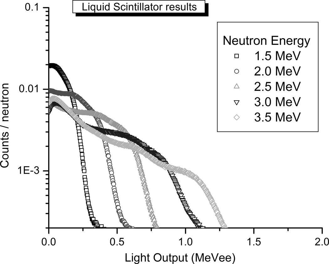
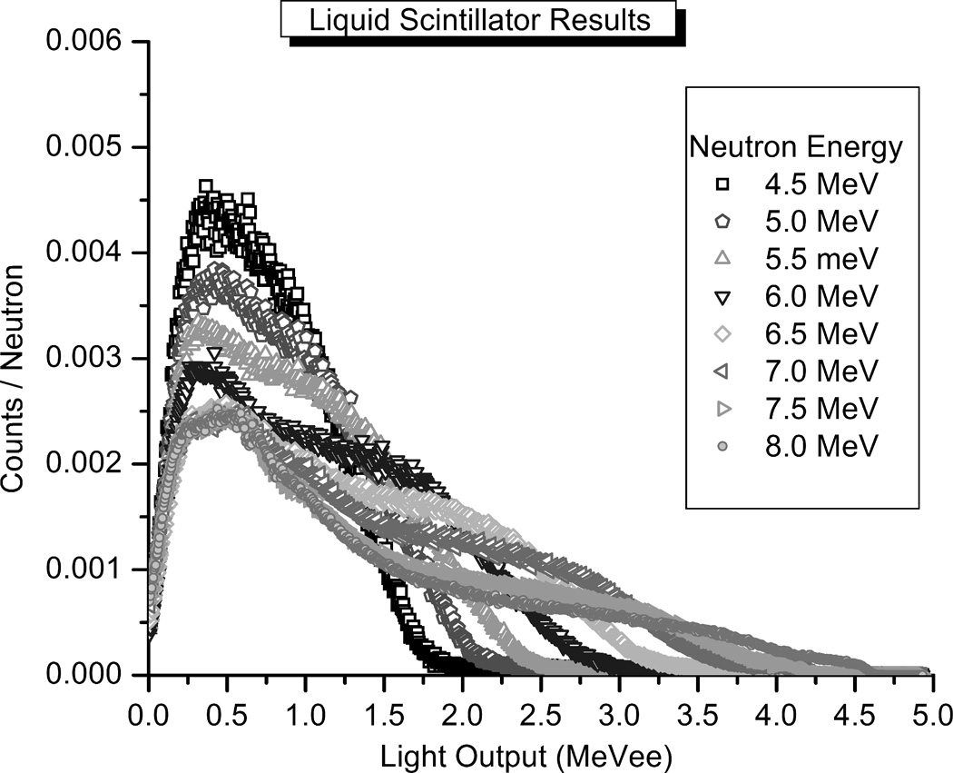
The detector was positioned at either 15 or 70 degrees from the direction of the initial ion beam. Based on the energy of the incident particle beam, and the neutron emission angle relative to the particle beam, the energy that will be observed by our detector can be calculated (Table 1), making this setup useful for detector calibration. The measured pulse height spectra from the T(p,n)3He reaction and D(d,n)3He reaction are given in Figs. 2 and 3. The beam current was monitored using a Keithley 617 electrometer (Keithley Instrument Inc., Cleveland, Ohio) and a custom voltage/frequency (V/F) converter. The recoil spectra were normalized to the total integrated beam charge on the target.
Table 1.
Parameters for quasi-monoenergetic neutron spectra measured using the liquid scintillation detector.
| Particle Energy (MeV) |
Production Reaction | Angle (Deg.) |
Neutron Energy (MeV) |
|---|---|---|---|
| 5.00 | D(d,n)3He | 15 | 8.0 |
| 4.44 | D(d,n)3He | 15 | 7.5 |
| 3.90 | D(d,n)3He | 15 | 7.0 |
| 3.39 | D(d,n)3He | 15 | 6.5 |
| 2.87 | D(d,n)3He | 15 | 6.0 |
| 2.36 | D(d,n)3He | 15 | 5.5 |
| 1.87 | D(d,n)3He | 15 | 5.0 |
| 4.08 | D(d,n)3He | 70 | 4.5 |
| 2.92 | D(d,n)3He | 70 | 4.0 |
| 4.37 | T(p,n)3He | 15 | 3.5 |
| 3.87 | T(p,n)3He | 15 | 3.0 |
| 3.35 | T(p,n)3He | 15 | 2.5 |
| 2.85 | T(p,n)3He | 15 | 2.0 |
| 2.34 | T(p,n)3He | 15 | 1.5 |
| 1.85 | T(p,n)3He | 15 | 1.0 |
| 1.35 | T(p,n)3He | 15 | 0.5 |
Figure 2.
Figure 3.
3.2. Hydrogen gas detector
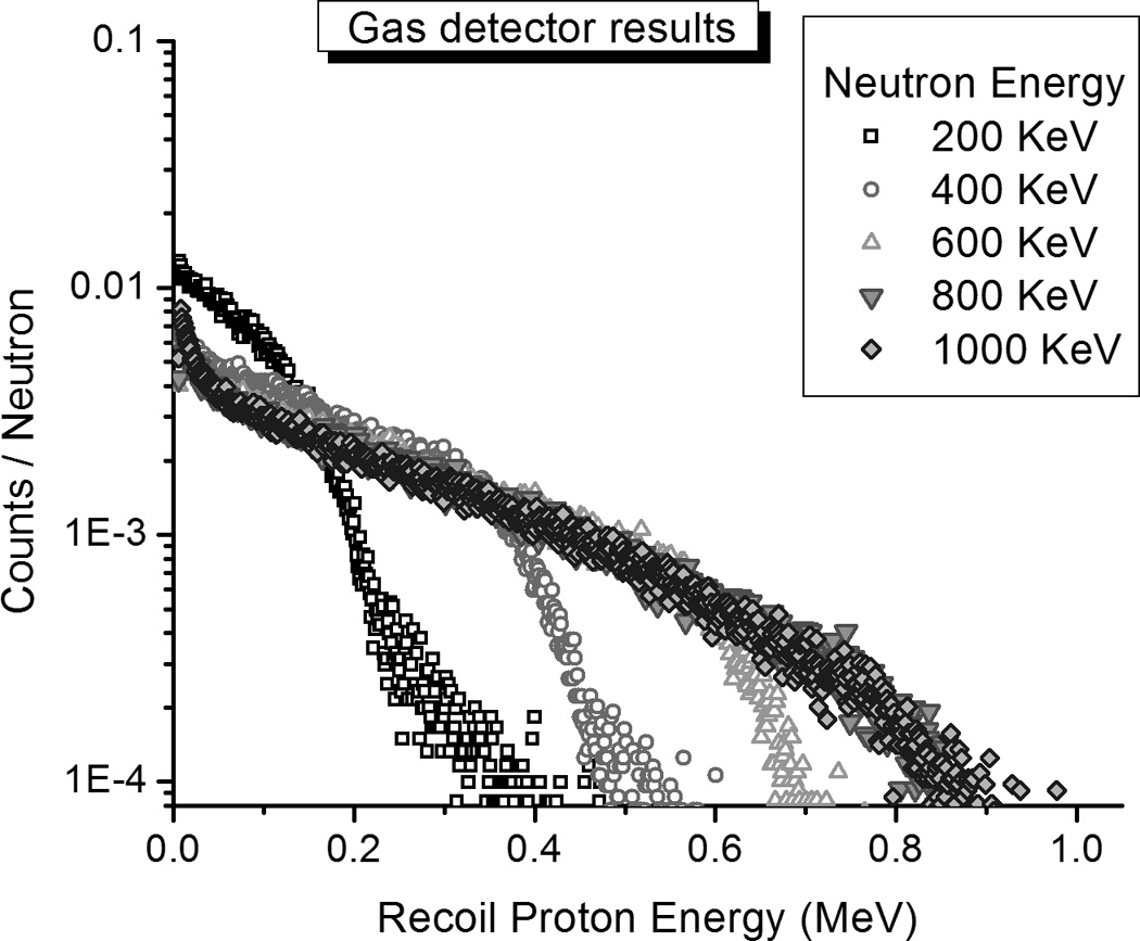
The hydrogen gas-filled detector was used to observe lower energy neutrons from the T(p,n)3He reaction. To reduce neutron energy broadening at small emission angles caused by low-energy protons passing through the relatively thick target (2 mg/cm2 Ti), the measurements were conducted at a large angle (100 degrees), which requires using higher energy protons and therefore less proton energy loss. The beam current monitor and neutron/gamma discrimination system were the same as used for the EJ-301 liquid scintillator measurements. The measured pulse height spectra from the T(p,n)3He reaction are given in Fig. 4.
Figure 4.
3.3. Quasi-monenergetic spectrum unfolding
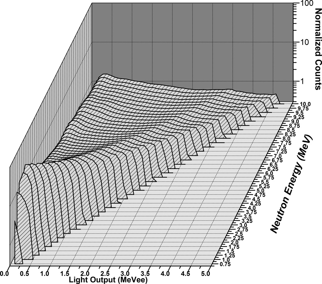
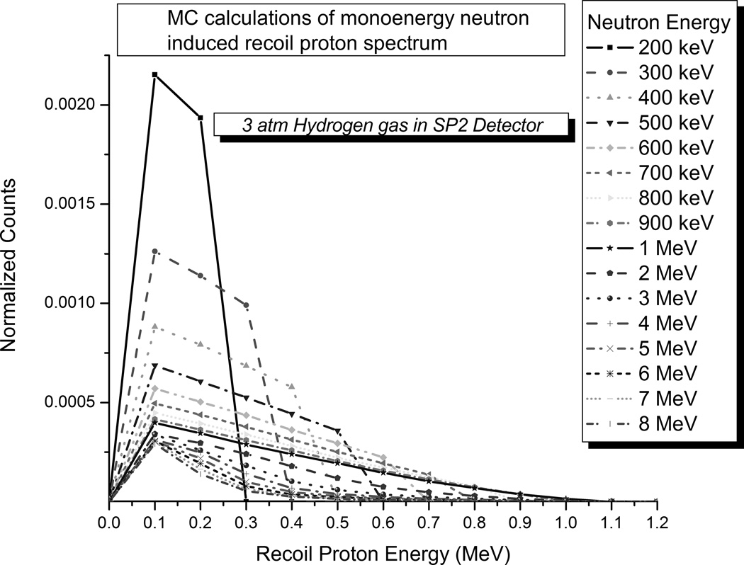
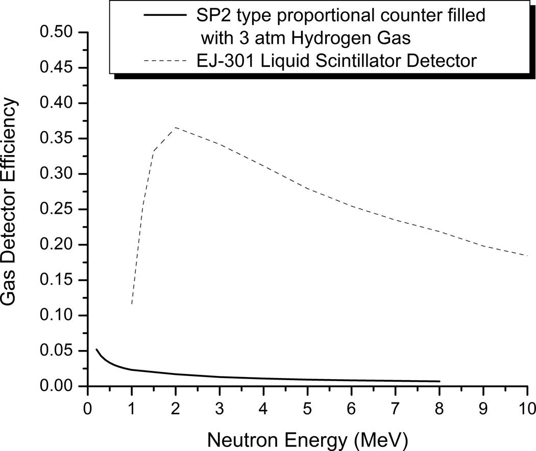
Monte Carlo simulations were used to calculate detector responses to monoenergetic neutrons. The neutron response matrix of the EJ-301 scintillator was obtained with MCNPX-Polimi program [5]. It has a feature with secondary photon production made to correlate to each neutron collision. The simulated responses for the EJ-301 scintillator at 38 quasi-monoenergetic energies are shown in Fig. 5. The neutron response matrix of the 3 atm hydrogen-filled proportional counter was calculated with MCNP6.1/MCNPX [9]. The calculated responses for 16 quasi-mono-energetic energies are shown in Fig. 6. Pulse-height tally was used for obtaining the energy distribution of pulses created in the radiation detectors with an FT8 special tally treatment option as an anticoincidence tally (FT8 PHL). The EJ-301 liquid scintillator detector and 3 atm hydrogen-filled gas proportional counter detection efficiencies also were calculated (Fig. 7).
Figure 5.
Figure 6.
Figure 7.
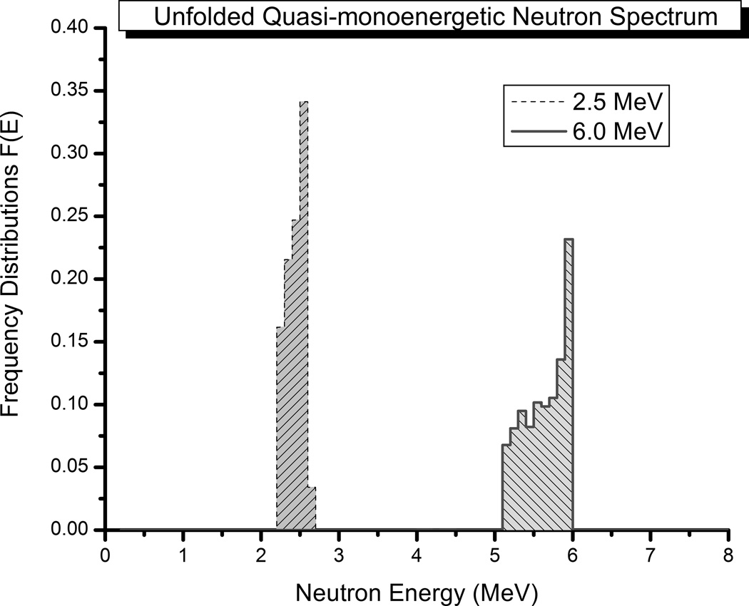
The UMG3.3 unfolding code (MAXED + GRAVEL) [6] provided by the Radiation Safety Information Computational Center (RSICC) at Oak Ridge National Laboratory was used with the calculated mono-energetic neutron response functions. Since it was known in advance that there would be negligible response in the lower channels, these channels were removed from the response matrix, thus reducing the possibility that errors in the response matrix would corrupt the final spectrum, resulting in a more accurate calculation. Two of the mono-energetic proton recoil pulse height spectra (2.5 MeV and 6.0 MeV neutrons) and the unfolded neutron energy spectrum are shown in Fig. 8.
Figure 8.
4. Deuterium neutron generation detection using the liquid scintillator detector
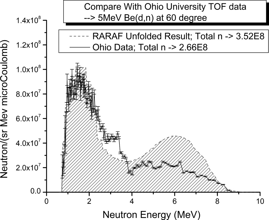
A measurement of the neutron spectrum due to 5 MeV deuterons impinging on a thick beryllium target (9Be(d,n) reaction) was conducted to verify the higher-energy portion of the full neutron spectrum. The liquid scintillation detector was setup vertically at ~20 inches from the center of the Be target and at a 60 degree forward angle to the incident beam which is the same angle for biological sample irradiation [1]. The total detected neutron yield was estimated using the calculated detector efficiency and normalized to MeV/Sr/uC. The unfolded data were compared with time of flight measurement results obtained at Ohio University [1] as seen in Fig. 9. The unfolded RARAF neutron spectrum (especially below 5 MeV) matches the Ohio data. The uncertainty caused by weight or thickness of the foils is about ±10%. A systematic error is about 5%. And the detector efficiency error is estimated about 5%. So the total measured flux has a 20% uncertainty. Although the differences between 6 and 7 MeV are quite large but the total flux is comparable to the Ohio data with the uncertainty estimation.
Figure 9.
5. Spectrum Measurement of RARAF IND-like neutron facility
To measure the full neutron energy spectrum of RARAF IND-like neutron radiation facility, spectroscopy measurements were performed at the sample position: a 60° forward angle relative to the ion beam direction (angle at which the biological samples are irradiated). The liquid scintillator detector was positioned ~34 cm away from the Be target while the gas proportional counter was positioned at ~22 cm due to its lower efficiency and smaller diameter. The ion beam current incident on the target (on the order of 10−7 A) was measured through a long Faraday cup-like isolated section of beam pipe connected to the Keithley 617 electrometer. The voltage output from the electrometer was integrated using a voltage/frequency converter and all spectrum were normalized to the total beam charge on target.
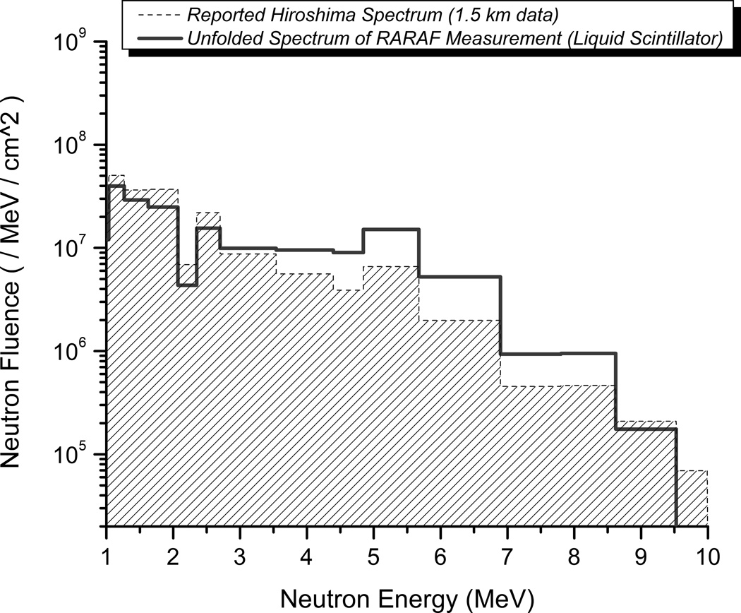
The higher-energy portion of the full energy neutron spectrum was measured with the liquid scintillator using the same setup as the deuterium test. The unfolded spectrum from the EJ-301 scintillator detector with the evaluated Hiroshima Spectrum at 1.5 km [8] is shown in Fig. 10. The spectrum was normalized with the total beam charge deposited on the Be target in units of neutron/MeV/cm2. The total number of neutrons/pC was estimated with the calculated average neutron detection efficiency of the EJ-301 liquid scintillator.
Figure 10.
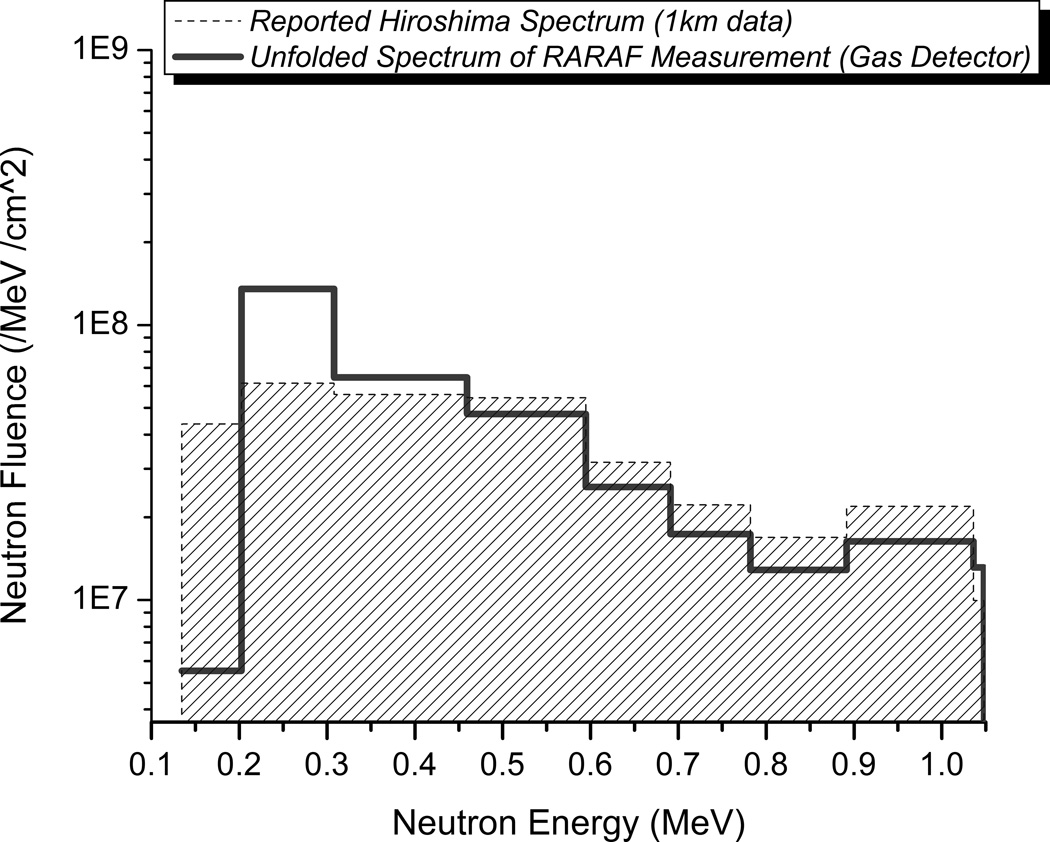
The gas proportional counter filled with 3 atm hydrogen gas was used for the lower-energy neutron spectrum measurements. The calculated gas counter detection efficiency indicates that this counter is good for neutrons with energies <1 MeV. However, when it was placed in the wide-energy range neutron field, the proton recoils from the higher-energy neutrons were still registered, although they were distorted by wall effects (truncated recoil tracks). This means the number of proton recoils detected by the gas counter is, in fact, from all the neutrons generated at the sample positon by the target through the full energy range of neutrons. The total neutron fluence was estimated using the calculated gas detector efficiency and was normalized to the same beam charge on target. Because the number of neutrons from the higher-energy portion of spectrum had been evaluated using the liquid scintillator, the total number of neutrons from the lower-energy portion of spectrum could be extracted. Comparing the number ratio from the scintillator (above 1 MeV) and the gas counter (both above and below 1 MeV). The contribution to the raw data from the higher-energy portion of spectrum, which was calculated and normalized to the number of neutrons, was subtracted. The lower-energy portion of the spectrum was then unfolded using the remaining raw recoil pulse height data, which is only from the low-energy portion of the full neutron spectrum. The result is shown in Fig. 11 in comparison to the Hiroshima data.
Figure 11.
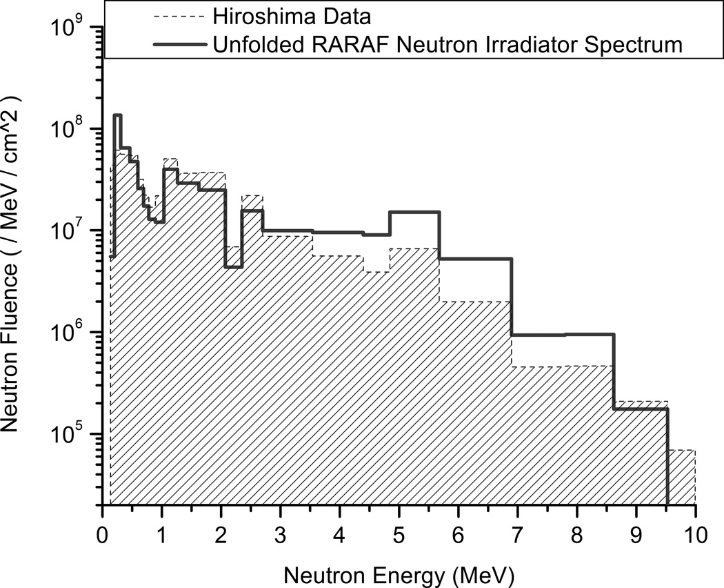
The two portions of the spectrum have been combined to give the full neutron spectrum from the RARAF IND-like neutron irradiator compared to the Hiroshima spectrum in Fig. 12. We have very good agreement between our IND-like irradiator and the Hiroshima neutron spectrum at 1.5 km from the epicenter.
Figure 12.
6. Summary
The RARAF broad-energy neutron irradiator spectrum measurement requires a combined detection method to obtain the low- (200 keV to 1 MeV) and high-energy (1MeV to 9 MeV) portions of the neutron spectrum. Monte Carlo simulations were used to generate response functions for two detectors: an EJ-301 liquid scintillator and a hydrogen gas proportional counter. The detectors were calibrated with quasi-monoenergetic neutrons to verify the detector response functions for unfolding complex spectra. The unfolded monoenergetic neutron spectra match well with the expectations. Furthermore, unfolded liquid scintillator data also fits the Ohio University time of flight measurement neutron spectrum very well for the 9Be(d,n) reaction.
The ongoing development of the Improvised Nuclear Device (IND)-like neutron radiator will experimentally simulate a broad-energy neutron spectrum from an IND (e.g. Hiroshima gun-style bomb), in order to characterize biodosimetry assays [10] for triage of individuals exposed to such a neutron source. In this case, a 5 MeV mixed beam (monatomic and polyatomic deuterons and protons) is used on a thick beryllium target with a ratio of 1:2 (H:D) recreates the Hiroshima neutron spectrum. The dosimetry measurements were performed at the same locations of the sample tubes which have a constant distance of 190 mm from the center of the target. The concrete shielding blocks are at least 1 meter away from the water cooled target [1]. The details of the measurements will be reported elsewhere. The neutron dose rate is ~79% of the total dose rate and the γ-ray dose rate is ~21% of the total dose rate. The detection technology described in this paper verifies and will support further tuning of the neutron spectrum generated for these important biodosimetry studies.
Table 2.
Parameters for quasi-monoenergetic neutron spectra measured using the hydrogen-filled proportional counter.
| Particle Energy (MeV) |
Production Reaction | Angle (Deg.) |
Neutron Energy (keV) |
|---|---|---|---|
| 3.485 | T(p,n)3He | 100 | 1000 |
| 3.025 | T(p,n)3He | 100 | 800 |
| 2.57 | T(p,n)3He | 100 | 600 |
| 2.105 | T(p,n)3He | 100 | 400 |
| 1.65 | T(p,n)3He | 100 | 200 |
Acknowledgments
This work is supported by grant number U19 AI067773 to the Center for High-Throughput Minimally Invasive Radiation Biodosimetry from the National Institute of Allergy and Infectious Diseases / National Institutes of Health. The content is solely the responsibility of the authors and does not necessarily represent the official views of the National Institute of Allergy and Infectious Diseases or the National Institutes of Health.
Footnotes
Publisher's Disclaimer: This is a PDF file of an unedited manuscript that has been accepted for publication. As a service to our customers we are providing this early version of the manuscript. The manuscript will undergo copyediting, typesetting, and review of the resulting proof before it is published in its final citable form. Please note that during the production process errors may be discovered which could affect the content, and all legal disclaimers that apply to the journal pertain.
References
- 1.Xu Y, Garty G, Marino SA, Massey TN, Randers-Pehrson G, Johnson GW, Brenner DJ. Novel neutron sources at the Radiological Research Accelerator Facility. J Instrum. 2012;7:C03031. doi: 10.1088/1748-0221/7/03/C03031. [DOI] [PMC free article] [PubMed] [Google Scholar]
- 2.Homeland Security Council. National Planning Scenarios (Final Version 21.3) Washington DC: 2006. [Google Scholar]
- 3.Klein H. Neutron spectrometry in mixed fields: NE213/BC501A liquid scintillation spectrometers. Radiation Protection Dosimetry. 2003;107:95–109. doi: 10.1093/oxfordjournals.rpd.a006391. [DOI] [PubMed] [Google Scholar]
- 4.Tagziria H, Hansen W. Neutron spectrometry in mixed fields: proportional counter spectrometers. Radiation Protection Dosimetry. 2003;107:73–93. doi: 10.1093/oxfordjournals.rpd.a006389. [DOI] [PubMed] [Google Scholar]
- 5.Pozzi SA, Flaska M, Enqvist A, Pázsit I. Monte Carlo and analytical models of neutron detection with organic scintillation detectors. Nuclear Instruments and Methods in Physics Research Section A: Accelerators, Spectrometers, Detectors and Associated Equipment. 2007;582:629–637. [Google Scholar]
- 6.Reginatto M, Goldhagen P, Neumann S. Spectrum unfolding, sensitivity analysis and propagation of uncertainties with the maximum entropy deconvolution code MAXED. Nuclear Instruments and Methods in Physics Research Section A: Accelerators, Spectrometers, Detectors and Associated Equipment. 2002;476:242–246. doi: 10.1016/s0168-9002(01)01386-9. [DOI] [PubMed] [Google Scholar]
- 7.Verbinski VV, Burrus WR, Love TA, Zobel W, Hill NW, Textor R. Calibration of an organic scintillator for neutron spectrometry. Nuclear Instruments and Methods. 1968;65:8–25. [Google Scholar]
- 8.Egbert SD, Kerr GD, Cullings HM. DS02 fluence spectra for neutrons and gamma rays at Hiroshima and Nagasaki with fluence-to-kerma coefficients and transmission factors for sample measurements. Radiat Environ Biophys. 2007;46:311–325. doi: 10.1007/s00411-007-0120-5. [DOI] [PubMed] [Google Scholar]
- 9.Hendricks JS, McKinney GW, Waters LS, Roberts TL, Egdorf HW, Finch JP, Trellue HR, Pitcher EJ, Mayo DR, Swinhoe MT, Tobin SJ, Durkee JW, Gallmeier FX, Lebenhaft J, Hamilton WB. Los Alamos National Laboratory Report LA-UR-03-2202. Los Alamos, NM: 2003. MCNPX, Version 2.5.c. [Google Scholar]
- 10.Garty G, Chen Y, Turner H, Zhang J, Lyulko OV, Bertucci A, Xu Y, Wang H, Simaan N, Randers-Pehrson G, Yao YL, Brenner DJ. The RABiT: A Rapid Automated BIodosimetry Tool For Radiological Triage II. Technological Developments International Journal of Radiation Biology. 2011;87:776–790. doi: 10.3109/09553002.2011.573612. [DOI] [PMC free article] [PubMed] [Google Scholar]


