Abstract
The transcription factor Runx3 promotes differentiation of naive CD4+ T cells into type-I effector T (TH1) cells at the expense of TH2. TH1 cells as well as CD8+ T cells express a subset-specific Runx3 transcript from a distal promoter, which is necessary for high protein expression. However, all T cell subsets, including naive CD4+ T cells and TH2 cells, express a distinct transcript of Runx3 that is derived from a proximal promoter and that produces functional protein in neurons. Therefore, accumulation of RUNX3 protein generated from the proximal transcript needs to be repressed at the post-transcriptional level to preserve CD4+ T cell capability of differentiating into TH2 cells. Here we show that expression of RUNX3 protein from the proximal Runx3 transcript is blocked at the level of translational initiation in T cells. A coding sequence for the proximal Runx3 mRNA is preceded by a nonoptimal context sequence for translational initiation, known as the Kozak sequence, and thus generates protein at low efficiencies and with multiple alternative translational initiations. Editing the endogenous initiation context to an “optimal” Kozak sequence in a human T cell line resulted in enhanced translation of a single RUNX3 protein derived from the proximal transcript. Furthermore, RUNX3 protein represses transcription from the proximal promoter in T cells. These results suggest that non-permissive expression of RUNX3 protein is restricted at the translational level, and that the repression is further enforced by a transcriptional regulation for maintenance of diverse developmental plasticity of T cells for different effector subsets.
Keywords: T Cells, Transcription Factors, Cell Differentiation, Gene Regulation
Introduction
The RUNX family transcription factors regulate differentiation of various cell types, including T lymphocytes (1). During T cell differentiation, RUNX3 plays important roles in the development and function of CD8+ cytotoxic T cells and type-I CD4+ (TH1) effector cells (2–8). RUNX3 directs the differentiation of MHC class I-selected thymocytes into the CD8+ T cell lineage via repression of ThPOK (9). RUNX3 activates expression of effector genes, Ifng, Eomes, and Gzmb and directly represses Il4 (2, 6, 10, 11). Consistent with these requirements, high RUNX3 protein expression is detected specifically in CD8+ T cells and TH1 cells, but RUNX3 is absent or expressed at low levels in their counterparts, naive CD4+ T cells and TH2 cells (2, 3, 6, 7, 12).
Although RUNX3 protein is expressed at high levels specifically in CD8+ T cells and TH1 cells, expression of its mRNA is broadly detected in developing thymocytes and T cells (3, 8). Runx3 mRNA is transcribed from two distinct promoters (proximal and distal) (13). The Runx3 isoform transcribed from the distal promoter (Runx3d) is required and sufficient for prominent expression of RUNX3 protein in CD8+ T cells and TH1 cells (3, 12). In postselection thymocytes and naive CD4+ T cells, Runx3d is negatively regulated by ThPOK, a crucial lineage commitment factor for the helper T cell lineage (12, 14, 15), resulting in specific expression of Runx3d in CD8+ T cells. In TH1 cells, Runx3d is upregulated in a T-BET-dependent manner (2), although it remains unknown how T-BET counteracts ThPOK-mediated repression of Runx3d. In addition to Runx3d, an isoform derived from the proximal promoter (Runx3p) is expressed in all subsets of developing thymocytes, naive CD4+ T cells and TH2 cells although protein expression derived from this transcript is substantially lower than that from Runx3d (3, 8). RUNX3 protein is also expressed in neural tissues and is essential for the development of TrkC+ proprioceptive neurons in dorsal root ganglia (DRG) (16, 17). Although germline deletion of the Runx3 gene results in perinatal lethality or growth retardation due to defective axonal projection of proprioceptive neurons, mice specifically lacking Runx3d show normal survival without neurological symptoms (12, 17, 18). These findings indicate that functional RUNX3 protein is expressed from Runx3p in TrkC+ proprioceptor neurons in DRG. Therefore, it remains unclear how RUNX3 protein expression from the proximal transcript is restricted in lymphocytes, although it is expressed at a requisite level in neurons. Since ectopic expression of an open reading frame of Runx3p in transgenic mice or retrovirally transduced cells allows T cells to express RUNX3p protein (3, 4), the low protein expression is caused by translational regulation rather than regulation via degradation of translated protein.
Ectopic expression of RUNX3 results in downregulation of CD4 in pre-selection CD4+ CD8+ double positive thymocytes and their subsequent development into the CD4+ T cell lineage due to inhibition of positive selection of MHC class II-restricted thymocytes (3, 4, 19). Furthermore, expression of RUNX3 protein in unpolarized CD4+ T cells results in skewed differentiation towards the IFN-γ+ TH1 lineage at the expense of TH2 differentiation (2, 5, 6). Therefore, expression of RUNX3 protein from Runx3p needs to be restricted in thymocytes and activated CD4+ T cells to assure normal development of CD4+ mature thymocytes and TH2 cells.
By using cell-number normalized quantitation of Runx3 transcript levels and measurement of translational efficiency, we show that translation of Runx3p is restricted due to lack of an efficient Kozak sequence for translational initiation. Introduction of an optimal Kozak sequence into the endogenous locus by genome editing enhanced the translation efficiency of RUNX3 in a human T cell line. Compared to T cells, DRG neurons express a substantially higher level of Runx3p and thereby achieve the requisite level of RUNX3 protein. These results provide a unique mechanism by which transcription factor expression is regulated between different cell types.
Materials and Methods
Mice
Runx3dYFP and Runx3-flox (Runx3F) alleles were described (6, 12) and have been maintained in the C57BL6 background. The Runx3F allele was crossed to Vav1-iCre (20) to delete Runx3 in all hematopoietic cells. C57BL6 mice were purchased from National Cancer Institute and Charles River, and used as control. All mice were maintained in a specific pathogen-free facility at Washington University in St. Louis, and all experiments were performed according to a protocol approved by Washington University's Animal Studies Committee.
Flow cytometry
Single-cell suspensions were prepared by mechanical disruption of spleens. The following monoclonal antibodies were purchased from Biolgend: PerCP-Cy5.5- or APC-conjugated anti-CD4 (RM4-5), PE-conjugated anti-CD25 (PC61), PE-Cy7-conjugated anti-CD8alpha (53-6.7), Pacific Blue-conjugated anti-CD44 (IM7), APC-conjugated anti-CD62L (MEL-14), FITC- or Alexa Fluor 647–conjugated anti-B220 (RA3-6B2) and PE-conjugated anti-IFN-γ (XMG1.2). For sorting of naive T cells, splenocyte samples were initially depleted of B220+ cells with Dynabeads Mouse Pan B (B220) magnetic beads (Life Technologies), stained with monoclonal antibodies, and sorted as CD62L+CD44− CD4−CD8+ naive CD8+ T cells or CD62L+CD44−CD25−CD8−CD4+ naive CD4+ T cells on a FACSAria II (BD Biosciences). Purity of sorted populations was 95–99%. For intracellular cytokine staining, cells were restimulated for 5 h with 50 ng/ml of phorbol 12-myristate 13-acetate (PMA, Sigma) and 500 ng/ml of ionomycin (Sigma) in the presence of brefeldin A (5 µg/ml, Biolegend), fixed with 2% paraformaldehyde, made permeable with 0.03% saponin and stained for IFN-γ in 0.3% saponin. Dead cells were excluded with staining with DAPI (Sigma) or Aqua LIVE/DEAD staining dye (Life Technology). Data were acquired with LSR Fortessa (BD Biosciences) and analyzed using Flowjo (Treestar).
In vitro stimulation of T cells
Naive T cells were cultured in RPMI medium supplemented with 10% FBS (Life Technologies) and 50 µM 2-mercaptoethanol in the presence of soluble anti-CD3 (0.1 µg/ml, 145-2C11; Biolegend) and anti-CD28 (1 µg/ml, 37.51; BioXCell) in multiwell tissue culture plates coated with rabbit antibody to hamster IgG (0855395; MP Biomedicals). CD4+ T cells were polarized to TH1 or TH2 as follows: for TH1 polarization, 20 ng/ml of murine IL-12 (R&D) and 5 µg/ml anti-IL-4 (11B11, Biolegend); for TH2 polarization, 20 ng/ml of murine IL-4 (eBioscience), 2 µg/ml anti-IL-12/23 p40 (C17.8, Biolegend) and anti-IFN-γ (XMG1.2, Biolgend). For non-polarizing culture (TH0) of CD4+ T cells and CD8+ T cells, no additional cytokines or antibodies added. Following culture for 3 days with TCR stimulation and polarization, the activated cells were rested for 3 days in the presence of IL-2 (25 U/ml, eBioscience). For retroviral transduction, viral supernatants were prepared by transfection of PlatE packaging cells (21) with TransIT 293 (Mirus Bio). After priming overnight under TH0 conditions, activated T cells were transduced by spin infection at 1,200 × g at 30 °C for 90 min in the presence of 10 µg/mL polybrene (Sigma).
RNA isolation and real-time quantitative RT-PCR
Cellular RNA was extracted with Trizol (Life Technologies) and reverse-transcribed with qScript Supermix (Quanta Bio). DyNAmo ColorFlash SYBR green qPCR mix (Thermofisher) and a LightCycler 480 (Roche) were used for real-time quantitative RT-PCR. Quantities of transcripts were normalized to that of Gapdh mRNA unless specified otherwise. For quantification of cell-number normalized gene expression, 1 µl of 1:1,000 dilution of ERCC (External RNA Controls Consortium) RNA Spike-In Control Mixes (Ambion) was added to cell lysates from 2 × 10⁵ cells in Trizol before RNA isolation and reverse transcription. The following primers were used: ERCC-00108, CTATCAGCTT GCGCCTATTAT and GTTGAGTCCACGGGATAGAGTC; Gapdh, CTCACAATTT CCATCCCAGACC and CATCAATGGTGCAGCGAACTTTATTG ATG; total Runx3, AGTGGGCGAGGGAAG AGTTTC and GCCTTGGTCTGGTCTTC TATCT; distal Runx3, CAAAACAGCAGCC AACCAAGT and AGATGCTGTTGG AAGCCATGT; proximal Runx3, CGTATTCCC GTAGACCCGAG and AGGGGAAGG CCGTGGAG; firefly luciferase, CGCAGGTGT CGCAGGTCTTC and CCGTCATCGTCTTTCCGT GCTC; human GAPDH, CCAGCA AGAGCACAAGAGGAAGAG and AGGAGGGGA GATTCAGTGTGGTG; human RUNX3, CTCAGCACCACAAGCCACTT and GGGTC GGAGAATGGGTTCAG; human RUNX3 3’UTR, TCAAGACCAGTGATGGGCCG and GGAGCGCAGGTCCCATTC; human RUNX3p, TATTCCCGTAGACCCAAGC ACC and CCGGGGAGGGAGGTGTGA.
Immunoblot analysis
Cell extracts were lysed cells for 10min at 4 °C in RIPA buffer (50 mM Tris-HCl (pH 8.0), 150 mM NaCl, 1% NP-40, 0.5% sodium deoxycholate and 0.1% SDS (sodium dodecyl sulfate)) with a protease inhibitor cocktail (P8340, Sigma). Lysates were cleared by centrifugation at 21,000 × g for 10 min at 4 °C. Lysates from equal numbers of cells were separated by 8% SDS PAGE and transferred to PVDF membranes (GE Healthcare). The membranes were incubated with primary antibodies (identified below), followed by detection with a horseradish peroxidase–conjugated antibody against rabbit immunoglobulin light chain (211-032-171; Jackson ImmunoResearch) and a Luminata HRP substrate (Millipore). Rabbit anti-RUNX3 monoclonal antibody (#9647, clone D6E2), which recognizes a peptide containing Alanine at position 40 of human RUNX3, and rabbit anti-HDAC1 (ab7028) polyclonal antibody were purchased from Cell Signaling Technology and Abcam, respectively. Protein expression was quantitated with an ImageJ program (NIH).
Plasmid construction and luciferase assay
A coding sequence of firefly luciferase ligated with a 5’ UTR and/or Kozak sequences of Runx3d or Runx3p was inserted into a murine stem cell virus (MSCV)-based retroviral backbone containing ires EGFP. The 1200M thymoma cells (22) were spin-infected as described above and were harvested for analyses 48 h after infection. The translational efficiency of the luciferase was assessed by calculating ratios of luciferase activity to a quantity of the luciferase transcript determined by quantitative RT-PCR.
Genome editing using a CRISPR/Cas9 system
A guide RNA targeting the sequence around the canonical initiation codon of RUNX3p (5’- TACGGGAATACGCATAACAG -3’) was designed using the CRISPR design tool (23), and inserted into pQCiG (24). A single strand oligonucleotide (ssODN), which contains gccaccATGGC flanked by 60 nucleotide homology sequences on both sides was synthesized (Sigma) and used as a homology directed repair template. 5 × 10⁵ of Jurkat cells in 10 µl of R buffer (supplied as part of the Neon Transfection System, Life Technologies), were mixed with 1 µg of pQCiG containing the guide RNA and 20 pmol of ssODN, and electroporated using the Neon Transfection System (Life Technologies) under the following conditions: pulse voltage, 1,400 V; pulse width, 10 msec; and pulse number, 3. GFP-positive cells were single cell sorted two days after electroporation. Correctly edited clones were screened for by genomic PCR using primers (TTCTGCTTTCCCGCTTCTCGCGGCAG and AGCACGTCCACCATCGAGCGCA CCTC) located outside of the homology regions, followed by NcoI digestion.
Statistical analysis
P values were calculated with an unpaired two-tailed Student's t-test for two-group comparisons and by one-way ANOVA for multiple-group comparisons with the Tukey post-hoc test in Prism 6 software (Graphpad). P values of <0.05 were considered significant.
Results
Endogenous Runx3p is inefficiently translated into protein
To determine whether the cell type-specific expression of RUNX3 is regulated at a translation level, we measured protein to transcript ratios by quantitating cell number-normalized levels of RUNX3 protein and transcripts. As a control, we used cells from Runx3dYFP/YFP mice, in which the first exon for Runx3d was replaced with an EYFP reporter followed by a polyadenylation sequence (12). As a result, all Runx3 protein in cells from these animals is derived exclusively from Runx3p.
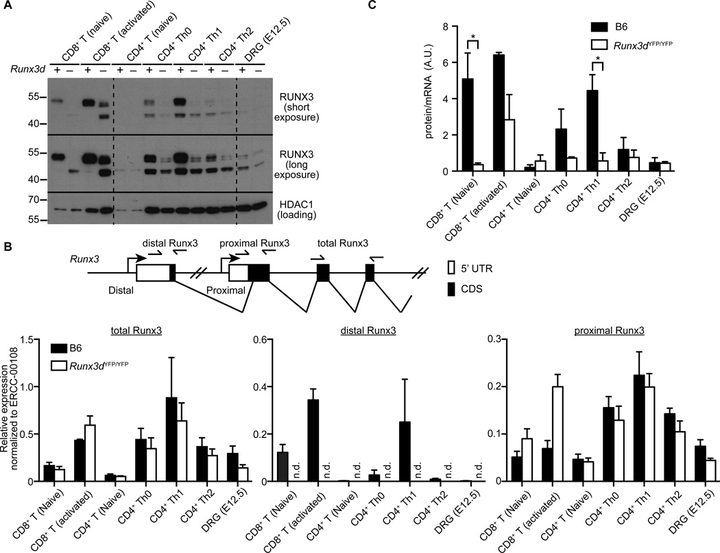
As previously reported (3, 7), expression of full-length 48 kDa RUNX3 protein was detected at high levels specifically in CD8+ T cells and TH1 cells (Fig. 1A). Expression of the 48 kDa product was also detected at an intermediate level in TH0 cells, whereas its expression in TH2 cells was substantially lower compared to other subsets. Expression of 48 kDa RUNX3 was severely reduced in CD8+ T cells, TH0 and TH1 cells derived from Runx3dYFP/YFP mice, indicating that a large proportion of this full-length product is derived from Runx3d, as previously demonstrated (12). In addition, all WT CD4+ T cell subsets expressed a form of RUNX3 protein that was approximately 42 kDa in its molecular weight, as well as two additional 46–48 kDa isoforms, both at substantially lower amounts compared to the 42 kDa isoform. Since CD8+ T cells lacking Runx3d also expressed RUNX3 proteins of similar molecular weights, the observed products must be derived from Runx3p.
Figure 1. Translation of RUNX3 protein from the endogenous proximal Runx3 transcript is inefficient.
(A) Immunoblot analysis of RUNX3 protein in CD8+ T cells, CD4+ T cells, and DRG from wild-type (+) and Runx3dYFP/YFP (−) mice. Lysates of 1 × 10⁵ cells for indicated populations were loaded per lane and expression of RUNX3 (top) and HDAC1 (bottom, loading control) was detected. Data shown are representative of three experiments. (B) qRT-PCR quantitation of expression of total and promoter-specific Runx3 transcripts. cDNA from indicated cell subsets was subjected to qRT-PCR using primers indicated in the schematic (top). Levels of expression were normalized to those of 'spiked-in' control RNA added in proportion to cell numbers prior to RNA preparation. Data are shown as mean and SD from three independent samples. n.d.: not detectable. (C) Ratios between RUNX3 protein and total Runx3 mRNA presented as a surrogate for translation efficiency in cells shown in (A) and (B). Values were calculated by dividing density of total Runx3 protein bands in (A) by levels of the total Runx3 transcript in (B) determined by qRT-PCR. Data are shown as mean and SD from three independent samples. *P < 0.05.
Levels of total Runx3 transcript, as normalized to fixed cell numbers using exogenous control RNA, were similar among the subsets of activated T cells or between naive CD4+ and CD8+ T cells (Fig. 1B). High RUNX3 protein levels correlated with elevated expression of Runx3d and with T cell activation, while levels of the 42 kDa product was linked to Runx3p expression. Ratios between steady-state levels of RUNX3 protein and total Runx3 transcripts in WT or Runx3dYFP/YFP CD8+ T cells suggested a low translation efficiency of Runx3p in lymphocytes (Fig. 1C). RUNX3 protein was expressed in pooled DRG neurons predominantly from Runx3p and the protein to transcript ratio was similar to that of TH2 cells (Fig. 1A and C), even though only 10–15% of DRG neurons express Runx3 at an mRNA or protein level (25). Therefore, a subset of DRG neurons appeared to express Runx3p mRNA at substantially higher levels than T cells to achieve high RUNX3 protein expression. These results collectively suggest that translation of Runx3p is substantially less efficient compared to that of Runx3d.
An inefficient Kozak sequence contributes to reduced translation of Runx3p
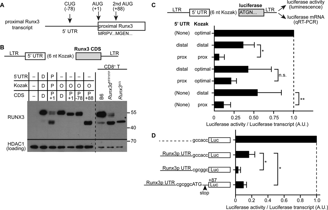
Runx3p and Runx3d transcripts have distinct 5’ UTRs and sequence contexts for translation initiation (Kozak sequences (26)). To determine whether the 5’UTR, Kozak sequences, or both, contribute to translational restriction of Runx3, we first generated retroviral constructs, in which a Runx3 coding sequence (CDS) was inserted downstream of the corresponding sequences from the Runx3d or Runx3p transcripts. These expression vectors were introduced into a thymoma cell, 1200M, which expresses only Runx3p and little endogenous Runx3 protein (Fig. 2A, B). The Runx3d construct was translated into a single 48 kDa protein. By contrast, Runx3p was translated into three proteins when expressed from a construct with its own 5’ UTR and Kozak sequence, recapitulating endogenous protein expression from Runx3p (Fig. 2B). Based on sequence predictions, translation of the Runx3p transcript could be initiated at two alternative nucleotide positions, −78 and +88, in addition to the canonical AUG (+1) that generates a 409 amino acid RUNX3 protein starting with an MPIPV sequence (Fig. 2A). The molecular weights of each product expressed from Runx3p corresponded to isoforms initiated at −78 (referred to as Runx3p (−26–409)), +1 and +88 (Runx3p (+30–409)) positions (Fig. 2B). Furthermore, consistent with endogenous protein patterns, the amounts of RUNX3 expressed from the Runx3p retroviral construct harboring its 5’UTR and Kozak sequence were lower than those of the Runx3d construct and Runx3p expression constructs with an optimal Kozak sequence (Fig. 2B). These data suggest that the restricted translation of Runx3p results from impaired translational initiation by 5’ UTR or Kozak sequence, which causes translational initiation at two additional alternative sites and reduced overall translation.
Figure 2. Protein expression of Runx3p is restricted by translational initiation.
(A) Schematic representation of alternative translational initiation sites of the Runx3p transcript. Numbers indicate positions (nucleotide) relative to the canonical AUG (+1) of Runx3p. (B) Immunoblot analysis of Runx3 proteins expressed from retroviral constructs with or without their 5’UTRs and/or initiation context sequences (Kozak) in 1200M thymoma cell line (left). Lysates of CD8+ T cells from wild-type (B6), Runx3dYFP/YFP and Runx3f/f: Vav1-iCre (Runx3Δ/Δ) mice were used as control (right). CDS: coding sequence, D: distal, P: proximal, O: optimal Kozak sequence (gccacc), *: truncated RUNX3 protein from the Runx3Δ allele. Data are representative of two experiments. (C) Translational efficiency of a luciferase reporter retrovirally expressed with or without 5’UTR with an endogenous or optimal Kozak sequence in 1200M cells. (D) Translational efficiency of a luciferase reporter placed at the alternative initiation site at +87 in retrovirally transduced 1200M cells. Translational efficiencies in (C) and (D) were calculated by luciferase activities normalized by the luciferase mRNA levels determined by qRT-PCR. Data are normalize to a value of a control construct without a 5’UTR and with an optimal Kozak sequence and shown by mean and SD from three experiments. *P < 0.01 and **P < 0.0001.
To quantitatively assess contribution of the Runx3 5’UTR and Kozak sequences in translation in T cells, we generated retroviral luciferase reporter constructs, containing a 5’ UTR and a Kozak sequence from Runx3d or Runx3p (Fig. 2C). The translational efficiency for luciferase transcripts was assessed by calculating ratios of luciferase activity to its transcript as determined by quantitative RT-PCR of transduced cells (27). Consistent with our experiments using the Runx3 CDS, the translational efficiency of luciferase was reduced by 5-fold when luciferase was expressed in the presence of the 5’UTR and Kozak sequence from Runx3p compared to those from Runx3d (Fig. 2C). Although we observed a trend for reduced translation efficiency of luciferase with the Runx3p 5’UTR alone compared to the Runx3d 5’UTR, this property was significantly reduced when the proximal rather than the distal Kozak sequence was employed (Fig. 2C). Furthermore, luciferase translation efficiency was also reduced in the context of Runx3p (+30–409) compared to that of the Runx3d 5’UTR and Kozak sequence (Fig. 2D), suggesting that translation of Runx3p from all three initiation sites was less efficient compared to that of Runx3d. These results collectively indicate that protein expression from Runx3p is compromised due to inefficient translational initiation.
Low translation efficiency and alternative initiation of Runx3p are resolved by editing the endogenous Kozak sequence
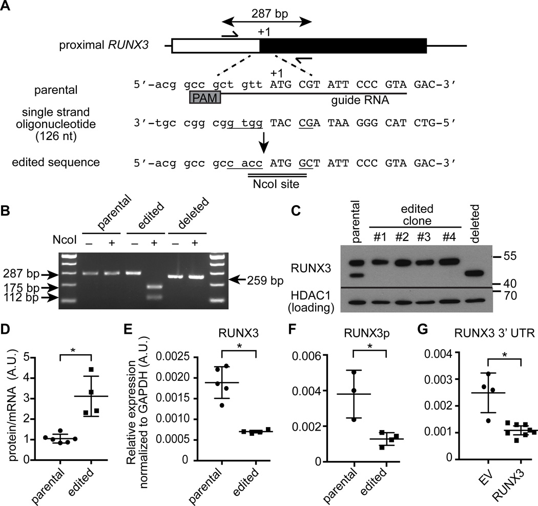
To determine whether the ineffcient Kozak sequence of Runx3p is the primary determinant of low translation efficiency and alternative initiation, we used the CRISPR/Cas9 system to edit this proximal element in Jurkat cells (Fig. 3A). We isolated clones with bi-allelic editing of the initiation context sequence into an optimal Kozak sequence (“edited”) and a clone with deletion of a 28-bp sequence containing the putative translation initiation site and a branch point for splicing of RUNX3d (“deleted”), as confirmed by restriction enzyme digestion of PCR products and DNA sequencing (Fig. 3B, and data not shown). In Jurkat cells, the endogenous Runx3 was expressed as proteins with two distinct molecular weights, 48 kDa and 42 kDa (Fig. 3C). While translation of RUNX3p is potentially initiated at an alternative CUG codon located −167 nucleotides upstream of the canonical AUG, this alternative product would be terminated by a stop codon after being translated into a 12 amino acid peptide even if it is expressed (data not shown). The deleted clone, therefore, served as a control for the 42 kDa isoform since RUNX3 protein could be expressed only from the alternative initiation site downstream of the canonical AUG of the RUNX3p transcript. In all four clones containing the edited Kozak sequence, RUNX3 protein was expressed exclusively as a 48 kDa protein (Fig. 3C), indicating that alternative translational initiation resulted from an inefficient Kozak sequence preceding the RUNX3p coding sequence, which may be poorly recognized by the initiation complex. The edited Kozak sequence increased the protein to mRNA ratios, surrogates for translational efficiency, by 3-fold (Fig. 3D). These data suggest that the low translational efficiency of RUNX3p resulted, at least in part, from the inefficient Kozak sequence upstream of its putative initiation codon. Contrary to our prediction, however, the levels of RUNX3p protein did not change substantially between the parental cells and the edited clones even though RUNX3p was expressed as a single protein from the edited Kozak sequence (Fig. 3C). Instead, levels of both total RUNX3 mRNA and RUNX3p in the edited Jurkat clones decreased by approximately 3-fold (Fig. 3E, F). Consistent with this observation, levels of endogenous RUNX3 mRNA in Jurkat cells transduced with a RUNX3-expressing retrovirus were also reduced by 2.5 to 3-fold (Fig. 3G). These findings also suggest that expression of RUNX3 protein derived from RUNX3p is tightly regulated by an additional feedback mechanism at the mRNA level.
Figure 3. Editing the translational initiation context of Runx3p increases translation efficiency and prevents alternative initiation.
(A) Strategy to edit the Kozak sequence of RUNX3p using the CRISPR/Cas9 technology in Jurkat cells. Locations of the primers used in (B), the guide RNA target sequence and the protospacer adjacent motif (PAM) and a repair template sequence are shown. (B) Detection of homology-mediated repair by an NcoI digestion of the PCR product encompassing the targeted sequence. (C) Immunoblot analysis and quantitative RT-PCR analysis of RUNX3 in parental Jurkat cells and edited clones. A clone with a deletion of a sequence including the canonical ATG was used as a negative control for RUNX3p (1–409). (D–F) RUNX3 protein to transcript ratios (D) and RT-PCR analysis of the total RUNX3 (E) and RUNX3p transcripts (F) in parental Jurkat cells and edited clones. (G) RT-PCR analysis of endogenous RUNX3 expression in Jurkat cells infected with an empty or RUNX3 retrovirus. Data are shown by mean and SD from two experiments. *P < 0.001.
Runx3 isoforms with alternative translational initiations are functionally intact
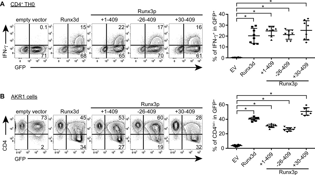
To test the functionality of the RUNX3 isoforms translated from Runx3p, a CDS corresponding to each was placed downstream of the optimized Kozak element and retrovirally expressed in primary CD4+ T cells (Fig. 4A). Retroviral expression of Runx3d in CD4+ T cells cultured under neutral TH0 conditions increased the percentage of IFN-γ+ cells compared to empty virus-infected control. CD4+ T cells expressing each of the three Runx3p-derived isoforms also expressed IFN-γ at frequencies comparable to those of Runx3d expressing CD4+ T cells. Next, to test the repressor activity associated with each Runx3 isoform, we retrovirally expressed these proteins in the AKR1 CD4+ CD8+ thymoma cell line and assessed downregulation of CD4 expression. As shown in Fig. 4B, Runx3d and all three alternatively initiated Runx3p isoforms induced CD4 downregulation with comparable efficiency. These results suggest that differences in protein sequences at the N-terminus of RUNX3 had little effect on their functionality and that distinct contribution to gene regulation between Runx3d and Runx3p transcripts is primarily dependent on their expression levels.
Figure 4. Alternatively initiated Runx3 isoforms are functionally intact.
(A) Intracellular staining for IFN-γ in CD4+ T cells transduced with retrovirus expressing each Runx3 isoform after 6 days of culture under neutral conditions. Percentages of GFP+ IFN-γ+ and GFP+ IFN-γ− cells in viable cells, as determined by LIVE/DEAD Aqua staining are shown in representative plots from two independent experiments with more than two biological replicates each. Statistical analysis of percentages of IFN-γ+ cells in GFP+ cells is shown in the right panel with mean and SD. (B) Surface CD4 staining of the AKR1 thymoma cell line transduced with retrovirus expressing each Runx3 isoform at 72 hours after infection. Percentages of GFP+ CD4+ and GFP+ CD4lo/− cells in DAPI-negative viable cells are shown in representative plots from two independent experiments. Statistical analysis of percentages of CD4lo/− cells in GFP+ cells is shown on the right panel with mean and SD. *P < 0.0001.
Discussion
Regulation of RUNX3 protein levels is critical to CD4 versus CD8 and TH1 versus TH2 lineage decisions. Whereas high RUNX3 protein expression is essential for the development and function of CD8+ T cells and TH1 differentiation (2, 3, 5–8, 10–12), expression of RUNX3 protein needs to be restricted during CD4+ T cell development and TH2 differentiation despite constitutive expression of Runx3 mRNA. Runx3 is expressed as two major transcripts originating from distinct promoter regions (3, 17). As we previously demonstrated, the high RUNX3 expression is dependent on transcription of Runx3d, which is efficiently translated into protein (12). By contrast, naive CD4+ T cells and TH2 cells express only Runx3p (3, 6), which is translated inefficiently. Our data suggested that the initiation context sequence of Runx3p contributes to low protein translation. Editing the endogenous Kozak sequence in a T cell line resulted in increased translation efficiency from the Runx3p transcript. Furthermore, levels of the Runx3p transcript are negatively regulated directly or indirectly by RUNX3 protein, which further provides tighter regulation of RUNX3 levels in these cell types. Even though the protein to transcript ratio increased by editing the Kozak sequence, levels of both RUNX3p and total RUNX3 mRNA level were reduced, resulting in a modest change in the RUNX3 protein level. These results suggest that transcription of the proximal message is also regulated by a negative feedback loop, which keeps RUNX3 protein levels sufficiently low to maintain T cell plasticity for non-TH1 lineages. This interpretation is also consistent with increased Runx3p in the presence of reduced RUNX3 protein in Runx3dYFP/YFP CD8+ T cells and the decreased RUNX3 transcript in Jurkat cells expressing retroviral RUNX3. RUNX3 binds to a few regions in the Runx3 locus based on ChIP-seq analysis using CD8+ T cells (28). However, all binding sites were located in the distal promoter and its upstream region, while there is no binding site near the proximal promoter or downstream region. Therefore, it seems likely that the feedback repression of the proximal transcript is mediated through indirect mechanisms. However, we cannot rule out the possibility that reduced RUNX3p levels in the presence of RUNX3 protein result from cleavage or degradation of the transcript rather than diminished transcription. In contrast to T cells, DRG neurons express requisite levels of RUNX3 protein from the proximal transcript. This seems to be achieved by substantially higher levels of Runx3p in TrkC+ DRG neurons. Amounts of the Runx3p transcript may thus be regulated differently in neurons from lymphocytes possibly independent of the negative feedback mechanism.
In summary, the results from this study reveal a unique mechanism by which expression of a transcription factor is regulated at the post-transcriptional level and provide insights into lineage- or cell type-specific transcription factor expression.
Acknowledgements
We thank Chun Chou and Kenneth M. Murphy for discussion, Sunnie Hsiung for technical assistance, Dan R. Littman (New York University) and Jerry Pelletier (McGill University) for providing reagents, and Chyi-song Hsieh and Eugene M. Oltz for critical reading of the manuscript.
This work was supported by a National Institutes of Health grant AI097244 and a grant from the Mallinckrodt Jr. Foundation to T.E. B.K. was supported in part by a fellowship from the National Research Foundation of Korea (2013R1A6A3A03058430).
Abbreviations
- CDS
coding sequence
- DRG
dorsal root ganglion
- Runx3d
distal Runx3 transcript
- Runx3p
proximal Runx3 transcript
- UTR
untranslated region
Footnotes
The authors declare no conflict of interest.
Authors Contribution
B.K. and T.E. designed research. B.K. and Y.S. performed research. B.K. and T.E. analyzed data. T.E. wrote the paper.
References
- 1.Collins A, Littman DR, Taniuchi I. RUNX proteins in transcription factor networks that regulate T-cell lineage choice. Nature reviews. Immunology. 2009;9:106–115. doi: 10.1038/nri2489. [DOI] [PMC free article] [PubMed] [Google Scholar]
- 2.Djuretic IM, Levanon D, Negreanu V, Groner Y, Rao A, Ansel KM. Transcription factors T-bet and Runx3 cooperate to activate Ifng and silence Il4 in T helper type 1 cells. Nature immunology. 2007;8:145–153. doi: 10.1038/ni1424. [DOI] [PubMed] [Google Scholar]
- 3.Egawa T, Tillman RE, Naoe Y, Taniuchi I, Littman DR. The role of the Runx transcription factors in thymocyte differentiation and in homeostasis of naive T cells. The Journal of experimental medicine. 2007;204:1945–1957. doi: 10.1084/jem.20070133. [DOI] [PMC free article] [PubMed] [Google Scholar]
- 4.Grueter B, Petter M, Egawa T, Laule-Kilian K, Aldrian CJ, Wuerch A, Ludwig Y, Fukuyama H, Wardemann H, Waldschuetz R, Moroy T, Taniuchi I, Steimle V, Littman DR, Ehlers M. Runx3 regulates integrin alpha E/CD103 and CD4 expression during development of CD4−/CD8+ T cells. Journal of immunology. 2005;175:1694–1705. doi: 10.4049/jimmunol.175.3.1694. [DOI] [PubMed] [Google Scholar]
- 5.Kohu K, Ohmori H, Wong WF, Onda D, Wakoh T, Kon S, Yamashita M, Nakayama T, Kubo M, Satake M. The Runx3 transcription factor augments Th1 and down-modulates Th2 phenotypes by interacting with and attenuating GATA3. Journal of immunology. 2009;183:7817–7824. doi: 10.4049/jimmunol.0802527. [DOI] [PubMed] [Google Scholar]
- 6.Naoe Y, Setoguchi R, Akiyama K, Muroi S, Kuroda M, Hatam F, Littman DR, Taniuchi I. Repression of interleukin-4 in T helper type 1 cells by Runx/Cbf beta binding to the Il4 silencer. The Journal of experimental medicine. 2007;204:1749–1755. doi: 10.1084/jem.20062456. [DOI] [PMC free article] [PubMed] [Google Scholar]
- 7.Sato T, Ohno S, Hayashi T, Sato C, Kohu K, Satake M, Habu S. Dual functions of Runx proteins for reactivating CD8 and silencing CD4 at the commitment process into CD8 thymocytes. Immunity. 2005;22:317–328. doi: 10.1016/j.immuni.2005.01.012. [DOI] [PubMed] [Google Scholar]
- 8.Taniuchi I, Osato M, Egawa T, Sunshine MJ, Bae SC, Komori T, Ito Y, Littman DR. Differential requirements for Runx proteins in CD4 repression and epigenetic silencing during T lymphocyte development. Cell. 2002;111:621–633. doi: 10.1016/s0092-8674(02)01111-x. [DOI] [PubMed] [Google Scholar]
- 9.Setoguchi R, Tachibana M, Naoe Y, Muroi S, Akiyama K, Tezuka C, Okuda T, Taniuchi I. Repression of the transcription factor Th-POK by Runx complexes in cytotoxic T cell development. Science. 2008;319:822–825. doi: 10.1126/science.1151844. [DOI] [PubMed] [Google Scholar]
- 10.Cruz-Guilloty F, Pipkin ME, Djuretic IM, Levanon D, Lotem J, Lichtenheld MG, Groner Y, Rao A. Runx3 and T-box proteins cooperate to establish the transcriptional program of effector CTLs. The Journal of experimental medicine. 2009;206:51–59. doi: 10.1084/jem.20081242. [DOI] [PMC free article] [PubMed] [Google Scholar]
- 11.Yagi R, Junttila IS, Wei G, Urban JF, Jr, Zhao K, Paul WE, Zhu J. The transcription factor GATA3 actively represses RUNX3 protein-regulated production of interferon-gamma. Immunity. 2010;32:507–517. doi: 10.1016/j.immuni.2010.04.004. [DOI] [PMC free article] [PubMed] [Google Scholar]
- 12.Egawa T, Littman DR. ThPOK acts late in specification of the helper T cell lineage and suppresses Runx-mediated commitment to the cytotoxic T cell lineage. Nature immunology. 2008;9:1131–1139. doi: 10.1038/ni.1652. [DOI] [PMC free article] [PubMed] [Google Scholar]
- 13.Levanon D, Groner Y. Structure and regulated expression of mammalian RUNX genes. Oncogene. 2004;23:4211–4219. doi: 10.1038/sj.onc.1207670. [DOI] [PubMed] [Google Scholar]
- 14.Vacchio MS, Wang L, Bouladoux N, Carpenter AC, Xiong Y, Williams LC, Wohlfert E, Song KD, Belkaid Y, Love PE, Bosselut R. A ThPOK-LRF transcriptional node maintains the integrity and effector potential of post-thymic CD4+ T cells. Nature immunology. 2014;15:947–956. doi: 10.1038/ni.2960. [DOI] [PMC free article] [PubMed] [Google Scholar]
- 15.Wang L, Wildt KF, Castro E, Xiong Y, Feigenbaum L, Tessarollo L, Bosselut R. The zinc finger transcription factor Zbtb7b represses CD8-lineage gene expression in peripheral CD4+ T cells. Immunity. 2008;29:876–887. doi: 10.1016/j.immuni.2008.09.019. [DOI] [PMC free article] [PubMed] [Google Scholar]
- 16.Inoue K, Ozaki S, Shiga T, Ito K, Masuda T, Okado N, Iseda T, Kawaguchi S, Ogawa M, Bae SC, Yamashita N, Itohara S, Kudo N, Ito Y. Runx3 controls the axonal projection of proprioceptive dorsal root ganglion neurons. Nature neuroscience. 2002;5:946–954. doi: 10.1038/nn925. [DOI] [PubMed] [Google Scholar]
- 17.Levanon D, Bettoun D, Harris-Cerruti C, Woolf E, Negreanu V, Eilam R, Bernstein Y, Goldenberg D, Xiao C, Fliegauf M, Kremer E, Otto F, Brenner O, Lev-Tov A, Groner Y. The Runx3 transcription factor regulates development and survival of TrkC dorsal root ganglia neurons. The EMBO journal. 2002;21:3454–3463. doi: 10.1093/emboj/cdf370. [DOI] [PMC free article] [PubMed] [Google Scholar]
- 18.Li QL, Ito K, Sakakura C, Fukamachi H, Inoue K, Chi XZ, Lee KY, Nomura S, Lee CW, Han SB, Kim HM, Kim WJ, Yamamoto H, Yamashita N, Yano T, Ikeda T, Itohara S, Inazawa J, Abe T, Hagiwara A, Yamagishi H, Ooe A, Kaneda A, Sugimura T, Ushijima T, Bae SC, Ito Y. Causal relationship between the loss of RUNX3 expression and gastric cancer. Cell. 2002;109:113–124. doi: 10.1016/s0092-8674(02)00690-6. [DOI] [PubMed] [Google Scholar]
- 19.Kohu K, Sato T, Ohno S, Hayashi K, Uchino R, Abe N, Nakazato M, Yoshida N, Kikuchi T, Iwakura Y, Inoue Y, Watanabe T, Habu S, Satake M. Overexpression of the Runx3 transcription factor increases the proportion of mature thymocytes of the CD8 single-positive lineage. Journal of immunology. 2005;174:2627–2636. doi: 10.4049/jimmunol.174.5.2627. [DOI] [PubMed] [Google Scholar]
- 20.de Boer J, Williams A, Skavdis G, Harker N, Coles M, Tolaini M, Norton T, Williams K, Roderick K, Potocnik AJ, Kioussis D. Transgenic mice with hematopoietic and lymphoid specific expression of Cre. European journal of immunology. 2003;33:314–325. doi: 10.1002/immu.200310005. [DOI] [PubMed] [Google Scholar]
- 21.Morita S, Kojima T, Kitamura T. Plat-E: an efficient and stable system for transient packaging of retroviruses. Gene therapy. 2000;7:1063–1066. doi: 10.1038/sj.gt.3301206. [DOI] [PubMed] [Google Scholar]
- 22.Sawada S, Scarborough JD, Killeen N, Littman DR. A lineage-specific transcriptional silencer regulates CD4 gene expression during T lymphocyte development. Cell. 1994;77:917–929. doi: 10.1016/0092-8674(94)90140-6. [DOI] [PubMed] [Google Scholar]
- 23.Hsu PD, Scott DA, Weinstein JA, Ran FA, Konermann S, Agarwala V, Li Y, Fine EJ, Wu X, Shalem O, Cradick TJ, Marraffini LA, Bao G, Zhang F. DNA targeting specificity of RNA-guided Cas9 nucleases. Nature biotechnology. 2013;31:827–832. doi: 10.1038/nbt.2647. [DOI] [PMC free article] [PubMed] [Google Scholar]
- 24.Malina A, Mills JR, Cencic R, Yan Y, Fraser J, Schippers LM, Paquet M, Dostie J, Pelletier J. Repurposing CRISPR/Cas9 for in situ functional assays. Genes & development. 2013;27:2602–2614. doi: 10.1101/gad.227132.113. [DOI] [PMC free article] [PubMed] [Google Scholar]
- 25.Yoshikawa M, Murakami Y, Senzaki K, Masuda T, Ozaki S, Ito Y, Shiga T. Coexpression of Runx1 and Runx3 in mechanoreceptive dorsal root ganglion neurons. Developmental neurobiology. 2013;73:469–479. doi: 10.1002/dneu.22073. [DOI] [PubMed] [Google Scholar]
- 26.Kozak M. Structural features in eukaryotic mRNAs that modulate the initiation of translation. The Journal of biological chemistry. 1991;266:19867–19870. [PubMed] [Google Scholar]
- 27.Lee MS, Kim B, Oh GT, Kim YJ. OASL1 inhibits translation of the type I interferon-regulating transcription factor IRF7. Nature immunology. 2013;14:346–355. doi: 10.1038/ni.2535. [DOI] [PubMed] [Google Scholar]
- 28.Lotem J, Levanon D, Negreanu V, Leshkowitz D, Friedlander G, Groner Y. Runx3-mediated transcriptional program in cytotoxic lymphocytes. PloS one. 2013;8:e80467. doi: 10.1371/journal.pone.0080467. [DOI] [PMC free article] [PubMed] [Google Scholar]


