Abstract
Hantaviruses are zoonotic viruses harbored by rodents, bats, and shrews. At present, only rodent-borne hantaviruses are associated with severe illness in humans. New species of hantaviruses have been recently identified in bats and shrews greatly expanding the potential reservoirs and ranges of these viruses. Brazil has one of the highest incidences of hantavirus cardiopulmonary syndrome in South America, hence it is critical to know what is the prevalence of hantaviruses in Brazil. Although much is known about rodent reservoirs, little is known regarding bats. We captured 270 bats from February 2012 to April 2014. Serum was screened for the presence of antibodies against a recombinant nucleoprotein (rN) of Araraquara virus (ARAQV). The prevalence of antibody to hantavirus was 9/53 with an overall seroprevalence of 17%. Previous studies have shown only insectivorous bats to harbor hantavirus; however, in our study, of the nine seropositive bats, five were frugivorous, one was carnivorous, and three were sanguivorous phyllostomid bats.
Hantaviruses (family Bunyaviridae) are present throughout the globe in rodents, bats, and shrews.1 Humans exposed to rodent excreta from hantaviral reservoirs may develop life-threatening diseases. However, none of the other reservoirs are associated with human illness presently.1,2 Bats (order Chiroptera) are known to harbor a broad diversity of emerging zoonotic pathogens.2 Their ability to fly and social behavior favors maintenance, evolution, and spread of pathogens.1,2 The prevailing hypothesis has been that hantaviruses have coevolved with their rodent reservoirs over millions of years.1,3 With the recognition of new species of hantavirus in bats in Africa and Asia,4 Guo and others5 hypothesized that hantaviruses originated primarily in bats and then spilled over into rodents and shrews, but it seems that shrews are the original hosts from which the viruses jumped into both rodents and bats.3 To determine if New World bats in Brazil may harbor hantaviruses, we screened bat sera for antibodies that react against the recombinant nucleoprotein (rN) of Araraquara hantavirus (ARAQV).
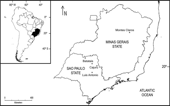
Bats were collected at five ecologically distinct sites in the northeast region of São Paulo state (sites 1–3) and north region of Minas Gerais state (sites 4 and 5), southeastern Brazil (Figure 1 and Table 1). Field collections were conducted from February 2012 to April 2014. Trap sites were visited twice: one in the dry season (April–September) and once in the rainy season (October–March). We used 12 mist nets (model 716/12P, 12 × 2.5 m; denier 75/2, mesh 16 × 16 mm; Ecotone Inc., Gdynia, Poland) in sites 1, 2 and 3; and six mist nets in sites 4 and 5 with a total sampling effort of 56,160 m2h. Captured bats were identified following Gardner,9 and one specimen per species by trap-night was anesthetized to collect blood by cardiac puncture; blood samples were stored in cryovials and flash-frozen in liquid nitrogen. At sites 4 and 5, five specimens per trap-night were randomly selected for blood collection. All bats were handled and sampled according to Sikes and others10 guidelines. This research project, along with its procedures and protocols, is in accordance with Brazilian environment and wildlife protection laws and regulations, and have been approved by the Chico Mendes Institute of Biodiversity Conservation (Ministry of Environment, Brasília, Distrito Federal, Brazil.), protocols nos. 19838-1 and 41709-3. It has also been approved by the Ethics Committee for Animal Research of University of São Paulo and Federal University of Minas Gerais (nos. 020/2011 and 333/2013, respectively). From 270 captured bats, 53 were bled for detection of immunoglobulin G (IgG) antibodies to rN-ARAQV by indirect enzyme-linked immunosorbent assay (ELISA) using anti-bat (Bethyl Laboratories, Inc., Montgomery, TX) secondary antibody. This ELISA, as previously described, showed 97.2% sensitivity, 100% specificity, 100% positive predictive value, and 98.1% negative predictive value when compared with an IgG-ELISA using rN antigen of Andes virus, which is the serological test for hantavirus most used in South America.11,12
Figure 1.
Study areas, highlighting the states of São Paulo and Minas Gerais in southeastern Brazil. The map shows cities where bats have been captured.
Table 1.
Trap sites general features6
| Trap sites/altitude (m) | City/state | Main vegetation | Secondary vegetation | Features | |
|---|---|---|---|---|---|
| 1 | JES/600 | Luis Antonio/SP | Cerrado* | Semideciduous forest | Continuous Cerrado |
| 2 | NEF/775 | Cajuru/SP | Grassland | Cerrado | Monocultures |
| 3 | SGF/860 | Batatais/SP | Sugarcane | Cerrado | Monocultures |
| 4 | SEP/872 | Montes Claros/MG | Dry forest†7 | Cerrado | Karst topography |
| 5 | LGEP/1,009 | Montes Claros/MG | Cerrado8 | Gallery forest | Caves and shelters |
JES = Jatai Ecological Station; LGEP = Lapa Grande Ecological Park; MG = Minas Gerais state; NEF = Nova Esperança Farm; SEP = Sapucai Ecological Park; SGF = Santa Gabriela Farm; SP = Sao Paulo state.
Cerrado = Brazilian savanna-like biome.
Dry forest = deciduous seasonal forest.
Nine bats had IgG antibodies to ARAQV, which represents an overall seroprevalence of 17%. Five of these bats were from São Paulo state and four were from Minas Gerais state. Of these, five were frugivorous, one was carnivorous, and three were sanguivorous (Table 2). From these infected bats, seven were males and two were females. We found more infected bats in the rainy season (N = 6) than in the dry season (N = 3).
Table 2.
Infected and tested bats for antibodies against rN-ARAQV
| Family | Species | Captured | Infected/tested | Main feeding items |
|---|---|---|---|---|
| Phyllostomidae | Artibeus lituratus | 41 | 1/6 | Fruits |
| Phyllostomidae | A. obscurus | 2 | 1/2 | Fruits |
| Phyllostomidae | A. planirostris | 41 | 1/3 | Fruits |
| Phyllostomidae | Carollia perspicillata | 43 | 1/10 | Fruits and insects |
| Phyllostomidae | Chiroderma villosum | 1 | 1/1 | Fruits |
| Phyllostomidae | Chrotopterus auritus | 1 | 1/1 | Small vertebrates |
| Phyllostomidae | Desmodus rotundus | 11 | 3/5 | Mammals blood |
| Phyllostomidae | Glossophaga soricina | 22 | 0/5 | Nectar and pollen |
| Phyllostomidae | Lonchophylla spp. | 1 | 0/1 | Nectar and pollen |
| Phyllostomidae | Micronycteris minuta | 1 | 0/1 | Insects |
| Molossidae | Molossops neglectus | 1 | 0/1 | Insects |
| Molossidae | Molossops temminckii | 2 | 0/1 | Insects |
| Vespertilionidae | Myotis nigricans | 13 | 0/5 | Insects |
| Vespertilionidae | Myotis albescens | 4 | 0/1 | Insects |
| Phyllostomidae | Platyrrhinus lineatus | 23 | 0/4 | Fruits |
| Phyllostomidae | Sturnira lilium | 38 | 0/6 | Fruits |
rN-ARAQV = recombinant nucleoprotein of Araraquara virus.
Main feeding items are shown according to Gardner.9
Bats evolution is dated around 50 million years ago, and they are distributed widely in the world, on all continents, except Antarctica.2,13 Perhaps, because of their ancient origin certain viruses seem to be coevolved with them. Thus, maintenance and transmission of these viruses crossed species barriers to infect wild and domestic mammals and also humans.2,13,14 Antibodies to viruses such as Hendra, Ebola, and severe acute respiratory syndrome (SARS)-like coronavirus (CoV) have been detected in wild bats, demonstrating that these animals are able to mount an antibody response, including IgM, IgE, IgA, and multiple IgG classes.14 Although bats may be persistently infected with many viruses, evidence from experimental and naturally infected bats has shown that they rarely produce an antibody response, probably because they are able to control viral replication via the innate immune antiviral response, and therefore, show a low viremia.13,14 However, here we were capable to show bats with IgG antibodies against the rN-ARAQV. The ELISA essays using rN-ARAQV as antigen have been previously used in hantavirus serologic surveys in rodents.15,16 Previous studies with bats of the Old World showed that only insectivorous bats are infected with hantavirus.5 Our study emphasizes that hantaviruses are infecting bats of several species and of different trophic groups in Brazil (Table 2). We also observed that the hantavirus prevalence in infected bats was higher when compared with that observed in rodent reservoirs from the same region.15,16 Despite, we have found antibodies against hantavirus, our results only support the idea that these bats become infected in some moment of their lifetime. Further studies in bats are necessary to detect the species and genotype of the infecting hantavirus and then determine the viral load in distinct organ tissues of these animals. Therefore, virus isolation followed by infection experiments could provide additional information if bats actually play a role as reservoirs of hantaviruses. Regardless of the negative public impression of bats, they possess important roles on insect control,17 reseeding forests, and pollinate plants that provide human and animal food.18 Bat guano is used as a fertilizer and for manufacturing soaps, gasohol, and antibiotics. Besides, bat echolocation and the infrared radiation of vampire bats (Desmodus rotundus) have provided models for sonar and infrared systems, respectively.13,19
Our study gives insights into ecology, conservational biology, and public health. These data may be useful to understand patterns of hantavirus evolution, in bats and other reservoirs, and to understand the virus dynamics and their potential public health importance. It is also important to preserve the native environment of these animals. Hence, this is the first report of the presence of hantavirus antibodies in phyllostomid bats in southeastern Brazil and also the first report of hantavirus antibodies among bats in the Americas.
ACKNOWLEDGMENTS
We thank specially Thiago Neves for all the support in Montes Claros city and field guidance at Sapucaia and Lapa Grande Ecological Parks. We are also grateful to Vinicius Kavagutti, Márcio Schafer, Milene Eigenher, Ariane and Gustavo Crepaldi de Morais for their help in field collections. We appreciate all the support from the Jatai Ecological Station Manager Edison Montilha, Armando Nascimento who supported us on his farm Santa Gabriela (Batatais city), and José Teotônio (Zezinho) for all support on his farm in Cajuru city and the Secretary of Health from Cajuru city through the official Toninho. We also thank Patricio Hernaez for the help in the map construction and Luciano Luna for the comments in the final version of the manuscript.
Footnotes
Financial support: This work was supported by the São Paulo State Research Foundation (FAPESP) grants 2011/06810-9 to Gilberto Sabino-Santos Jr and 2008/50617-6 to Luiz Tadeu Moraes Figueiredo.
Authors' addresses: Gilberto Sabino-Santos Jr. and Luiz Tadeu Moraes Figueiredo, Center for Virology Research, School of Medicine in Ribeirão Preto, University of São Paulo, Ribeirão Preto, São Paulo, Brazil, E-mails: sabinogsj@usp.br and ltmfigue@fmrp.usp.br. Felipe Gonçalves Motta Maia, Center for Virology Research, School of Medicine in Ribeirão Preto, University of São Paulo, Ribeirão Preto, São Paulo, Brazil, and Department of Microbiology, Institute of Biomedical Sciences, University of São Paulo, São Paulo, Brazil, Miranda Lima, Cristieli Barros Gonçalves, Patricia Doerl Barroso, and Maria Norma Melo, Department of Parasitology, Institute of Biological Sciences, Federal University of Minas Gerais, Belo Horizonte, Brazil, E-mails: thallytabio@gmail.com, sabrina_mlima@hotmail.com, criscat_87@hotmail.com, patydoerl@hotmail.com, and normamello@gmail.com. Renata de Lara Muylaert, Department of Ecology, São Paulo State University, Rio Claro, São Paulo, Brazil, E-mail: renatamuy@gmail.com. Colleen B. Jonsson, Department of Microbiology and Immunology and Center for Predictive Medicine for Biodefense and Emerging Infectious Diseases, University of Louisville, Louisville, KY, and Department of Microbiology, National Institute for Mathematical and Biological Synthesis, Knoxville, TN, E-mail: cbjons01@louisville.edu. Douglas Goodin, Department of Geography, Kansas State University, Manhattan, KS, E-mail: dgoodin@ksu.edu. Jorge Salazar-Bravo, Department of Biological Sciences, Texas Tech University, Lubbock, TX, E-mail: j.salazar-bravo@ttu.edu.
References
- 1.Holmes EC, Zhang YZ. The evolution and emergence of hantaviruses. Curr Opi Virol. 2015;10:27–33. doi: 10.1016/j.coviro.2014.12.007. [DOI] [PubMed] [Google Scholar]
- 2.Brook CE, Dobson AP. Bats as ‘special’ reservoirs for emerging zoonotic pathogens. Trends Microbiol. 2015;23:172–180. doi: 10.1016/j.tim.2014.12.004. [DOI] [PMC free article] [PubMed] [Google Scholar]
- 3.Bennett SN, Gu SH, Kang HJ, Arai S, Yanagihara R. Reconstructing the evolutionary origins and phylogeography of hantaviruses. Trends Microbiol. 2014;22:473–482. doi: 10.1016/j.tim.2014.04.008. [DOI] [PMC free article] [PubMed] [Google Scholar]
- 4.Gu SH, Lim BK, Kadjo B, Arai S, Kim JA, Nicolas V, Lalis A, Denys C, Cook JA, Dominguez SR, Holmes KV, Urushadze L, Sidamonidze K, Putkaradze D, Kuzmin IV, Kosoy MY, Song J-W, Yanagihara R. Molecular phylogeny of hantaviruses harbored by insectivorous bats in Côte d'Ivoire and Vietnam. Viruses. 2014;6:1897–1910. doi: 10.3390/v6051897. [DOI] [PMC free article] [PubMed] [Google Scholar]
- 5.Guo W-P, Lin X-D, Wang W, Tian J-H, Cong M-L, Zhang H-L, Wang M-R, Zhou R-H, Wang J-B, Li M-H, Xu J, Holmes EC, Zhang Y-Z. Phylogeny and origins of hantaviruses harbored by bats, insectivores, and rodents. PLoS Pathog. 2013;9:e1003159. doi: 10.1371/journal.ppat.1003159. [DOI] [PMC free article] [PubMed] [Google Scholar]
- 6.Ribeiro JF, Walter BMT. As principais fitofisionomias do Bioma Cerrado. In: Sano SM, Almeida SP, Ribeiro JF, editors. Cerrado: Ecologia e Flora. Planaltina, Brazil: Embrapa Cerrados; 2008. pp. 152–212. (In Portuguese) [Google Scholar]
- 7.Santos RM, Vieira FA, Gusmão E, Nunes F, Roberta Y. Florística e estrutura de uma floresta estacional decidual, no Parque Municipal da Sapucaia, Montes Claros (MG) Cerne. 2007;13:248–256. (In Portuguese) [Google Scholar]
- 8.Oliveira GL, Moreira DL, Mendes ADR, Guimarães EF, Figueiredo LS, Kaplane MAC, Martins ER. Growth study and essential oil analysis of Piper aduncum from two sites of Cerrado biome of Minas Gerais State, Brazil. Rev Bras Farmacog. 2013;23:743–753. [Google Scholar]
- 9.Gardner AL. Mammals of South America: Marsupials, Xenarthrans, Shrews, and Bats. Volume 1. Chicago, IL: University of Chicago Press; 2007. p. 669. [Google Scholar]
- 10.Sikes RS, Gannon WL. the Animal Care and Use Committee of the American Society of Mammalogists Guidelines of the American Society of Mammalogists for the use of wild mammals in research. J Mammal. 2011;92:231–253. doi: 10.1093/jmammal/gyw078. [DOI] [PMC free article] [PubMed] [Google Scholar]
- 11.Figueiredo LTM, Moreli ML, Borges AA, de Figueiredo GG, Badra SJ, Bisordi I, Suzuki A, Capria S, Padula P. Evaluation of an enzyme-linked immunosorbent assay based on Araraquara virus recombinant nucleocapsid protein. Am J Trop Med Hyg. 2009;81:273–276. [PubMed] [Google Scholar]
- 12.Figueiredo LT, Moreli ML, Borges AA, Figueiredo GG, Souza RL, Aquino VH. Expression of a hantavirus N protein and its efficacy as antigen in immune assays. Braz J Med Biol Res. 2008;41:596–599. doi: 10.1590/s0100-879x2008000700008. [DOI] [PubMed] [Google Scholar]
- 13.Moratelli R, Calisher CH. Bats and zoonotic viruses: can we confidently link bats with emerging deadly viruses? Mem Inst Oswaldo Cruz. 2015;110:1–22. doi: 10.1590/0074-02760150048. [DOI] [PMC free article] [PubMed] [Google Scholar]
- 14.Baker ML, Schountz T, Wang LF. Antiviral immune responses of bats: a review. Zoo Pub Health. 2013;60:104–116. doi: 10.1111/j.1863-2378.2012.01528.x. [DOI] [PMC free article] [PubMed] [Google Scholar]
- 15.De Sousa RLM, Moreli ML, Borges AA, Campos GM, Livonesi MC, Figueiredo LTM, Pinto A. Natural host relationship and genetic diversity of rodent associated hantaviruses in southeastern Brazil. Intervirol. 2008;51:299–310. doi: 10.1159/000171818. [DOI] [PubMed] [Google Scholar]
- 16.Figueiredo GG, Borges AA, Campos GM, Machado AM, Saggioro FP, Sabino Júnior Gdos S, Badra SJ, Ortiz AA, Figueiredo LT. Diagnosis of hantavirus infection in humans and rodents in Ribeirão Preto, State of São Paulo, Brazil. Rev Soc Bras Med Trop (Impresso) 2010;43:348–354. doi: 10.1590/s0037-86822010000400002. [DOI] [PubMed] [Google Scholar]
- 17.Boyles JG, Cryan PM, Mccracken GF, Kunz TH. Economic importance of bats in agriculture. Science. 2011;332:41–42. doi: 10.1126/science.1201366. [DOI] [PubMed] [Google Scholar]
- 18.Dirzo R, Young HS, Galetti M, Ceballos G, Isaac NJ, Collen B. Defaunation in the Anthropocene. Science. 2014;345:401–406. doi: 10.1126/science.1251817. [DOI] [PubMed] [Google Scholar]
- 19.Campbell AL, Naik RR, Sowards L, Stone MO. Biological infrared imaging and sensing. Micron. 2002;33:211–225. doi: 10.1016/s0968-4328(01)00010-5. [DOI] [PubMed] [Google Scholar]


