Abstract
Primary mucinous cystic cystadenomas of the retroperitoneum are very rarely encountered, and there have been only about 30 cases reported in the literature. The histogenesis of primary mucinous cystadenomas is unclear. Most authors suggested that it develops through mucinous metaplasia in a pre-existing mesothelium-lined cyst. Complete surgical excision is the only treatment and it is required for the final diagnosis and cure. We present here a case report of a 38-year-old Korean woman with primary retroperitoneal cystadenoma. It was a thin-walled, multilocular cyst with a dominant loculus that measured 10.0×7.5×5.5 cm3 in size, and to the best of our knowledge, this is the first such case to be reported in in Korea.
Keywords: Cystadenoma, Cystadenoma/pathology, Retroperitoneal neoplasms, Retroperitoneal neoplasms/surgery, Female, Tomography, X-ray computed
INTRODUCTION
Retroperitoneal mucinous cystadenomas are grossly and microscopically similar to benign mucinous cystadenoma of the ovary, which are clinically and pathologically common tumors. The primary mucinous cystadenomas of the retroperitoneum, however, are extremely rare, and their histogenesis remains unclear1,2). We report a case of a 38-year-old Korean woman with retroperitoneal cystadenoma. As far as we know, this report for primary retroperitoneal mucinous cystadenoma is the first such case in Korea.
CASE REPORT
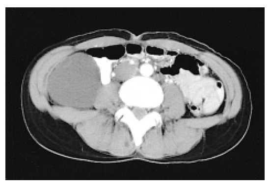
A 38-year-old, gravida 4, para 2, female Korean patient presenting with indigestion and vague abdominal discomfort of several months’ duration was referred from a private clinic for a large retroperitoneal mass that was discovered on abdomen computer tomography. She had suffered from cervical tuberculosis 7 years ago, and this had been confirmed by excisional biopsy at a private clinic, and it was eradicated after a 1 year treatment with anti-tuberculosis medications. The large cystic abdominal mass was palpated on her right flank and in the RLQ abdomen on the physical examination. It was mobile, non-tender and estimated to be about 8 cm in diameter. The rest of her pelvic examination was unremarkable. The laboratory findings including colonoscopy and serum tumor markers (CEA, CA 125, and CA 19-9) were within the normal limits. Imaging studies on abdomen and pelvis computer tomography showed an unilocular cystic mass, 10×8×6 cm3 in dimensions, was located at the inferior aspect of the liver and posterior along the right colon, and the right retroperitoneal mass had displaced the right colon ventromedially. It was an encapsulated cystic mass that contained fluids, and there was no lesion in the pelvic organs (Figure 1).
Figure 1.

Abdominal CT showed the retroperitoneal mass had displaced the right colon medially and this encapsulated cystic mass contained fluids.
A celiotomy was performed; there was a large cystic retroperitoneal mass adhered to the right colon and its mesocolon, right kidney and duodenum, but it had not invaded to the adjacent organs. Macroscopically there was no lesion observed in the ovaries and fallopian tubes, appendix, right colon, kidneys and pancreas. The retroperitoneal tumor was completely removed with no spillage of its contents, and no combined resection with the associated organs was performed.
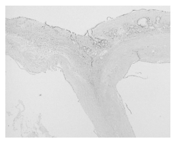
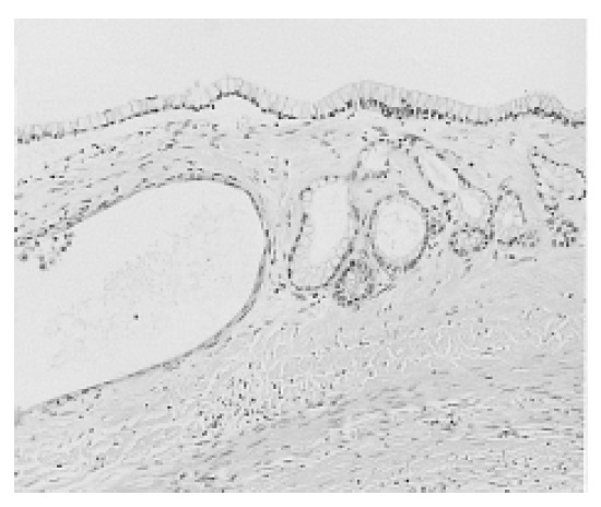
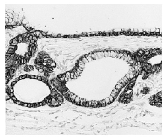
Upon gross examination, the specimen was a thin-walled, multilocular cyst with a dominant loculus that measured 10.0×7.5×5.5 cm3. The external surface is smooth and pinkish. Upon opening it, it contained mucinous fluids and inner surface is smooth. The wall of the cyst contained smaller cysts; the largest of these measured 0.8 cm in diameter. The thickness of the walls was less than 0.1 cm. There are no solid areas or papillary projections. On the histologic examination, the cyst was lined by a single layer of tall columnar epithelium with clear cytoplasm and small nuclei that were basely located in the cells (Figure 2). There was no papillary growth or stromal invasion of epithelium. The cyst wall consisted of fibrocollagenous tissue of varying cellularity and vascularity. No pancreatic or ovarian components were found (Figure 3). Immunohistochemical staining showed that the lining cells were positive for cytokeratin, cytokeratin 7, CAM 5.2, and carcinoembryogenic antigen, but the cells were negative for cytokeratin 19, epithelial membrane antigen, estrogen and progesterone (Figure 4).
Figure 2.

Multilocular cyst lined by a single layer of tall columnar cells. The stroma consisted of fibrocollagenous tissue with no pancreatic or ovarian components (H & E, ×40).
Figure 3.

The cells had clear cytoplasm and basely located small nuclei with no cytologic atypia (H & E, ×200).
Figure 4.

On immunohistochemical staining, the lining cells showed positive reactions to CAM 5.2 (×200).
The final diagnosis was primary mucinous cystadenoma arising from the retroperitoneum. The patient had an uneventful postoperative recovery, and there was no lesion or recurrence on the follow-up abdomen and pelvis computer tomography at postoperative one year.
DISCUSSION
Primary mucinous cystadenomas of the retroperitoneum are extremely rare lesions. Retroperitoneal mucinous neoplasms can be classified into 3 types according to the clinicopathologic features. Benign mucinous cystadenoma, the most common type, are benign cystic tumors with no recurrence following curative resection3). In the second type, borderline mucinous cystadenoma, the lining epithelium contains foci of proliferative columnar epithelium in addition to columnar epithelium, and these tumors resemble the ovarian mucinous tumors having a low malignant potential4,5). The third one is malignant mucinous cystadenocarcinoma, which can be recurrent or metastaic6–8).
The genesis of this tumor remains unclear; however there are 2 main theories for the histogenesis of retroperitoneal mucinous cystadenoma. Because they have resemblance to ovarian mucinous cystadenomas, they are thought to arise from heterotrophic ovarian tissue, which may explain why they occur only in women; however, ovarian remnants have not been identified in the wall of the cysts1,2). The second theory is that these tumors arise from an invagination of multipotential mesothelium with subsequent mucinous metaplasia of the mesothelial lining cells, and this giving rise to a mucinous cyst that enlarges to form a cystic tumor. To support this theory, there has been no evidence of ovarian tissue in these tumors, the immunohistochemical staining for estrogen or progesterone is negative and there has been no gut-like muscle in the pathologic specimens, thus these tumors are not from the ectopic ovarian tissue and intestinal duplication8–10).
Preoperatively differential diagnosis from other primary retroperitoneal mucinous neoplasms such as cystic lymphangoma, cystic mesothelioma, lymphocele, cystic teratoma, urinoma, or cyst of parasitic origin is not possible. Careful examination of the wall of the cyst and adequate sampling of the lining epithelium for microscopic examination must be done to exclude any foci of adenocarcinoma9). A careful and complete surgical excision with no spillage from the retroperitoneal cystic neoplasms would be most important factor for cure11).
REFERENCES
- 1.Pennell TC, Gusdon JP., Jr Retroperitoneal mucinous cystadenoma. Am J Obstet Gynecol. 1989;160:1229–1231. doi: 10.1016/0002-9378(89)90201-9. [DOI] [PubMed] [Google Scholar]
- 2.Balat O, Aydin A, Sirikci A, Kutlar I, Aksoy F. Huge primary mucinous cystadenoma of the retroperitoneum mimicking a left ovarian tumor. Eur J Gynaecol Oncol. 2001;22:454–455. [PubMed] [Google Scholar]
- 3.Yunoki Y, Oshima Y, Murakami I, Takeuchi H, Yasui Y, Tanakaya K, Konaga E. Primary retroperitoneal mucinous cystadenoma. Acta Obstet Gynecol Scand. 1998;77:357–358. [PubMed] [Google Scholar]
- 4.Motoyama T, Chida T, Fujiwara T, Watanabe H. Mucinous cystic tumor of the retroperitoneum: a report of two cases. Acta Cytol. 1994;38:261–266. [PubMed] [Google Scholar]
- 5.Papadogiannakis N, Gad A, Ehliar B. Primary retroperitoneal mucinous tumor of low malignant potential: histogenetic aspects and review of the literature. APMIS. 1997;105:483–486. doi: 10.1111/j.1699-0463.1997.tb00597.x. [DOI] [PubMed] [Google Scholar]
- 6.Roth LM, Ehrlich CE. Mucinous cystadenocarcinoma of the retroperitoneum. Obstet Gynecol. 1977;49:486–488. [PubMed] [Google Scholar]
- 7.Fujii S, Konishi I, Okamura H, Mori T. Mucinous cystadenocarcinoma of the retroperitoneum: a light and electron microscopic study. Gynecol Oncol. 1986;24:103–112. doi: 10.1016/0090-8258(86)90013-2. [DOI] [PubMed] [Google Scholar]
- 8.Kehagias DT, Karvounis EE, Fotopoulos A, Gouliamos AD. Retroperitoneal mucinous cystadenoma. Eur J Obstet Gynecol Reprod Biol. 1999;82:213–215. doi: 10.1016/s0301-2115(98)00254-1. [DOI] [PubMed] [Google Scholar]
- 9.Subramony C, Habibpour S, Hashimoto LA. Retroperitoneal mucinous cystadenoma. Arch Pathol Lab Med. 2001;125:691–694. doi: 10.5858/2001-125-0691-RMC. [DOI] [PubMed] [Google Scholar]
- 10.Erdemoglu E, Aydogdu T, Tokyol C. Primary retroperitoneal mucinous cystadenoma. Acta Obstet Gynecol Scand. 2003;82:486–487. doi: 10.1034/j.1600-0412.2003.00158.x. [DOI] [PubMed] [Google Scholar]
- 11.Chen JS, Lee WJ, Chang YJ, Wu MZ, Chiu KM. Laparoscopic resection of a primary retroperitoneal mucinous cystadenoma: report of a case. Surg Today. 1998;28:343–345. doi: 10.1007/s005950050137. [DOI] [PubMed] [Google Scholar]


