Abstract
Carcinosarcoma is defined as a malignant tumor with an admixture of carcinoma and sarcoma. Pulmonary carcinosarcoma accounts for about 0.27 percent of all lung neoplasms. It occurs frequently in males, particularly in smokers between 50 and 80 years of age. Preoperative diagnostic tests, such as sputum cytology, percutaneous fine needle biopsy and bronchoscopy, have a low yield in detection of pulmonary carcinosarcoma. The diagnosis is verified by postoperative pathologic findings and by immunohistochemical investigations in many cases. Surgical resection is the treatment of choice. As the metastasis to regional lymph nodes and distant organ is common at diagnosed time, the prognosis is quite poor.
We report a case of pulmonary carcinosarcoma presented with persistent mild fever and blood-tinged sputum in a 66-year-old male.
Keywords: Carcinosarcoma, Lung neoplasms
INTRODUCTION
The first reported case of pulmonary carcinosarcoma was attributed to Kika in 1908, as noted by Herxheimer and Reinke in 19121). In 1951, Bergmann et al. reported two cases of the pulmonary carcinosarcoma and established this unusual malignant lesion as a distinct entity2). It is reported to constitute 0.2% or 0.27% of primary pulmonary malignancies3,4).
Pulmonary carcinosarcma is reported to occur most frequently in male patients between 50 and 80 years of age, predominantly affecting the upper lobe and/or the principal bronchi5–8) and is associated with a history of smoking8,9). There is no specific clinical presentation but typical symptoms are chest pain, cough, hemoptysis, dyspnea, fever and weight loss9).
Preoperative diagnostic tests, such as sputum cytology, percutaneous fine needle biopsy and bronchoscopy, have a low yield in detection of pulmonary carcinosarcoma. Thus, the diagnosis is verified by postoperative pathologic and immunohistochemical findings in many cases. The most frequent epithelial component was squamous cell carcinoma, adenocarcinoma and adenosquamous carcinoma, whereas the mesenchymal component most frequently was rhabdomyosarcoma, chondrosarcoma, osteosarcoma or combinations of these elements10). Surgical resection is the treatment of choice. The 2-year survival rate in patients with this cancer has been reported to be 9.5%11).
We report a case of pulmonary carcinosarcoma with persistent mild fever in a 66-year-old male.
CASE
A 66-year-old man was referred to our hospital for evaluation of mild fever, blood-streaked sputum and chest discomfort for 7 days prior to admission. At five months before admission, a cough with minimal episodes of bloody sputum had developed. At that time, chest radiographs were said to be unremarkable. He denied having shortness of breath, wheezes, weight loss or night sweats. He works as a fisherman and has not smoked for 1 year but has a 30-pack-year history of tobacco use. His medical history was significant for cholecystectomy and laminoplasty for herniation of nucleus pulposus five years previously. His family history was noncontributory.
On presentation, the patient appeared to be in no distress, relatively. His blood pressure was 110/70 mmHg; heart rate 78 beats /min; respiratory rate 23 breaths /min; body temperature 37.60°C. Examination of the lung revealed slightly reduced breath sounds over the right lower lung. Cardiac examination revealed a regular rhythm without murmurs. Findings of the abdominal examination were normal and the extremities were without clubbing, cyanosis or edema.
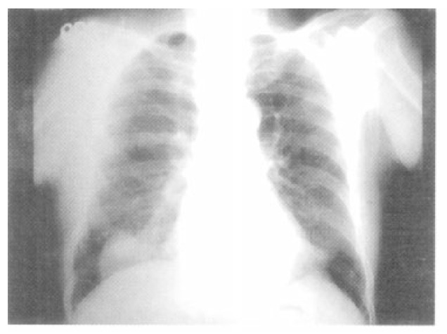
The laboratory tests were as follows: WBC count, 15.7×10³/uL with 78% neutrophils, 13% lymphocytes, 7% monocytes, 1% eosinophils and others 1%; Erythrocyte sedimentation rate, 44 mm/h; hemoglobin, 14.4 g/dL; hematocrit, 41.4%; platelet count, 313×103/uL. Other laboratory findings including liver function, blood glucose level, renal function, electrolytes and urine microscopy, were all within normal limits. Arterial blood gas analysis on room air revealed a pH of 7.459, Pco2 of 36.8 mmHg and Po2 of 76.9 mmHg. The chest radiograph obtained at the time of admission showed a round mass in the right lower lobe (Figure 1). The chest CT scan performed at the referring hospital confirmed the presence of a 4×4 cm-sized, smoothly marginated, soft tissue density with central lower density in the right lower lobe.
Figure 1.

The chest PA shows a round mass with central low density in the right lower lobe.
An initial diagnosis of subclinical infectious disease or necrotizing solitary mass was made; the patient was treated with empirical antibiotics. However, mild fever and blood-tinged sputum persisted despite the use of broad-spectrum antibiotics. The lesion looked larger on a chest radiograph. The patient still felt well, apart from the mild fever. Sputum culture findings were negative for bacteria and fungus. Tuberculosis smear was negative and cultures were pending. Sputum cytology testing results were also negative.
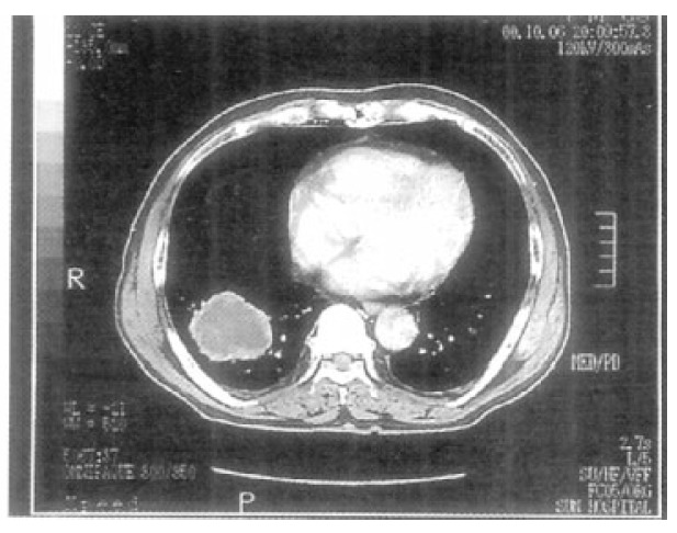
The chest CT scan documented a slightly increased size of the mass, with more increased extent of the central low density compared with the previous chest CT and no mediastinal or hilar lymphadenopathy (Figure 2). The patient declined bronchoscopy and therefore, percutaneous fine needle aspiration was performed. Only blood without pus was aspirated and the results of aspirates were negative for routine bacterial Gram’s stain and culture, including acid-fast bacilli and fungi, thus ruling out infectious process.
Figure 2.

The chest CT shows a large cystic mass with central low density and no mediastinal or hilar lymphadenopathy.
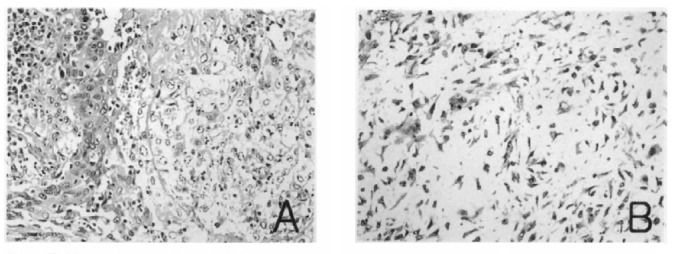
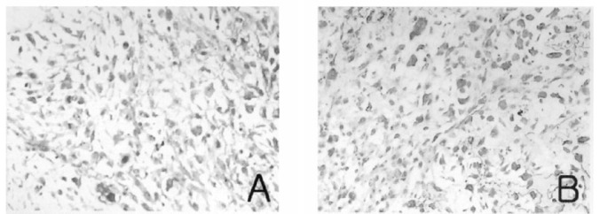
To define the exact etiology of the radiologic findings, thoracoscopic wedge biopsy of the left lower lobe demonstrated the mass to be contained within the lateral basal segment and the medial basal segment of the left lower lobe. On gross section, the mass was 10×9×7 cm in size, sharply circumscribed and smoothly contoured. It had no involvement to the chest wall and no endobronchial component, but extended centrally to hilar areas. Light microscopic examination demonstrated a tumor composed of malignant epithelial components and mesenchymal components. The carcinomatous components consisted of poorly differentiated squamous cell carcinoma and the sarcomatous components were considered chondroid sarcoma in the light of microscopic findings (Figure 3). Light microscopic immunohistochemical analysis showed that the tumor cells in the carcinomatous component reacted with anti-epithelial membrane antigen (EMA) antibody and the tumor cells in the sarcomatous component reacted with anti-vimentin antibody (Figure 4). He underwent workup for distant metastasis, the findings of which were unremarkable. Therefore, left lower lobectomy was performed with lymph node dissection. There was no microscopic evidence of lymph node metastases making this the international TNM stage lb (T2N0M0) lesion.
Figure 3.

Microscopic views. The epithelial component shows poorly differentiated squamous cell carcinoma (A) and the mesenchymal component shows chondrosarcoma (B). (H & E stain, × 400)
Figure 4.

Immunohistochemical stainings. The poorly differentiated squamous cell carcinoma shows focal positive for epithelial membrane antigen (A). The chondrosarcoma shows positive for vimentin (B). (× 400)
After surgical resection, mild fever and blood-tinged sputum disappeared. His postoperative course was uneventful without major complication. At 6-month follow-up, there remains no evidence of metastasis or tumor recurrence
DISCUSSION
Carcinosarcoma is a biphasic malignant tumor with an admixture of the epithelial and mesenchymal tissue. This neoplasm has been reported in most of the organs in which carcinoma can occur, most frequently including uterus, breast, thyroid gland, esophagus and lung12,13). It has been given names such as carcinosarcoma, carcinoma with sarcomalike stroma, malignant mixed tumor, blastoma, spindle cell carcinoma and heterologous biphasic sarcomatoid carcinoma.
The frequency of carcinosarcoma in the lung is 0.2% or 0.27% of all lung tumors3,4). Pulmonary carcinosarcoma is reported to occur most frequently in males between 50 and 80 years of age, especially smokers5,8,9).
There can be symptoms such as chest pain, cough, hemoptysis, wheezing and dyspnea by bronchial irritation or occlusion, fever and weight loss9). These are divided into two clinicopathologic groups: endobronchial or central type and solid parenchymal or peripheral type on the basis of the location of pulmonary carcinosarcomas14). Endobronchial type is frequently pedunculated with limited extension into the surrounding lung. This type involves lobar or segmental bronchi and frequently produces symptoms such as cough, fever, dyspnea and hemoptysis due to bronchial irritation or occlusion. Thus endobronchial type has a relatively good prognosis due to early detection by these symptoms. Parenchymal type is mainly asymptomatic in early stages and is reported to have a poor prognosis due to metastasis to lymph nodes or distant organs at detected time. Macroscopically, the tumor in this case has no endobronchial involvement and extended centrally to hilar area.
Cohen Salmon et al.6) described the histogenesis of pulmonary carcinosarcoma as follows: 1) malignant change in hamartoma 2) simultaneous epithelial and stromal malignancy 3) malignant change in the stroma induced by carcinoma 4) connective tissue metaplasia of epithelial cells 5) carcinomatous change in a sarcoma.
The most frequent epithelial component was squamous cell carcinoma (46%), followed by adenocarcinoma (31%) and adenosquamous carcinoma (19%), whereas sarcomatous elements most frequently included rhabdomyosarcoma, chondrosarcoma, osteosarcoma or combinations of these elements10). However, some characteristics may need reconfirmation because confusion between the diagnosis of carcinosarcomas and spindle cell carcinomas is undeniable in light microscopic studies.
Pulmonary carcinosarcoma is rarely diagnosed preoperatively. If the tumor is located centrally, in most cases biopsies show only one component and peripheral tumors cannot be often reached endoscopically. Sputum cytology, bronchoscopy and transthoracic fine needle biopsy can sometimes help to diagnose it. Since blastoma, metastatic sarcoma to lung, spindle cell carcinoma is difficult to discriminate from pulmonary carcinosarcoma by light microscopic study15), immunohistochemical studies and/or electron microscopic ultrastructural findings play an important role in differential diagnosis of pulmonary carcinosarcoma. In immunohistochemical studies, the carcinamatous component shows positive for pankeratin, epithelial membrane antigen (EMA), carcinoembryonic antigen (CEA), B72.3, or neuron-specific enolase and sarcomatous components can show positive for actin, S-100 protein, lysosome, desmin, vimentin or factor VIII according to a kind of differentiation in case of specific differentiation. Thus, the special stains for these antibodies can help to diagnose it7,16,17). We performed immunohistochemical stains for cytokeratin, EMA, S-100 protein and vimentin in this case. The results of the stains were positive for vimentin and focal positive for EMA.
Although surgical resection, such as pneumonectomy or lobectomy when possible is the treatment of choice, multiagent chemotherapy consisting of cyclophosphamide, doxorubicin and cisplatin seems to be a reasonable consideration for use in patients with metastatic carcinosarcoma. If there is uncertainty regarding prognostic factors, there is general agreement that the outcome of pulmonary carcinosarcoma is quite poor due to the marked tendency of the tumor to metastasize at the distant sites and the high rate of local recurrence7,18). The average postoperative survival is nine months and 9.5% of patients survive two years11).
REFERENCES
- 1.Kika Pathologie des Krebses. Ergeb Allg Pathol Anat. 1912;16:1. cited by Herxheimer G, Reinke. [Google Scholar]
- 2.Bergmann M, Ackerman LV, Kemler RL. Carcinosarcoma of the lung. Review of the literature and report of two cases treated by pneumo-nectomy. Cancer. 1951:4919–929. doi: 10.1002/1097-0142(195109)4:5<919::aid-cncr2820040505>3.0.co;2-w. [DOI] [PubMed] [Google Scholar]
- 3.Davis MP, Eagan RT, Weiland LH, Pairolero PC. Carcinosarcoma of the lung. Mayo Clinic experience and response to chemotherapy. Mayo Clin Proc. 1984;59:598–603. doi: 10.1016/s0025-6196(12)62410-0. [DOI] [PubMed] [Google Scholar]
- 4.Diaconita G. Bronchopulmonary carcinosarcoma. Thorax. 1975;30:682–686. doi: 10.1136/thx.30.6.682. [DOI] [PMC free article] [PubMed] [Google Scholar]
- 5.Cabarcos A, Dorronsoro MG, Beristain JLL. Pulmonary carcinosarcoma, a case study and review of the literature. Br J Dis Chest. 1985;79:319–323. doi: 10.1016/0007-0971(85)90011-7. [DOI] [PubMed] [Google Scholar]
- 6.Cohen-Salmon D, Michel RP, Wang NS, Eddy D, Hanson R. Pulmonary carcinosarcoma and carcinoma: Report of a case studied by eletron microscopy, with critical review of the literature. Ann Pathol. 1985;5:115–124. [PubMed] [Google Scholar]
- 7.Ishida T, Tateishi M, Kaneko S, Yano T, Mitsudomi T, Sugimachi K, Hara N, Ohta M. Carcinosarcoma and spindle cell carcinoma of the lung. J Thorac Cardiovasc Surg. 1990;100:844–852. [PubMed] [Google Scholar]
- 8.Bull JC, Grimes OF. Pulmonary carcinosarcoma. Chest. 1974;65:9–12. doi: 10.1378/chest.65.1.9. [DOI] [PubMed] [Google Scholar]
- 9.Davis MP, Eagen RT, Weiland LH, Pairolero PC. Carcinosarcoma of the lung : Mayo Clinic Experience and Response to Chemotherapy. Mayo Clin Proc. 1984;59:598–603. doi: 10.1016/s0025-6196(12)62410-0. [DOI] [PubMed] [Google Scholar]
- 10.Koss MN, Hochholzer L, Frommelt RA. Carcinosarcomas of the lung: A Clinicopathologic Study of 66 Patients. Am J Surg Pathol. 1999;23(12):1514–1526. doi: 10.1097/00000478-199912000-00009. [DOI] [PubMed] [Google Scholar]
- 11.Gebauer C. The postoperative prognosis of primary pulmonary sarcomas: A review with a comparison between the histological forms and the other primary endobronchial sarcomas based on 474 cases. Scand J Thorac Cadiovasc Surg. 1982;16:91–97. doi: 10.3109/14017438209100617. [DOI] [PubMed] [Google Scholar]
- 12.Guarino M, Tricomi P, Giordano F, Cristofori E. Sarcomatoid carcinomas : pathological and histopathogenetic considerations. Pathology. 1996;44:295–305. doi: 10.1080/00313029600169224. [DOI] [PubMed] [Google Scholar]
- 13.Saphir O, Vass A. Carcinosarcoma. Am J Cancer. 1938;33:331–361. [Google Scholar]
- 14.Moore TC. Carcinosarcoma of the lung. Surgery. 1961;50:886–936. [PubMed] [Google Scholar]
- 15.The World Health Organization histological typing of lung tumor. Am J Clin Pathol. (2nd ed) 1982;77(2):123–136. doi: 10.1093/ajcp/77.2.123. [DOI] [PubMed] [Google Scholar]
- 16.Addis BJ, Corrin B. Pulmonary blastoma, carcinosarcoma and spindle cell carcinoma: An Immunohistochemical study of keratin intermediate filaments. J Pathol. 1985;147:291–301. doi: 10.1002/path.1711470407. [DOI] [PubMed] [Google Scholar]
- 17.Cupples J, Wright J. An immunohistochemical comparison of primary lung carcinosarcoma and sarcoma. Pathol Res Pract. 1990;186(3):326–329. doi: 10.1016/S0344-0338(11)80290-6. [DOI] [PubMed] [Google Scholar]
- 18.Sümmermann E, Huwer H, Seitz G. Carcinosarcoma of the lung, a tumor which has a poor prognosis and is extremely rarely diagnosed preoperatively. J Thorac Cardiovasc Surg. 1990;38:247–250. doi: 10.1055/s-2007-1014027. [DOI] [PubMed] [Google Scholar]


