Abstract
Objectives
Long standing ulcerative colitis (UC) has been known to be one of the precancerous diseases of colorectal cancer. Although the frequent loss of p53 allele (LOH) and aneuploidy were reported as the molecular events in carcinoma and dysplasia known as the precursor of UC, p53 genetic alteration was not reported in indefinite dysplasia and UC involved mucosa in long standing UC. Therefore, we investigated the mutational inactivation of the p53 gene in UC patients who showed dysplastic mucosa, as well as non-dysplastic mucosa on H & E stain and, secondly, if there is p53 mutation, we examined the relationship between p53 alteration and clinical data.
Method
Sixteen patients with UC who had different duration of colitis were studied by endoscopic examination with rectal mucosal biospies. p53 gene alterations were detected by PCR-SSCP for exon 4–8 and immunohistochemical staining with p53 monoclonal antibody.
Results
Among 16 patients, 2 patients (12%) showed dysplasia on H-E stain. The p53 point mutations were detected in 4 (two dysplasia and 2 normal looking mucosa) on PCR-SSCP. 4 patients who had p53 gene mutation were positive in immunohistochemical staining. With regard to clinical characteristics, these patients with p53 point mutation showed poor resoponse to medical treatment.
Conclusion
These results suggest that the p53 mutation may be an early molecular event of cancerous change in UC.
Keywords: ulcerative colitis, p53 mutation, PCR-SSCP
INTRODUCTION
Chronic, extensive ulcerative colitis (UC) is well established as a predisposing lesion for the development of colorectal adenocarcinoma1–3). There is compelling evidence that most carcinomas occuring in UC do not arise de novo but are preceded by and evolve from dysplastic mucosa, analogously to the development of sporadic colorectal carcinomas from adenomas4,5) However, the molecular basis of neoplasia in UC is not well understood at the present time.
In sporadic colon cancer, one copy of p53 is often inactivated by point mutation whereas the other copy is frequently deleted6). This inactivation and allelic loss has been known to be a late molecular event in adenoma — cancer sequence.
UC colons with dysplasia, carcinoma or both provide a valuable model for the study of genetic events during tumorigenesis. Histological progression towards cancer occurs in a stepwise fashion in UC (negative — indefinite for dysplasia-dysplasia-cancer)4). The p53 gene mutation has been demonstrated in dysplasia and cancer developed from UC. But there is no report that the mutation of p53 tumor suppressor gene is occured in normal looking mucosa (negative or indefinite for dysplasia).
Recently, there was an interesting report conducted by Robert et al7). They found the chromosomal alterations which precede the histological phenotype of dysplasia with the use of FISH and comparative genomic hybridization. But it is not certain whether the molecular event is preceded in the histological evolution. Therfore, we have conducted this study to determine the mutation of p53 gene which might be occurred in the early stage of tumorigenesis of UC.
MATERIAL AND METHOD
1. Patient and Specimens
Subjects consisted of 16 patients (12 male, 4 female), aged 44.6 on the average, and the period ranged from 1 month to 27 years in this study. Patients showing endoscopic signs of inflammation were classified as normal (N=0), mildly active (N=6), moderlately active (N=2), severe (N=8) according to Sninsky et al8). The 6 pieces of mucosal biopsy specimens in each patient were obtained from the rectal mucosa approximately 15 cm from the anal verge by sigmoidoscopy or colonoscopy. All the patients had no evidence of dysplasia-associated lesions or masses on endoscopic finding. Among the 6 specimens in each patient, 4 specimens were used for DNA extraction, 1 specimen for H&E stain and 1 specimen for immunohistochemical stainning of p53 protein. The normal mucosal specimens (control) for PCR-SSCP were obtained from the colonic mucosa which showed no evidence of inflammation on endoscopic examination.
2. Histological Evaluation
The histopathoiogy of all biopsies taken from UC and normal subjects was blindly evaluated by two gastrointestinal pathologists. Each biopsy specimen was classified as negative for dysplasia, indefinite for dysplasia, low-grade dysplasia, high-grade dysplasia according to the microscopic criteria of the Inflammatory Bowel Disease Dysplasia Morphology Study Group4).
3. DNA extraction
For analsysis of mutational assay of p53 locus, DNA was extracted from 4 specimens of normal and UC mucosa in each patient. Each specimen was put in a bath containing liquid nitrogen and homogenized finely. Powdered tissue product was added with 1 ml of STE (0.1 M Tris-HCL, 40 mM EDTA, 20% sodium dodecyl sulfate) and with proteinase K 10μl (50 mg/ml) in a microcentrifuge tube.
This mixture was reacted in a 55°C tremulous cistern for 10 to 12 hours for digesting the protein. The same volume of phenol/chloroform/isoamyl alcohol (Sigma Chemical Co, Saint Louis Mo. USA) was added to it and centrifuged at 12,000 rpm for 15 minutes. After the centrifuging, the supernatant was taken. This process was repeated 3 times. The supernatant was mixed with 3 M sodium acetate (pH 5.2, 1/10 of the supernatant) and then 100% ethanol were added with the mixture. Finally, genomic DNA was picked up and cleaned with 75% ethanol, dried and dissolved in TE (10 mM Tris pH 8.0, 1 mM EDTA pH 7.2) solution. The DNA content was measured by a spectrophotometer at 260 nm and 280 nm and then used after the confirmation of the absorbancy ratio.
4. PCR
0.5 μg genomic DNA was used for the PCR amplification. The used primer sequence was displayed in Table 1. 10 pmol of primer with a 50 μl capacity was used. The Taq DNA polymerase, 10× PCR buffer (Mg2+, 100 mM Tris-HCl pH 8.3, 500 mM KCl, 15 mM MgCl2), and dNTP mixture (2.5 mM) was used. The PCR products were pre-denaturated at 94°C for 5 minutes, annealed at 57°C for 1 minute, at 72°C for 1 minute and 30 seconds, and then at 72°C for 7 minutes. This process was repeated for 35 cycles. This PCR process was done by mastercycler 5330 (Eppedorf, Germany), and PCR products were confirmed with 2% agarose gel (Sigma, Chemical Co.). Gels were stained with ethidium bromide, photographed under UV light and we evaluated the differences in band intensities between normal and tumor DNAs.
Table 1.
The Sequence of Primer
| Amplified region | Primer sequence | Location of codon | ||||||
|---|---|---|---|---|---|---|---|---|
| Exon 4 | AGT | CCC | CCT | TGC | CGT | CCC | AA | 33–125 |
| CGT | GCA | AGT | CAC | AGA | CTT | GG | ||
| Exon 5 | TTC | CTC | TTC | CTG | CAG | TAC | TC | 126–201 |
| GCA | AAT | TTC | CTT | CCA | CTC | GG | ||
| Exon 6 | ACC | ATG | AGC | GCT | GCT | CAG | AT | 179–224 |
| AGT | TGC | AAA | CCA | GAC | CTC | AA | ||
| Exon 7 | GTT | ATC | CCC | TAG | GTT | GGC | TCT | 226–261 |
| CCT | AGC | CTG | GGA | TCT | TCC | AG | ||
| Exon 8 | CCT | ATC | CTG | AGT | AGT | GGT | AA | 282–331 |
| CCA | AGA | CTT | AGT | ACC | TGA | AG | ||
5. PCR-SSCP
Single-strand conformational polymorphism (SSCP) analysis was used to detect for mutations with exons 4–8 of the p53 gene. We used modified cold SSCP method which did not require radioisotope and the adjustment of temperature.
15 μl of loading mixture was composed of PCR product (50 ng DNA) with 0.2% glycerol, 1 N NaOH, TBE (4°C) buffer.
The mixture was kept at 75°C for 4 minutes and we put it into ice and rapidly performed electrophoresis in the gel. A 4–20% gradient polyacrylamide gel (8.0 × 8.0 × 0.1cm, 39°C)(Novex, San Dieg, CA) was used. Electrophoresis lasted at 280 volts 25 amphere for 1 hour 45 minutes. The staining of gel was carried out by ethidium bromide (0.5 μg/ml) for 10 minutes. Destaining with the third distilled water was done for 20 minutes, and we observed the band in 340 nm UV box.
6. Silver Sequencing
The 16 μl of the mixture composed of 6% polyacrylamide gel (7 M urea), 30 ng template DNA (PCR products), and DNA sequencing buffer (10 mM Tris-HCl, pH 8.0, 1 mM EDT pH 8.0) was put into the tubes containing GATC annealing buffers and then sequencing of the PCR products was done. Initially, the mixture was predenaturated at 95°C for 2 minutes, denaturated at 94°C for 10 seconds, annealed at 42°C for 30 seconds, at 72°C for 55 seconds, and amplified at 72°C for 55 cycles. After this process, addition of 3 μl of DNA sequencing solution (10 mM NaOH, 95% formide, 0.05% bromophenol blue, 0.05% xylene cyanol) was done. This solution was kept at 70°C for 3 minutes, stuck into ice for 3 minutes. It was electrophoresised in 6% urea gel, with 60 W/1500 V for 1½ hour until bromophenol blue perished. Then, the eletrophoresised gel was kept in stop solution (10% glacial acetic acid) for 20 minutes and cleansed with the third distilled water for 4 minutes. This cleansing process was done 3 times. After cleansing, gel was put in a flask shaker with staining soution (1 g/L silver nitrate, 0.056% formaldehyde) and was shaken slowly for 30 minutes. It was kept in distilled water for 5 seconds and then kept in developing solution (30 g/L sodium carbonate, 0.056% formaldehyde, 2 mg/L sodium thiosulfate) until the band was visible. After visuallization of the band, gel was kept in stop soultion for 10 minutes, and dried at 55°C for 2 to 3 hours. We observed the sequence.
Immunohistochemical analysis
Immunostaining of the p53 protein was performed by the avidin-biotin peroxidase complex method. Briefly, 4–5 μl frozen sections were cut, mounted on polylysine-coated slides, air dried and fixed in cold (4°C) acetone for 10 minutes before being subsequently incubated with:(1) monoclonal antibody 1801 (Ab-2; Oncogene Science. Vector, Burlingame, CA.)(diluted 1:100 overnight at 4°C) which recognizes a denaturation-resistant epitope from amino acid 32 to 79 of the p53 protein;(2) a biotinylated horse antimouse IgG antibody (diluted 1:500, 30 minutes at room temperature); (3) avidin-peroxidase complexs (1 hour). Finally, the slides were developed with 0.5% diaminobenxidine in 0.05 M Tris buffer pH 7.4 containing 0.5% hydrogen peroxide, rinsed in tap water, counterstained with 5% haematoxylin, dehydrated, cleared in xylene and mounted in permanent coverslipping medium. Positive controls were used with two known positive cases of human colon cancer. Negative controls were obtained by replacement of primary antiserum with Tris buffer.
RESULTS
1. Characteristics of patients
Eight patients (50%) had proctosigmoiditis, 6 patients had proctitis (37%) and the other 2 had pancolitis (12%). The p53 mutated patients were composed of 3 proctosigmoiditis and 1 pancolitis. The sex (M:F) ratio in p53 mutated patients was 3:1. Non-mutated group showed 9:3 (M:F). The mean age of patients group of positive for p53 gene mutation was 44±11.5 years, the patients of negative for p53 gene mutation was 44 ± 16.4 years. The mean duration of colitis in p53 mutated group was 6 ± 1.7 years and non-mutated group was 8 ± 7 years. All patients who showed p53 gene mutation were poor response to conventional medical treatment such as 5-ASA and/or steroids. But all patients belonging to non-mutated group were good responders except one patient. All the characteristics of patients are displayed in Table 2.
Table 2.
The Charateristics of Patients
| Sex/Age | Disease Duration |
Disease Extent | Response to medical therapy |
Dysplasia | p53 immnohistochemical stain |
p53 mutation by PCR-SSCP |
|
|---|---|---|---|---|---|---|---|
| case 1 | m/59 | 4yr | proctosimoiditis | poor | no | positive | positive |
| case 2 | m/32 | 8yr | pancolitis | poor | yes | positive | positive |
| case 3 | f/42 | 7yr | proctosimoiditis | poor | yes | positive | positive |
| case 4 | m/45 | 6yr | proctosimoiditis | poor | no | positive | positive |
| case 5 | m/40 | 3yr | proctitis | good | no | negative | negative |
| case 6 | m/34 | 2yr | proctitis | good | no | negative | negative |
| case 7 | f/23 | 1yr | proctosimoiditis | good | no | negative | negative |
| case 8 | f/53 | 4yr | proctosimoiditis | good | no | negative | negative |
| case 9 | m/44 | 10yr | proctosimoiditis | good | no | negative | negative |
| case 10 | m/73 | 7yr | pancolitis | poor | no | negative | negative |
| case 11 | m/37 | 10yr | proctosimoiditis | good | no | negative | negative |
| case 12 | m/26 | 1mon. | proctosimoiditis | good | no | negative | negative |
| case 13 | m/40 | 1mon. | proctitis | good | no | negative | negative |
| case 14 | m/72 | 27yr | proctitis | good | no | negative | negative |
| case 15 | f/29 | 10yr | proctitis | good | no | negative | negative |
| case 16 | m/54 | 4yr | proctitis | good | no | negative | negative |
2. Histology and immunohistochemical staining of p53 protein
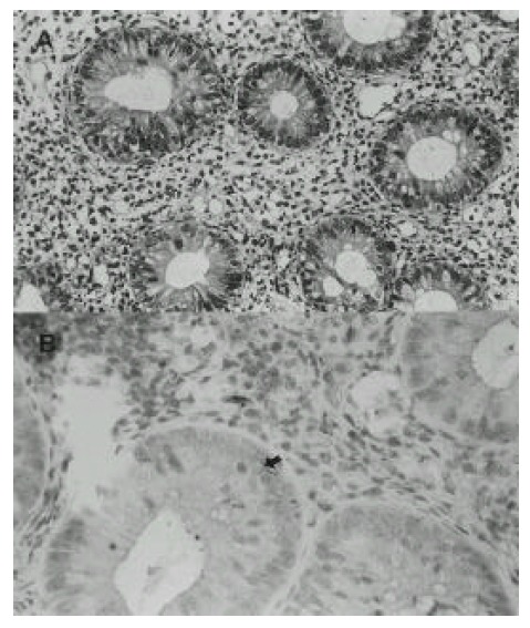
Among 16 UC patients, two patients (2%) with mild inflammation on colonoscopy showed low grade dysplasia on H&E stain. In addition, there were 4 patients who showed p53 overexpression in immunohistochemical stain (Fig. 1) and all of them were also shown to be positive for p53 mutation.
Fig. 1.

A: The lining epithelium of intestinal gland show low-grade dysplasia. (H&E, x400) B: The arrow shows positive immunohistochemical staining with p53 antibody.
3. PCR-SSCP analysis
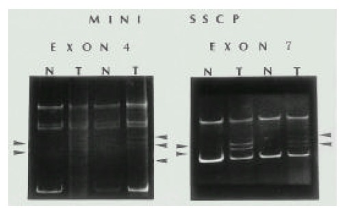
Positive results of SSCP assays in 4 patients performed on exon 4, 5, 6, 7, 8 of p53 are displayed in Table 3. Examples are shown in Figure 2. In the other 12 patients and in normal looking mucosal specimens, no mutations were found by SSCP. Point mutations were observed in 2 histologically normal tissues and 2 patients who showed dysplastic mucosal change.
Table 3.
p53 Mutated Cases: the Results of PCR-SSCP and DNA Sequencing
| Case 1(Pt. 88) | Case 2 | Case 3 | Case 4(Pt. 95) | |
| Exon | 4 | 7 | 7 | 4 |
| Codon | 121 | 249 | 260 | 123 |
| Amino acid | TCT→TGAT | AGG→AGT | TCC→TAC | ACT→ACC+T |
Fig. 2.

The results of PCR-SSCP. In case 1, 4 (exon 4) and case 2, 3 (exon 7) left (exon 4): comparing with N (normal) column, abnormal 2 bands (case 1) were shown in first T (patient) column, 3 bands in second T column, right (exon 7): abnormal 2 bands were shown in first and second T column.
4. DNA Sequencing analysis
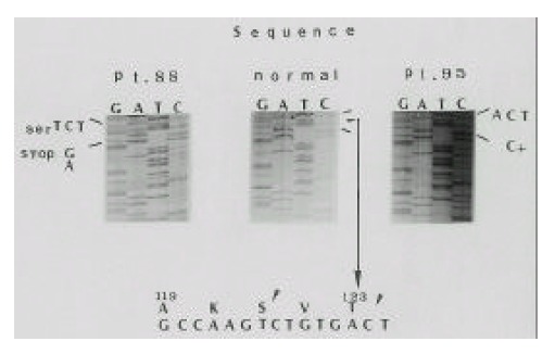
Findings of SSCP were confirmed and further characterized by DNA sequencing in the four SSCP positive patients. All samples had been sequenced repeatedly. Sequence abnormalities included single-base mis-sense mutation in codons 249 (AGG to AGT), 260 (TCC:arginine to TAC:methionine) and 123 (ACT:stop codon to ACC+T) and non-sense mutation in codon 121 (TCT: serine to TGAT: stop codon) (Table 3), (Fig. 3).
Fig. 3.

DNA sequencing of cloned PCR products. Positions of mutated nucleotides are noted. At Pt. 88 (case 1), codon 121, TCT to TGAT (stop codon). At Pt. 95 (case 4), codon 123, ACT to ACC+T.
DISCUSSION
Dysplastic lesion is a marker of colon cancer risk in longstanding ulcerative colitis 9–12). So, many investigators have tried to demonstrate the molecular events in dysplastic mucosa. Among these events, the studies for p53 gene have been undertaken actively. As has been previously reported by a number of investigators, p53 alteration is evidently related to certain steps of the multi-step carcinogenesis, because mutations of p53 gene are proven to be present in a variety of types of human cancer, neurofibrosarcoma, brain tumor, osteosarcoma and colon cancer13).
Most tumors undergo inactivation of both copies of p53, one by allelic deletion and the other by point mutation13). This mechanism appears to have been operative in most of the UC associated neoplastic lesions, suggesting that in UC associated neoplasia, as in sporadic colorectal carcinoma, mutation and allelic deletion may occur closely spaced in time5). p53 LOH appears to be a relatively late genetic event in tumorigenesis in sporadic colon cancers14). On the other hand, the timing of p53 mutation appears to be different between the two diseases. Previous work by Baker et al6). indicates that although p53 mutation does precede LOH in sporadic dolon cancer, it is likely to be a relatively late event in the process of tumorigenesis, occurring during the transition from adenoma to carcinoma5,14–16). Allelic deletion of the p53 gene precedes cancer in UC and can be present in its earliest histologically identifiable precursor such as dysplastic mucosa17). And also, p53 gene mutation was demonstrated in non-invasive dysplasia and dysplasia associated lesion or mass in UC18). The recent report7) that chromosomal abnormality is proceded to the histological phenotype in UC dysplasia, this study suggested that molecular event happens earlier than dysplasia in UC dysplasia-cancer sequence.
In this study, 2 patients whose tissue showed low-grade dysplastic mucosa had p53 genetic alteration. Although the number of cases are limited, this finding corresponds with other recent reports that p53 mutation may occur earlier in UC associated colon cancer than in sporadic colon cancer. The different timing of p53 gene alteration in both diseases may potentially illustrate different roles for p53 mutation in various types of cancer. When p53 mutation occurs as a relatively early event, as appears to be the the case in UC, it may an initial growth advantage.
However, when p53 mutation occurs as a be later event, as appears to be the case in sporadic colon cancer, it may contribute to conversion to malignancy19).
Until now, there have been few studies for detecting the p53 genetic alteration in histologically indefinite or negative for dysplasia tissue. Teresa et al19). reported p53 mutation in histologically indefinite or negative tissue in colectomy specimens. However, these specimens were obtained from regions immediately adjacent to histologically abnormal mucosa. Our study showed that 2 patients, whose tissue has no evidence for dysplasia, have p53 genetic alteration. Overall, 4 cases with p53 genetic alterations were demonstrated in our study, two were negative and two were as two low-grade dysplasia. So, p53 gene mutation may appear to be a relatively early event in carcinogenesis in UC.
Interestingly, all p53 gene mutated cases in this study have shown to be a poor responder group with conventional medical treatment. But, non-mutated cases were relatively good responders to medical treatment, except one patient. However, the duration of this study was too short to evautate the possibility that p53 mutated mucosa would develop to the cancerous change. At any rate, we suggested judiciously that poor response to medical treatment may be an additional clinical risk factor of carcinogenesis in UC. Our results showed that the duration of colitis and the extent did not appear to be related with p53 mutation. But the study population was too small to postulate this assumption.
Wang et al. have reported that p53 protein had been overexpressed in 2 non-dysplastic mucosal specimens20). These 2 specimens were obtained from patients with invasive carcinoma. Also, we observed the p53 protein overexpression by immunohistochemical staining in all p53 gene mutated patients. So, our results support this data and may also support the demonstration of p53 mutations which are proven by PCR-SSCP method.
There is a limitation to our study in the collection of specimens. Many other studies have used microdissection to increase the percentage of epithelial cells to be analyzed. We think that if we had used this method, we may have obtained a higher frequency rate of mutation. In conclusion, we suggest judiciously that p53 gene mutation may play a role for the relatively early stage of carcinogenesis, and the factor of clinically poor response to medical treatment may be the predisposing factor of colon cancer.
REFERENCES
- 1.Lashner BA, Silverstein MD, Hanauer SB. Hazard rates for dysplasia and cancer in ulcerative colitis. Results from a surveillance program. Dig Dis Sci. 1989;34:1536–1541. doi: 10.1007/BF01537106. [DOI] [PubMed] [Google Scholar]
- 2.Gyde SN, Prior P, Allan RN, Stenvens, Jewell DP, Truelove SC, Lofberg R, Brostrom O, Hellers G. Colorectal cancer in ulcerative colitis: a cohort study of primary referrals from their centers. Gut. 1988;29:206–217. doi: 10.1136/gut.29.2.206. [DOI] [PMC free article] [PubMed] [Google Scholar]
- 3.Isbell G, Levin B. Ulcerative colitis and colon cancer. Clin Gastroenterol. 1988;17:773–791. [PubMed] [Google Scholar]
- 4.Riddell RH, Goldman H, Ranshohoff DF, Appelman HD, Fenoglio CM, Haggitt RC, Ahren C, Correa P, Hamilton SR, Morson BC, Sommers SC, Yardley JH. Dysplasia in inflammatory bowel disease: standardized classification with provisional clinical applications. Human Pathol. 1983;14:931–968. doi: 10.1016/s0046-8177(83)80175-0. [DOI] [PubMed] [Google Scholar]
- 5.Guido B, Giovanni B, Gian MP, Fracesco PR, Renat S, Guilio DF, Mario M, Mauro R, Antonio ML, Luigi B. Colorectal cancer in patients with ulcerative colitis A prospective cohort study in Italy. Cancer. 1995;75:2045–2050. doi: 10.1002/1097-0142(19950415)75:8<2045::aid-cncr2820750803>3.0.co;2-x. [DOI] [PubMed] [Google Scholar]
- 6.Baker SJ, Preisinger AG, Jessup JM, Paraskeva C, Markowitz S, Willson JKV, Hamilton S, Vogelstein B. p53 gene mutations occur in combination with 17p allelic deletions as late events in colorectal tumorigenesis. Cancer Res. 1990;50:7717–7722. [PubMed] [Google Scholar]
- 7.Robert FW, Suzane JW, Linda DF, Dan HM, Frederic MW. Chromosomal alterations in ulcerative colitis-related neoplastic progression. Gastroenterology. 1997;113:791–801. doi: 10.1016/s0016-5085(97)70173-2. [DOI] [PubMed] [Google Scholar]
- 8.Sninsky CA, Cort DH, Shanahan F, Powers BJ, Sessions JT, Pruitt RE, Jacobs WH, Lo SK, Targan SR, Cerda JJ, Gremillion DE, Snape WJ, Sabel J, Jinich H, Swinehart JM, DeMicco MP. Oral Mesalamine (Asacol) for mildly to moderately active ulcerative colitis. Ann Intern Med. 1991;115:350–355. doi: 10.7326/0003-4819-115-5-350. [DOI] [PubMed] [Google Scholar]
- 9.Lennard JE, Morson BC, Ritchie JK, Williams CB. Cancer surveillance in ulcerative colitis: Experience over 15 years. Lancet. 1983;ii:149–152. doi: 10.1016/s0140-6736(83)90129-0. [DOI] [PubMed] [Google Scholar]
- 10.Nugent FW, Haggitt RC. Result of long-term prospective surveillance program for dysplasia in ulcerative colitis. Gastroenetrology. 1984;86:1197–1202. [Google Scholar]
- 11.Brostrom O, Lofberg R, Ost A, Reichard H. Cancer surveillance in patients with longstanding ulcerative colitis: a clinical, endoscopical and histological study. Gut. 1986;27:1408–1413. doi: 10.1136/gut.27.12.1408. [DOI] [PMC free article] [PubMed] [Google Scholar]
- 12.Bernstein C, Shanahan F, Weinstein W. Are we telling patients the truth about surveillance colonoscopy in ulcerative colitis? Lancet. 1994;343:71–74. doi: 10.1016/s0140-6736(94)90813-3. [DOI] [PubMed] [Google Scholar]
- 13.Hollstein M, Sidransky D, Vogelstein B, Harris CC. p53 mutations in human cancers. Science. 1991;253:49–53. doi: 10.1126/science.1905840. [DOI] [PubMed] [Google Scholar]
- 14.Baker SJ, Fearon ER, Nigro JM, Hamilton SR, Preisinger AC, Jessup JM, Vantuinen P, Ledbetter DH, Barker DF, Nakamura Y, White R, Vogelstein B. Chromosome 17 deletions and p53 mutations in colorectal carcinomas. Science. 1989;244:217–221. doi: 10.1126/science.2649981. [DOI] [PubMed] [Google Scholar]
- 15.Nigro JM, Baker SJ, Preisinger JC, Jessup JM, Hostetter R, Cleary K, Bigner SH, Davidson N, Baylin S, Devilee P, Glover T, Collins FS, Weston A, Modali R, Harris CC, Vogelstin B. Mutations in the p53 gene occur in diverse human tumor types. Nature. 1989;342:705–708. doi: 10.1038/342705a0. [DOI] [PubMed] [Google Scholar]
- 16.Kern SE, Fearon ER, Termette KW, Enterline JP, Leppert M, Nakamura Y, Hite R, Vogelstein B. Allelic loss in colorectal carcinoma. J Am Med Assoc. 1989;261:3099–3103. doi: 10.1001/jama.261.21.3099. [DOI] [PubMed] [Google Scholar]
- 17.Glenna CB, David AC, Venkateswara RK, Rodger CH, Bruce GK, Cyrus ER, Peter SR. Frequent loss of a p53 allele in carcinomas and their precursors in ulcerative colitis. Cancer Communications. 1991;3:167–172. doi: 10.3727/095535491820873254. [DOI] [PubMed] [Google Scholar]
- 18.Jin YN, Noam H, Yi T, Ying H, Jacqueline L, Bruce DG, Maria H, Carnell N, Stepahen JM. p53 point mutations in dysplastic and cancerous ulcerative colitis lesions. Gastroenterology. 1993;104:1633–1639. doi: 10.1016/0016-5085(93)90639-t. [DOI] [PubMed] [Google Scholar]
- 19.Teresa AB, David AC, Peter SR, Rodger CH, Cyrus ER, Allyn CS, Glenna CB. Mutations in the p53 gene: An early marker of neoplastic progression in ulcerative colitis. Gastroenterology. 1994;107:369–378. doi: 10.1016/0016-5085(94)90161-9. [DOI] [PubMed] [Google Scholar]
- 20.Wang C, Cymes K, Young E, Meltzer SJ, Itzkowitz SH, Harpaz N. p53 overexpression in colonoscopic surveillance biopsies of patients with longstanding ulcerative colitis. Gastroenterology. 1996;4:A611. [Google Scholar]


