Abstract
Redox signaling is a critical component of cell signaling pathways that is involved in regulation of cell growth, metabolism, hormone signaling, immune regulation and variety of other physiological functions. Peroxiredoxin (Prx) is a family of thiol-based peroxidases that acts as a regulator of redox signaling. Members of Prx family can act as antioxidants and chaperone. Sulfiredoxin (Srx) is an antioxidant protein that exclusively reduces over-oxidized typical 2-Cys Prx. Srx have different affinities for individual Prx and it also catalyzes deglutathionylation of variety of substrates. Individual components of Srx-Prx system play critical roles in carcinogenesis by modulating cell signaling pathway involved in cell proliferation, migration and metastasis. Expression levels of individual components of Srx-Prx axis has been correlated with patient survival outcome in multiple cancer types. This review will summarize the molecular basis of differences in affinity of Srx for individual Prx and the role of individual components of Srx-Prx system in tumor progression and metastasis. This enhanced understanding of molecular aspects of Srx-Prx interaction and its role in cell signal transduction will help in defining Srx-Prx system as a future therapeutic target in human cancer.
Keywords: Sulfiredoxin, Peroxiredoxin, Redox signaling, Tumorigenesis, Oncogene
Introduction
Redox signaling is an essential component of various cellular processes that maintain physiological homeostasis in eukaryotes as well as prokaryotes. The intracellular activity regulated by the reactive oxygen/nitrogen species (ROS1/RNS) includes (but is not limited to) growth factor such as EGF [1] and IGF [2] signaling as well as important energy metabolism and hormonal signaling [3]. ROS/RNS have very short half-life partly due to their highly reactive nature and the presence of antioxidants in host organisms. Abnormal accumulation of ROS/RNA leads to oxidative stress, which is known to cause multiple disorders in human, such as diabetes, Alzheimer’s & Parkinson’s disease, hepatic diseases, and cancer [4] [5]. Antioxidants are internal housekeeping (expressed in intracellular or extracellular compartments of animal tissue) or external (part of daily diet or supplements) molecules that get preferentially oxidized under oxidative stress conditions. The biological system expresses multiple antioxidant molecules at intracellular as well as extracellular sites to protect it from oxidative damages. Thiol-based antioxidants are major internal housekeeping antioxidant molecules that acts as redox switches to modulate homeostasis [6]. Peroxiredoxins (Prx) as well as Sulfiredoxin (Srx) are part of thiol-based antioxidant system.
Prx was first discovered about 27 years ago in yeast [7]. These proteins were given multiple names, for example, ‘Protector protein’, ‘Thiol-specific antioxidants (TSA)’, ‘Thioredoxin-linked thiol peroxidase’ and ‘Thioredoxin peroxidase (TPx)’ before they are widely accepted as ‘Peroxiredoxin’ [7; 8; 9; 10; 11; 12]. Prx is a class of thiol-based peroxidases ubiquitously found in prokaryotes as well as eukaryotes. There are six different isoforms of Prx expressed in human [13]. These Prxs are involved in the regulation of cell proliferation, apoptosis, embryonic development, lipid metabolism, immune response etc. [14]. All human Prxs have the enzymatic cysteine called peroxidatic cysteine (CP) on its N-terminus. Five out of six human Prxs also contain one resolving cysteine (CR) on its C-terminus. Depending on the presence and behavior of resolving cysteine, human Prxs are classified into three classes i.e. (i) typical 2-Cys Prxs including Prx1-4, (ii) atypical 2-Cys Prx i.e. Prx5, and (iii) 1-Cys Prx i.e. Prx6 [15]. The Prx family of proteins reduces H2O2, alkyl hydroperoxides and peroxynitrite into water and other harmless metabolites. In this process, the thiol group of peroxidatic cysteine (Cp) is oxidized to sulfenic acid, which can be reduced back by glutaredoxin (Grx) or thioredoxin (Trx)-thioredoxin reductase system [16; 17]. The pKa of most biological cysteine is in the range of 8–9 if not stabilized by other factors, while the pKa of peroxidatic cysteine falls in a lower range of 5–6 due to the stabilization by neighboring conserved arginine and threonine residues in Prxs [18]. The lower pKa of Prxs facilitates their ability to scavenge ROS at very low levels [18]. There is no evidence that residues close to the resolving cysteine have similar function. Therefore, the higher pKa of resolving cysteine making it more resistant to oxidation compared with the peroxidatic cysteine. Since the rate constant of Prx-thiol oxidation is higher than most of thiol-based proteins, Prxs are approximately 105–107 times more efficient than other thiol-based antioxidants such as GSH, Thioredoxin, GAPDH, PTP1B etc [19]. Higher rate constant indicates the ability of Prx to reduce the ROS present even in minute amounts that cannot be eliminated by other antioxidants. Depending on the levels of oxidative stress and amount of Prxs present in the system, the peroxidatic cysteine can be over-oxidized to sulfinic or sulfonic acid, leading to the loss of their antioxidant activity [20]. This hyperoxidation of Prx is present in majority of eukaryotes and few prokaryotes such as cyanobacteria [21]. It is important to clarify that in some older literature the hyperoxidation of Prx was reviewed as unique to eukaryotes. However, the latest research have indicated occurrence of Prx hyperoxidation in prokaryotes too [21]. The hyperoxidation of Prxs helps them to function as molecular chaperone, adding additional role in protein folding besides their function as antioxidants [22]. However, the molecular basis of Prxs to function as chaperone is yet to be determined. More research needs to be carried out to identify proteins whose folding is assisted by Prx. Results of such research will further help to identify different signaling pathways that are modulated by the chaperone function of Prx. Classification of signaling pathways regulated by chaperone as well as antioxidant functions of Prx will help to design better targeting strategy against Prx in tumor cells. The chaperone function of Prxs was used to be considered only to eukaryotes, however, similar activity is also detected in the Prx homolog of prokaryotes such as Helicobacter pylori [23]. Experts were wondering for long time about existence of any enzyme having potential to reduce hyperoxidized Prx until Srx was identified in Saccharomyces cerevisiae [24] and later found to be conserved in higher eukaryotes and few species of cyanobacteria. Rate constants from two independent studies indicates that the reduction of oxidized Prx by Trx (rate constant 106 M−1s−1) is much faster than the rate of reduction of hyperoxidized Prx by Srx (rate constant approximately 2 M−1s−1) [25; 26]. Therefore, reduction of hyperoxidized Prx by Srx can be considered as a rate limiting step in reduction of hyperoxidized Prx. Closest prokaryotic counterpart of Srx is a functionally unrelated protein called ‘ParB’ in bacteria, which carries out function of chromosome partitioning [27]. Oncogenic suppressive activity or ‘Osa’ protein is probably a connecting link between ParB and Srx. Osa contains both DNAse [18] of ParB as well as ATPase domain of Srx [28]. In normal human tissues, Srx is present in kidney, lungs, and pancreas [29]. Srx is mainly a cytosolic protein that can be translocated into mitochondria under oxidative stress conditions [30]. In this manner, Prx along with Srx play an important role in the management of mitochondria redox balance.
The Srx-Prx axis can be explored as therapeutic target as well as therapeutic tools depending on their role in particular pathological condition. For example, individual Prx isoforms can be considered as good therapeutic targets in lung cancer [31], glioblastoma [32], colorectal cancer [33], prostate cancer [34] etc. where they protect tumor cells. It is important to evaluate the risk-benefit ratio of targeting the individual members of Srx-Prx axis as they also have protective role in normal (non-tumor) tissue. The Srx null mice have normal phenotype under laboratory conditions [33]. Prx3 knockout mice also born and mature normally [35]. Prx4 knockout mice have mild prostate atrophy [36]. Prx1 & Prx2 knockout mice are reported to have some issue with erythropoiesis [37; 38]. Hence, majority of proteins in Srx-Prx axis can be knocked-out without any life threatening issue. Considering the risk associated with cancer, it is worth exploring a target that can prolong the lives of patients by few extra years. Hence, benefits associated with targeting Srx or individual Prx outweighs the risk associated with it and Srx-Prx system can be considered a therapeutic target in cancer. On the other hand, individual Prx isoforms can be explored as therapeutic or diagnostic tools in Parkinson’s disease, Alzheimer’s disease, and diabetic complications [39; 40; 41]. These differential properties of individual components of the Srx-Prx system draw our attention towards differences in molecular properties of individual Prx isoforms that gives them ability to play such diverse roles. Improved understanding of these molecular differences will help us in therapeutic intervention of the Srx-Prx system.
Enzymatic roles of Srx
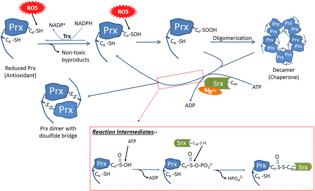
Human Srx has a length of 137 amino acids [42]. Srx is present in mammals, birds and multiple (not all) other eukaryotic organisms and few prokaryotes [43]. It is an exclusive enzyme that acts as an antioxidant to reduce sulfinic acid form of typical 2-Cys Prx [44]. Biteau B et al (2003) identified how ATP-bound yeast Srx in the presence of Mg2+ approaches the hyperoxidized Prx, phosphorylates it and form thiosulfinate intermediate, which can be further reduced by other thiol reducing enzymes [24]. Yeast Srx has two cysteines where the first cysteine (Cys48) helps the enzymatic cysteine (Cys84) by recycling the thiosulfinate intermediate [45]. However, human Srx have only one cysteine i.e. Cys99 (a homologue of Cys84 of yeast). Therefore, it needs an external source of thiol such as thioredoxin (Trx) or Glutathione (GSH) to reduce the thiosulfinate intermediate [45; 46]. The evolution of an ATP consuming process to reactivate Prx after deactivation of its peroxidase function by H2O2 have given a unique advantage to host organism where H2O2 and Srx acts as an On-Off switch for chaperone and peroxidase function of various Prxs. The excess of H2O2 enhances the chaperone function and reduces the peroxidase function of Prx whereas excess of Srx reverses this process [47]. Figure 1 depicts the mechanism by which Srx performs aforementioned antioxidant function. The Prx structure in this figure is designed to give rough idea about the positions of individual cysteines in a typical 2-Cys Prx. The C-terminal resolving cysteine is shown in C-terminal arm and the other cysteine in Prx indicates the N-terminal peroxidatic cysteine.
Figure 1.
Sulfiredoxin specifically reduces hyperoxidized form of typical 2-Cys peroxiredoxins and acts as an on-off switch to keep the balance between antioxidant and chaperone function of Prxs.
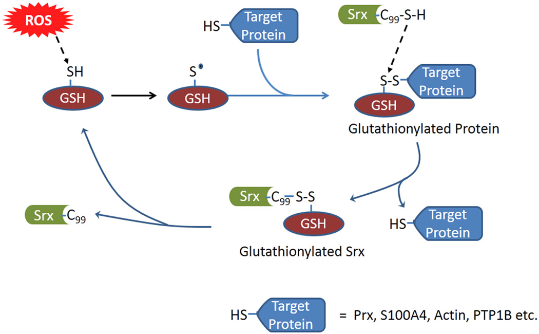
Another important action of Srx involves the deglutathionylation of several substrates in eukaryotes [42]. Most of the Prx-independent and few Prx-dependent functions of Srx is mediated by this mechanism. Figure 2 depicts role of Srx in deglutathionylation process. Srx can regulate the chaperone function of Prx1 by controlling its levels of glutathionylation. The glutathionylation of Cys83 of Prx1 favors formation of dimer over decamer, resulting in the loss of chaperone activity [48]. Although it is a general consensus that Prx-reducing activity of Srx is more important than its deglutathionylation function, more mechanistic studies are required to assess individual contribution of Prx reduction and deglutathionylation processes in regulating the chaperone function of Prx1 or another typical 2-Cys Prx. There is no evidence of tissue specific predominance of one function of Srx over the other. However, there is a great scope for exploration of Srx deglutathionylation function in more details and lack of extensive biochemical studies in this field may be a possible reason behind difficulty in ranking the importance of antioxidant Vs deglutathionylation functions of Srx. Unlike the antioxidant function of Srx that is exclusive to Prx, the deglutathionylation carried out by Srx seems not substrate specific. S100A4, Actin and PTP1B are examples of substrates other than Prx whose glutathionylation levels can be regulated by Srx [22; 49]. There may be other intracellular targets of Srx that can be deglutathionylated by Srx. Identification of such substrates will help to identify different mechanisms by which Srx regulates cell signaling.
Figure 2.
Sulfiredoxin catalyzes deglutathionylation of a variety of substrates.
The molecular characteristics of the Srx-Prx interaction and the substrate specificity of Srx
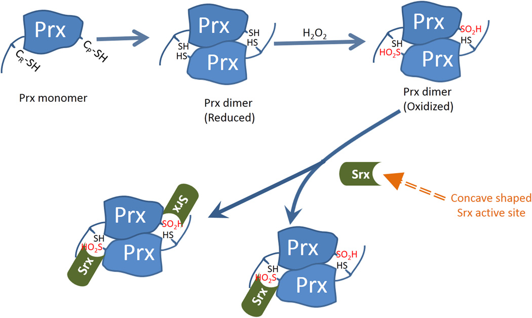
The Cys99 of human Srx is not involved in the Srx-Prx binding but it is directly involved in antioxidant as well as deglutathionylation functions of Srx [44; 47; 50]. Amino acids adjacent to Cys99 i.e. Gly97, Gly98, His100 & Arg101 are considered to be supportive and are also important for the enzymatic activity of Srx [51]. Pro52, Leu82, Phe96, Val118, Val127 and Tyr128 are amino acids that form a hydrophobic pocket in Srx that acts as the interface for Srx-Prx interaction [51; 52]. The hydrophobic pocket formed by the active site of Srx forms a depression that wraps around the slightly protruding active site of Prx [51]. This model of the Srx-Prx interaction is illustrated in figure 3.
Figure 3.
A model of Srx-Prx interaction showing how concave shaped active site of Srx interacts with Prx dimer.
The Prx family of proteins is one of the most abundant and most efficacious antioxidants in human body. The classification of Prx is based mainly on presence and behavior of the resolving cysteine in different Prx isoforms [15]. Individual Prx isoforms also contains few cysteines other than peroxidatic and resolving cysteine that may play some regulatory role in particular protein. For example, Cys83 of Prx1 mediates formation of decameric complex of Prx1 that differentiates the functions of Prx1 from Prx2 [53]. Despite of 78% sequence similarity, one individual cysteine (Cys83) of Prx1 plays such an important role which increases the efficiency of Prx1 to act as a chaperone [53]. Another report has indicated that the Cys83- Cys83 disulfide bond formation is not essential for rat Prx1 as it can form decameric structure through hydrophobic interactions and van der Waals bonds [54]. Glutathionylation of Cys83 has been reported to negatively affect the chaperone function of Prx1 [48]. However, how the glutathionylation impacts the chaperone activity of typical 2-Cys Prx remains to be understood. The number of amino acids between the peroxidatic and resolving cysteine is critical for the formation of the Prx dimer. All human typical 2-Cys Prx have 121 amino acids between the peroxidatic and resolving cysteine, whereas in atypical 2-Cys Prx it is reduced to only 104 amino acid [44]. Another highly conserved feature is the distance of two cysteines from the GGLG motif, which is located between the peroxidatic and resolving cysteines and is 42 amino acids downstream of the peroxidatic cysteine. The YF motif is another feature that localized between the resolving cysteine and the N-terminus, and is 20 amino acids downstream of the resolving cysteine. GGLG and YF motif bestows these Prx with unique ability to get hyperoxidized by H2O2 [55]. The YF motif interacts with the GGLG motif, which causes steric hindrance for the interaction between peroxidatic cysteine of oxidized Prx and resolving cysteine of other monomer. This allows the 2nd H2O2 molecule to react with the peroxidatic cysteine of the first Prx monomer in a timely manner, resulting in the formation of hyperoxidized Prx [56]. The hyperoxidation of typical 2-Cys Prx adds an extra chaperone function to these Prx [22]. In the absence of the GGLG and YF motifs, Prx will not become hyperoxidized, thus they are important for the chaperone function of Prx [55]. There are no reports indicating the involvement of the GGLG and/or YF motif for the Srx-Prx interaction. The GGLG and YF motifs were also identified in prokaryotic Prxs too [21]. The chaperone function is gained by formation of higher molecular weight complexes of Prx that looks like a stack or rings in transmission electron microscopy and X-ray crystallography studies [57]. In some species, hyperoxidation of the peroxidatic cysteine is not absolutely necessary for the gain of chaperone function, as their Prx can form similar structure in the absence of hyperoxidation [58]. However, human Prxs have been known to gain chaperone function only after the peroxidatic cysteine is hyperoxidized. Also, the loss of C-terminal arm of Prx results in the loss of chaperone function [59]. Even among the typical 2-Cys Prx, the susceptibility to hyperoxidation varies. Prx3 is considered more resistant to hyperoxidation than other isoforms [60]. The conservation of amino acids around the peroxidatic cysteine probably indicates their importance for the enzymatic activity of Prx or a particular behavior of a Prx isoform. For example, most Prxs have a Proline and a Threonine (occasionally Serine) before the peroxidatic cysteine, which results in a PXXXTXXC motif that may be importance for the enzymatic activity of Prx [61]. In human typical 2-Cys Prxs, amino acids around the peroxidatic cysteine (i.e. PLDFTFVCPTEI motif) and the resolving cysteine (i.e. HGEVCPAXW motif) are highly conserved [62], which may indicate their importance [63]. However, the significance of these amino acids has not been experimentally proved yet and it may be of interest for further studies..
Although all typical 2-Cys Prx are generally considered as substrate of Srx, the affinity of Srx to individual Prx is not the same [31]. Data from our lab suggest that the orientation of C-terminal arm of Prx may affect the affinity of Srx for individual Prx (unpublished). Srx have highest affinity for Prx4 among all the typical 2-Cys Prx [31]. However, it still needs to be studied how this high affinity of interaction affects the kinetics of Prx4 reduction compared to other Prx. Members of the Prx family may have different subcellular localization, and their abundance in different tissues also varies. The interaction between Srx and different isoform of Prx is thus also affected by their subcellular localization. For example, Srx-Prx3 interaction is not significant under low oxidative stress conditions due to mitochondrial location of Prx3, however, this interaction becomes significant under higher oxidative stress conditions where mitochondrial membrane is damaged and hence Srx gets a chance to translocate from cytosol to mitochondria [30]. An alternative explanation of this phenomena is that, Prx3 can get over-oxidized only at higher oxidative stress levels due to its high resistance to over-oxidation [60]. Probably some molecular characteristics of Prx3 do not allow Srx-Prx3 interaction under reduced conditions and interaction is possible only after molecular rearrangements during the oxidation or over-oxidation of Prx3. However, more mechanistic studies are required to clarify whether this is the case, or Srx can bind to Prx3 only in its oxidized/over-oxidized state. All these molecular factors affect the signaling of the Srx-Prx axis. Differential affinity of Srx for individual Prx as well as molecular characteristics of individual Prx allow them to regulate a myriad range of cell signaling.
The Srx-Prx axis in tumorigenesis and cancer progression
The main function of the Srx-Prx system is to protect host cells from oxidative damages. This property of the Srx-Prx system becomes harmful to host organism when it starts protecting the survival of tumor cells. As per the data from Oncomine (an online microarray database) [64] and other published literature, the Srx-Prx system is altered in multiple types of cancer. Table 1 summarizes different types of cancer in which expression of individual members of Srx-Prx system is altered. The information in Table 1 indicates changes in mRNA expression. The up-regulation indicates more than 1.5 fold increase in mRNA levels whereas down-regulation indicates more than 1.5 fold decrease in mRNA levels. Apart from table 1, we also notice the alterations at the protein level. The information about expression changes at places other than table 1 are mainly based on studies of their protein levels. The correlation between patient survival and protein expression changes has not been studied. From published data in literature, the Srx-Prx system predominantly functions as activators or enhancers of oncogenic signaling to promote cancer development. There are also studies reporting that members of Prxs repress cancer development by acting as tumor suppressors, suggesting that Prx may function as double-edged sword in tumorigenesis. Therefore, the exact role of individual component of the Srx-Prx system in cancer can be complicated, and should be considered under specified context of cancer and cell types..
Table 1.
Expression pattern of Srx-Prx system in different cancer types as evident from microarray data available at Oncomine online microarray dataset; Up-regulation is classified as more than 1.5 fold increase in expression compared to normal non-tumor cells; Down-regulation is classified as more than 1.5 fold decrease in expression compared to normal non-tumor cells. Data summarized here is the one that could be confirmed by other independent studies.
| Protein | Up-Regulation | Down-regulation |
|---|---|---|
| Srx | Breast cancer, Colorectal Cancer, Lung Cancer, Prostate Cancer, Skin cancer | Esophageal Cancer |
| Prx1 | Bladder cancer, Colorectal cancer, Gastric cancer, Leukemia, Liver Cancer, Lymphoma, Breast Cancer, Pancreatic cancer, Sarcoma | Esophageal Cancer, Head & Neck cancer, Myeloma |
| Prx2 | Colorectal cancer, Lung cancer, Lymphoma, Myeloma, Ovarian cancer | Brain & CNS cancer, Esophageal Cancer, Head & Neck cancer, Kidney cancer, Leukemia, Pancreatic cancer, Sarcoma |
| Prx3 | Gastric cancer, Head & Neck cancer, Lymphoma, Prostate Cancer | Bladder cancer, Brain & CNS cancer, Kidney cancer, Leukemia, Pancreatic cancer |
| Prx4 | Bladder cancer, Brain & CNS cancer, Breast cancer, Cervical cancer, Colorectal cancer, Head & Neck cancer, Kidney cancer, Lung cancer, Lymphoma, Melanoma, Prostate Cancer, Sarcoma | Leukemia, Liver cancer, Pancreatic cancer |
Srx in cell-signal transduction and tumorigenesis
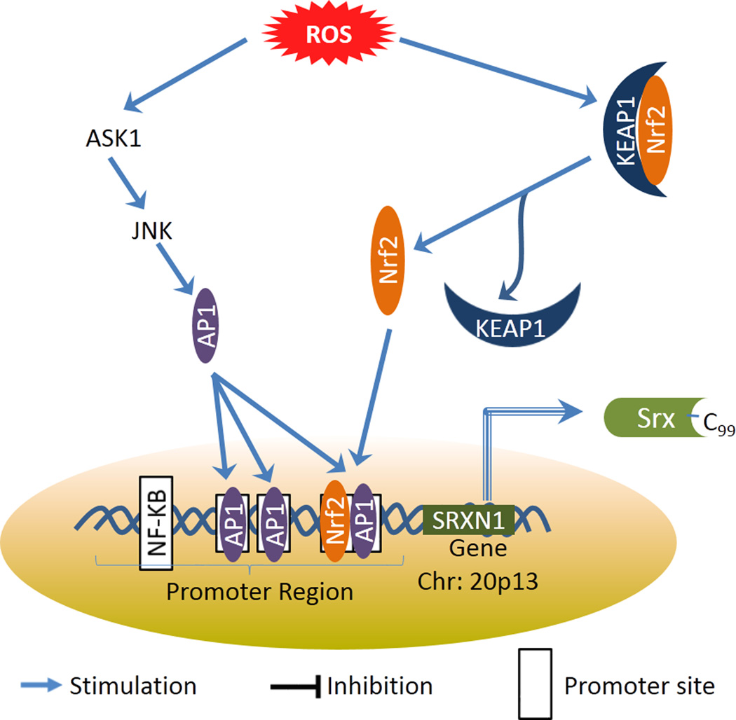
The expression of Srx is regulated by different factors at both transcriptional and translational levels. Redox signaling is the major component that activates Srx expression. Figure 4 summarizes how the expression of Srx is regulated by redox signaling. Activation of transcription factors, such as nuclear factor erythroid 2-related factor 2 (Nrf2), induce Srx expression [65]. Activator Protein-1 (AP-1) also up-regulates Srx expression [66]. c-Jun is a component of AP-1 complex and its activation stimulates Srx expression. TAM67 is an N-terminal deletion mutant of c-Jun and it acts as a c-Jun antagonist. Therefore, TAM67 can negatively regulate Srx expression by inhibiting the activity of AP-1 complex [67]. Multiple intracellular as well as extracellular factors such as nitric oxide (NO), cigarette smoke, dietary derived electrophiles and tumor promoters like 12-O-tetradecanoylphorbol-13-acetate (TPA) that lead to the activation of nrf2 or AP-1 have the potential to stimulate the expression of Srx [67; 68]. In mouse macrophages, treatment with lipopolysaccharide strongly induces Srx expression in an Nrf2 and AP1 dependent manner, and the absence of either significantly affect the levels of Srx induction [69]. Besides aforementioned transcriptional regulation, Srx expression is negatively regulated at translational level by cAMP-PKA (cyclic AMP-Protein kinase A) through the elF2 kinase Gcn2 [70].
Figure 4.
Oxidative stress stimulates Sulfiredoxin expression by regulating AP-1 and Nrf2 activity.
Srx is over-expressed in a variety of cancer and it may promote carcinogenesis in Prx-dependent as well as independent manner [31; 49]. It promotes tumor progression in lung cancer by enhancing intracellular phosphokinase signaling such as mitogen-activated protein kinase (MAPK) and AP-1/MMP9 (Matrix metalloproteinase 9) signaling in Prx4-dependent manner [31]. It may also enhance cell migration in lung cancer in a Prx-independent manner by interacting with S100A4 (a calcium binding protein) and non-muscle myosin IIA (NMIIA) [49]. Aberrant expression of Srx in lung squamous cell carcinoma, lung adenocarcinoma, and pancreatic cancer is correlated with poor survival in those patients [71; 72; 73]. Srx protein is also over-expressed in renal cell carcinoma where it is proposed to be a good antibody target that can result in tumor cell death [74]. Srx expression is stimulated by TPA via MAPK/JNK (c-Jun N-terminal kinase) pathway in skin carcinogenesis and Srx depletion at least partially protects mice against DMBA(7,12-dimethylbenz[a]anthracene)/TPA-induced skin carcinogenesis [75]. Srx is also necessary for colon carcinogenesis as it is highly over-expressed in colon tumor tissue compared to normal human colon, and Srx null mice are highly resistant to azoxymethane/dextran sulfate sodium-induced colon carcinogenesis [33]. Although the importance of Srx in various tumor types is well established, we still need a lot of research to understand the mechanism by which Srx plays its role in tumor progression and metastasis. Considering lung cancer as an example, the antioxidant and deglutathionylation activities of Srx may work in tandem to enhance the chances of tumor promotion and metastasis [31; 49]. However, more studies are required before we can rank their individual contribution towards cancer. Unraveling the mechanistic details of Srx signaling will further help us in designing a better approach to target tumors in which Srx plays an essential role.
Prx1 in cell-signal transduction and tumorigenesis
Prx1 is mainly localized in the cytoplasm, but can also be found in the nuclear [76]. The expression of Prx1 is regulated at both transcriptional as well as post-transcriptional levels. At the transcriptional level, Nrf2 directly activates its expression [77]. Focal Adhesion Kinase (FAK) is also reported to be involved in transcriptional regulation of Prx1 [78]. In one study, Prx1 null mice were shown to be prone to spontaneous tumor development [37], suggesting that Prx1 may function as a tumor suppressor However, Prx1 null mice developed in another lab are normal and free of tumor development [79]. The tumor suppressor function of Prx1 may be mediated by its regulation of PTEN levels as indicated in a mouse breast cancer model [80]. Also, PTEN null mouse embryonic fibroblasts are resistant to ROS mediated induction of Prx1/Prx2 expression [81]. Prx1 may also be required for the ROS mediated activation of the K-Ras/ERK pathway that contributes to lung tumorigenesis [82]. Moreover, Prx1 along with Prx4 play essential roles in the regulation of c-Jun and AP-1 mediated promoter activity in lung cancer cells [83], and activation of Prx1 by histone deacetylase inhibitor FK228 result in induction of apoptosis in esophageal tumor cells [84]. Furthermore, Prx1 helps reactivate DEP-1, a protein tyrosine phosphatase that functions as tumor suppressor,) by reducing the levels of ROS [85]. Aforementioned mechanisms are few example mechanisms by which Prx1 acts as a tumor suppressor.
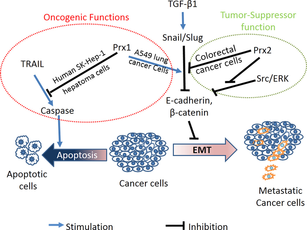
On the other hand, there are many reports indicating that Prx1 has an essential pro-oncogenic role in cancer. For example, Prx1 promotes the vascular endothelial growth factor (VEGF) expression in Toll-like receptor 4 (TLR4)-dependent manner. This effect of Prx1 enhances angiogenesis and results in an environment favorable for tumor cell proliferation and promotes tumor progression in prostate cancer [86; 87]. Prx1 is over-expressed in esophageal cancer cells and has an auto-immunogenic activity [88]. Prx1 protein is also found aberrantly increased in early stage endometrial cancer where its functional significance is yet to be established [89]. Prx1 induces TRAIL (tumor necrosis factor–related apoptosis-inducing ligand) resistance by suppressing the redox-dependent activation of caspase [90]. TRAIL is a biological agent that induces apoptosis of cancer cells and is considered a promising anticancer agent [91]. Down-regulation of Prx1 using RNA interference or chemical agents like dioscin results in the induction of apoptosis in tumor cells [92; 93]. Also, in A549 lung adenocarcinoma cells Prx1 enhances the TGF-β1 induced epithelial-mesenchymal transition (EMT) by stimulating the expression of snail and slug, two transcription factors that inhibits E-cadherin expression [94]. For this function, the Cys51 (peroxidatic cysteine) of Prx1 is essential as replacement of Cys51 by Ser nullifies such effects [94]. Another study using murine hepatocytes as well as human esophageal and lung cancer cell lines reports that the TGF-β1 enhances the ROS production by up-regulating the levels of ferritin heavy chain (FHC) and intracellular labile iron pool (LIP) [95]. It can be inferred from these studies that ROS produced by TGF-β1 signaling probably oxidizes the peroxidatic cysteine of Prx1 and this oxidation is essential for role of Prx1 in EMT. Therefore, higher levels of ROS may promote of the progress of EMT. On the other hand, oxidation of Prx1 will reduce the levels of ROS. Whether and how the hyperoxidation of Prx1 and its molecular chaperone activity are involved in the process of EMT are largely unknown. Figure 5 depicts how Prx can perform both tumor suppressor as well as oncogenic functions. However, the factors that determine the dominance of one role over other are yet to be studied in more details. It is possible that Prx1 functions as a tumor suppressor before the transformation of a normal cell to tumor, but after transformation it promotes tumor cell proliferation by protecting them from ROS-induced cell death. Other possible explanations may be related with the single nucleotide polymorphism (SNPs) or allelic variants of Prx1 but none of these factors have been investigated in detail in the literature.
Figure 5.
Peroxiredoxin may act as tumor-suppressor or oncogene depending on the context of tumor type.
Prx2 in cell-signal transduction and tumorigenesis
Prx2 is the 2nd member of the typical 2-Cys Prxs that are mainly present in cytosol [76]. It is one of the most efficient H2O2 scavenger in cell compared to majority of other antioxidants [96]. In red blood cells (RBCs), the oxidation-reduction cycle of Prx2 correlates with the circadian rhythm, which results in circadian rhythm dependent oligomerization of Prx2 [97]. This oscillation in levels of hyperoxidized Prx2 is not controlled at the transcriptional level since RBCs do not have a nucleus [97], and is not likely controlled by Srx as the oscillations existed in Srx null mice [98]. It is rather controlled by hemoglobin autoxidation and 20S proteasome in RBCs [98]. Extensive methylation of CpG islands in the promoter region of the Prdx2 gene is one of the mechanisms to control Prx2 expression in melanoma [99]. Prx2 expression is also regulated by transcription factor Hand1/Hand2 [100]. In mouse embryonic fibroblasts, Prx2 is induced by ROS in a PTEN dependent manner [81]. As mentioned earlier, the PTEN activation itself is regulated by Prx1, therefore, it can be assumed that Prx1 may have potential to affect Prx2 expression too. Prx2 is down-regulated in few cancers where Prx1 is up-regulated, but the exact mechanism behind these differential expression is not available yet [101; 102]. Whether or not PTEN is responsible for this relationship between Prx1 and Prx2 expression in those tissues, is still a question. Nitrosylation of Tyr193 in the YF motif of Prx2 is an important post-translational modification that plays a critical role in the regulation of disulfide bond formation under oxidative stress conditions [103]. Glutathionylation is another post-translational modification of Prx2, which may affect its localization to extracellular compartment [104]. The extracellular glutathionylated Prx2 induces the TNFα production and leads to oxidative stress dependent inflammatory reaction [104]. In this manner, Prx2 plays a role in cytokine mediated inflammatory signaling. The serum levels of Prx2 in colorectal cancer are correlated with the survival of patients [105]. In human papillomavirus (HPV) related cervical cancer, increased expression of Prx2 is proposed to mediate the carcinogenesis in cervical tissue [106; 107]. However, more studies are required to establish whether the alteration of Prx2 is a cause or effect of carcinogenesis. Prx2 is the main factor determining the metabolic stress and oxidative stress response of breast cancer cells metastasized to lung [108]. It also regulates the activation of transcription factor STAT3 by transferring the oxidative equivalents to later resulting in the generation of disulfide-linked inactive STAT3 oligomer [96]. Prx2 reduces the chances of metastasis by negatively regulating Src/ERK activation, resulting in increased E-cadherin expression and β-catenin retention [109]. Prx2 overexpression also reduces the chances of TGF-β1 induced EMT and cell migration in colorectal cancer cells [110]. It is interesting to note that the effect of Prx2 on TGF-β1 induced EMT in colorectal cancer cells is exactly opposite to the effect of Prx1 on same signaling pathway in A549 cells, which is discussed earlier in this review and depicted in figure 5. However, it is not clear yet whether these activities are regulated in a tissue-specific manner or they co-exist in same cancer type too.
Prx3 in cell-signal transduction and tumorigenesis
Prx3 is primarily a mitochondrial Prx. The expression of Prx3 is enhanced by SirT1 in partnership with FoxO3a and PGC1α, and the absence of either leads to its down-regulation [111]. SirT1 enhances the complex formation of FoxO3a with PGC1α and this complex regulates the Prx3 as well as multiple other antioxidant protein expressions [111]. Prx3 expression is also regulated by superoxide dismutase (SOD) through an unknown mechanism [112]. Prx3 is a downstream target of c-Myc transcription factor and it acts as a major mediator for the regulation of C-Myc functions in cell transformation, tumor progression and apoptosis [113]. In medulloblastoma, Prx3 is a target of MiR-383 (a microRNA), and its expression reduces cell proliferation [114]. In cervical cancer, Prx3 is over-expressed and its levels are correlated with increased rate of cell proliferation [115]. SNP RS7082598 of PRDX3 gene is correlated with a reduced risk of cervical cancer [116]. In lung squamous cell carcinoma, Prx3 is over-expressed along with increased Srx in an Nrf2 dependent manner, which indicates a potentially important role of the Srx-Prx3 axis in these tumors [71]..
Prx4 in cell-signal transduction and tumorigenesis
Prx4 is the 4th member of typical 2-Cys Prx family which resides mainly in endoplasmic reticulum (ER). There is also a low molecular weight secretory form of Prx4, which can be found in extracellular matrix and plasma. Although there are few reports about the post-transcriptional regulation of Prx4, how this protein is regulated at the transcriptional level is yet to be studied. Calpain (a calcium-dependent cysteine protease) can enhance the expression of Prx4 through post-transcriptional regulation [117].
Besides its regular antioxidant function, Prx4 also mediates the oxidative folding of various endoplasmic reticulum proteins through its chaperone function, which may accomplished through the cooperation of protein disulfide isomerase (PDI) [118]. Data in our lab indicate that Prx4 is susceptible to hyperoxidation at very low levels of oxidative stress (unpublished), which may facilitate its molecular chaperone function. Prx4 improves insulin synthesis by enhancing the endoplasmic reticulum folding of insulin and thus improves pancreas β-cell function [119]. In pancreatic cancer, Prx4 is reported to be downregulated [64]. However, it is not clear whether the Prx4 downregulation is a cause or effect of pancreatic cancer. Expression of Prx4 promotes the metastatic potential of lung adenocarcinoma cells [83]. Prx4 along with Srx increases RAS-RAF-MEK signaling by enhancing intracellular phosphokinase signaling [31]. RAS-RAF-MEK pathway is well known for controlling cancer cell proliferation and metastasis in various types of cancer. Therefore, the ability of Srx-Prx4 system to modulate this pathway indicates their importance in cancer development. The exact mechanism by which Srx or Prx4 carries out regulation of RAS-RAF-MEK pathway still needs to be identified. Theoretically, an ROS dependent mechanism may be involved since Srx restores the antioxidant function of Prx4 [31]. Moreover, Prx4 is a downstream mediator of Srx in lung cancer development, which is demonstrated by the recapitulation of reduced tumor phenotypes in Srx knockdown cells by knockdown of Prx4 (i.e. reduction in anchorage independent colony formation, cell migration, and invasion) [31]. There are other few typical 2-Cys Prx isoforms that may have similar effect in other pathological or physiological conditions, but such a strong relationship of Srx and Prx4 in lung cancer has not been reported before. Furthermore, Prx4 is over-expressed in the majority of cancers where Srx is overexpressed (Refer to Table 1) [64]. In prostate cancer, over-expressed Prx4 enhances the rate of cell proliferation [120]. In oral cavity squamous cell carcinoma, expression of Prx4 enhances cancer metastasis [121]. In colorectal cancer, high expression of Prx4 is correlate with poor survival of patients [122]. As mentioned before, Srx is also highly expressed in colon cancer and is required for chemical induced colon carcinogenesis [33]. Therefore, it may be of interest to study the significance of Srx and Prx4 in colon cancer.
Conclusions
Srx is an exclusive enzyme that reduces over-oxidized forms of typical 2-Cys Prxs. The Srx-Prx interaction plays critical roles in a variety of physiological as well as pathological conditions involving redox signaling. Molecular characteristics of Srx have been studied in great details as most of the important amino acids that are involved in the Srx-Prx interaction as well as deglutathionylation reaction are already known. However, the molecular structure of Prxs needs to be further explored to identify essential amino that impacts the formation of the Srx-Prx complex. Although some information is available about the cross-talk of the Srx-Prx axis in several signaling pathways, factors that affect these cross-talks are largely unknown and how individual isoform of Prxs contributes to different signaling pathways remains elusive. It is also necessary to differentiate the contribution of the antioxidant function of Prx and its molecular chaperone function in terms of impacting signaling transduction. Prx is clearly shown to play protective role in cardiovascular and neurological diseases. However, its role in cancer is still controversial due to both tumor-suppressor as well as oncogenic roles played by Prx isoforms in different cancer types. Special attention need to be paid to mechanism by which same Prx isoform can play different and sometimes opposite roles in different cancer types. Post-translational modifications of Prx may be one of the mechanisms that contribute to the dual behavior of Prx. Other possible explanations may include the presence of allelic variants or single nucleotide polymorphism of the Prx genes. More in-depth mechanistic studies in the future will help to unravel the interweaved behavior of Prxs and lead to the development of better therapeutic strategies for cancer prevention or treatment.
Highlights.
Specificity of individual Prx signaling is determined by minor molecular changes
Difference in Srx-individual Prx affinity is defined by their molecular differences
All enzymatic activities of Srx/Prx collaborates to maintain cellular homeostasis
Srx-Prx axis regulates carcinogenesis through modulation of cell-signaling pathways
Srx-Prx axis is a promising therapeutic target in variety of human cancers
Acknowledgement
This work was partially supported by the National Institutes of Health, with funding from the National Cancer Institute grant number R00-CA149144 to Q. Wei
Footnotes
Publisher's Disclaimer: This is a PDF file of an unedited manuscript that has been accepted for publication. As a service to our customers we are providing this early version of the manuscript. The manuscript will undergo copyediting, typesetting, and review of the resulting proof before it is published in its final form. Please note that during the production process errors may be discovered which could affect the content, and all legal disclaimers that apply to the journal pertain.
AP-1, Activator Protein-1; EGF, Epidermal growth factor; EMT, Epithelial-mesenchymal transition; ERK, Extracellular-signal-regulated kinases; MAPK, Mitogen-activated protein kinase; Nrf2, Nuclear factor erythroid 2-related factor 2; PRDX, Peroxiredoxin gene; Prx, Peroxiredoxin; PTEN, Phosphatase and tensin homolog; RNS, Reactive nitrogen species; ROS, Reactive oxygen species; Srx, Sulfiredoxin; TGF-β1, Transforming growth factor beta 1; TLR, Toll-like receptor; TPA, 12-O-tetradecanoylphorbol-13-acetate; Trx, Thioredoxin; TRAIL, Tumor necrosis factor–related apoptosis-inducing ligand.
Conflict of Interest Statement
None
References
- 1.Palanivel K, Kanimozhi V, Kadalmani B, Akbarsha MA. Verrucarin A induces apoptosis through ROS-mediated EGFR/MAPK/Akt signaling pathways in MDA-MB-231 breast cancer cells. Journal of cellular biochemistry. 2014 doi: 10.1002/jcb.24874. [DOI] [PubMed] [Google Scholar]
- 2.Oh YI, Kim JH, Kang CW. Protective effect of short-term treatment with parathyroid hormone 1–34 on oxidative stress is involved in insulin-like growth factor-I and nuclear factor erythroid 2-related factor 2 in rat bone marrow derived mesenchymal stem cells. Regulatory peptides. 2014;189:1–10. doi: 10.1016/j.regpep.2013.12.008. [DOI] [PubMed] [Google Scholar]
- 3.Diano S. Role of reactive oxygen species in hypothalamic regulation of energy metabolism. Endocrinology and metabolism. 2013;28:3–5. doi: 10.3803/EnM.2013.28.1.3. [DOI] [PMC free article] [PubMed] [Google Scholar]
- 4.Rochette L, Zeller M, Cottin Y, Vergely C. Diabetes, oxidative stress and therapeutic strategies. Biochimica et biophysica acta. 2014;1840:2709–2729. doi: 10.1016/j.bbagen.2014.05.017. [DOI] [PubMed] [Google Scholar]
- 5.Nakamura T, Cho DH, Lipton SA. Redox regulation of protein misfolding, mitochondrial dysfunction, synaptic damage, and cell death in neurodegenerative diseases. Experimental neurology. 2012;238:12–21. doi: 10.1016/j.expneurol.2012.06.032. [DOI] [PMC free article] [PubMed] [Google Scholar]
- 6.Groitl B, Jakob U. Thiol-based redox switches. Biochimica et biophysica acta. 2014;1844:1335–1343. doi: 10.1016/j.bbapap.2014.03.007. [DOI] [PMC free article] [PubMed] [Google Scholar]
- 7.Kim K, Kim IH, Lee KY, Rhee SG, Stadtman ER. The isolation and purification of a specific "protector" protein which inhibits enzyme inactivation by a thiol/Fe(III)/O2 mixed-function oxidation system. The Journal of biological chemistry. 1988;263:4704–4711. [PubMed] [Google Scholar]
- 8.Lim YS, Cha MK, Kim HK, Uhm TB, Park JW, Kim K, Kim IH. Removals of hydrogen peroxide and hydroxyl radical by thiol-specific antioxidant protein as a possible role in vivo. Biochemical and biophysical research communications. 1993;192:273–280. doi: 10.1006/bbrc.1993.1410. [DOI] [PubMed] [Google Scholar]
- 9.Cha MK, Kim HK, Kim IH. Thioredoxin-linked "thiol peroxidase" from periplasmic space of Escherichia coli. The Journal of biological chemistry. 1995;270:28635–28641. doi: 10.1074/jbc.270.48.28635. [DOI] [PubMed] [Google Scholar]
- 10.Ichimiya S, Davis JG, O'Rourke DM, Katsumata M, Greene MI. Murine thioredoxin peroxidase delays neuronal apoptosis and is expressed in areas of the brain most susceptible to hypoxic and ischemic injury. DNA and cell biology. 1997;16:311–321. doi: 10.1089/dna.1997.16.311. [DOI] [PubMed] [Google Scholar]
- 11.Stacy RA, Munthe E, Steinum T, Sharma B, Aalen RB. A peroxiredoxin antioxidant is encoded by a dormancy-related gene, Per1, expressed during late development in the aleurone and embryo of barley grains. Plant molecular biology. 1996;31:1205–1216. doi: 10.1007/BF00040837. [DOI] [PubMed] [Google Scholar]
- 12.Zhou Y, Wan XY, Wang HL, Yan ZY, Hou YD, Jin DY. Bacterial scavengase p20 is structurally and functionally related to peroxiredoxins. Biochemical and biophysical research communications. 1997;233:848–852. doi: 10.1006/bbrc.1997.6564. [DOI] [PubMed] [Google Scholar]
- 13.Dammeyer P, Arner ES. Human Protein Atlas of redox systems - what can be learnt? Biochimica et biophysica acta. 2011;1810:111–138. doi: 10.1016/j.bbagen.2010.07.004. [DOI] [PubMed] [Google Scholar]
- 14.Hanschmann EM, Godoy JR, Berndt C, Hudemann C, Lillig CH. Thioredoxins, glutaredoxins, and peroxiredoxins--molecular mechanisms and health significance: from cofactors to antioxidants to redox signaling. Antioxidants & redox signaling. 2013;19:1539–1605. doi: 10.1089/ars.2012.4599. [DOI] [PMC free article] [PubMed] [Google Scholar]
- 15.Rhee SG, Kang SW, Chang TS, Jeong W, Kim K. Peroxiredoxin, a novel family of peroxidases. IUBMB life. 2001;52:35–41. doi: 10.1080/15216540252774748. [DOI] [PubMed] [Google Scholar]
- 16.Arner ES, Holmgren A. Physiological functions of thioredoxin and thioredoxin reductase. European journal of biochemistry / FEBS. 2000;267:6102–6109. doi: 10.1046/j.1432-1327.2000.01701.x. [DOI] [PubMed] [Google Scholar]
- 17.Dietz KJ. Plant peroxiredoxins. Annual review of plant biology. 2003;54:93–107. doi: 10.1146/annurev.arplant.54.031902.134934. [DOI] [PubMed] [Google Scholar]
- 18.Zeida A, Reyes AM, Lebrero MC, Radi R, Trujillo M, Estrin DA. The extraordinary catalytic ability of peroxiredoxins: a combined experimental and QM/MM study on the fast thiol oxidation step. Chemical communications. 2014;50:10070–10073. doi: 10.1039/c4cc02899f. [DOI] [PMC free article] [PubMed] [Google Scholar]
- 19.Winterbourn CC. The biological chemistry of hydrogen peroxide. Methods in enzymology. 2013;528:3–25. doi: 10.1016/B978-0-12-405881-1.00001-X. [DOI] [PubMed] [Google Scholar]
- 20.Rabilloud T, Heller M, Gasnier F, Luche S, Rey C, Aebersold R, Benahmed M, Louisot P, Lunardi J. Proteomics analysis of cellular response to oxidative stress. Evidence for in vivo overoxidation of peroxiredoxins at their active site. The Journal of biological chemistry. 2002;277:19396–19401. doi: 10.1074/jbc.M106585200. [DOI] [PubMed] [Google Scholar]
- 21.Pascual MB, Mata-Cabana A, Florencio FJ, Lindahl M, Cejudo FJ. Overoxidation of 2-Cys peroxiredoxin in prokaryotes: cyanobacterial 2-Cys peroxiredoxins sensitive to oxidative stress. The Journal of biological chemistry. 2010;285:34485–34492. doi: 10.1074/jbc.M110.160465. [DOI] [PMC free article] [PubMed] [Google Scholar]
- 22.Jang HH, Lee KO, Chi YH, Jung BG, Park SK, Park JH, Lee JR, Lee SS, Moon JC, Yun JW, Choi YO, Kim WY, Kang JS, Cheong GW, Yun DJ, Rhee SG, Cho MJ, Lee SY. Two enzymes in one; two yeast peroxiredoxins display oxidative stress-dependent switching from a peroxidase to a molecular chaperone function. Cell. 2004;117:625–635. doi: 10.1016/j.cell.2004.05.002. [DOI] [PubMed] [Google Scholar]
- 23.Chuang MH, Wu MS, Lo WL, Lin JT, Wong CH, Chiou SH. The antioxidant protein alkylhydroperoxide reductase of Helicobacter pylori switches from a peroxide reductase to a molecular chaperone function. Proceedings of the National Academy of Sciences of the United States of America. 2006;103:2552–2557. doi: 10.1073/pnas.0510770103. [DOI] [PMC free article] [PubMed] [Google Scholar]
- 24.Biteau B, Labarre J, Toledano MB. ATP-dependent reduction of cysteine-sulphinic acid by S. cerevisiae sulphiredoxin. Nature. 2003;425:980–984. doi: 10.1038/nature02075. [DOI] [PubMed] [Google Scholar]
- 25.Tairum CA, Jr, de Oliveira MA, Horta BB, Zara FJ, Netto LE. Disulfide biochemistry in 2-cys peroxiredoxin: requirement of Glu50 and Arg146 for the reduction of yeast Tsa1 by thioredoxin. Journal of molecular biology. 2012;424:28–41. doi: 10.1016/j.jmb.2012.09.008. [DOI] [PubMed] [Google Scholar]
- 26.Roussel X, Bechade G, Kriznik A, Van Dorsselaer A, Sanglier-Cianferani S, Branlant G, Rahuel-Clermont S. Evidence for the formation of a covalent thiosulfinate intermediate with peroxiredoxin in the catalytic mechanism of sulfiredoxin. J Biol Chem. 2008;283:22371–22382. doi: 10.1074/jbc.M800493200. [DOI] [PubMed] [Google Scholar]
- 27.Basu MK, Koonin EV. Evolution of eukaryotic cysteine sulfinic acid reductase, sulfiredoxin (Srx), from bacterial chromosome partitioning protein ParB. Cell cycle. 2005;4:947–952. doi: 10.4161/cc.4.7.1786. [DOI] [PubMed] [Google Scholar]
- 28.Maindola P, Raina R, Goyal P, Atmakuri K, Ojha A, Gupta S, Christie PJ, Iyer LM, Aravind L, Arockiasamy A. Multiple enzymatic activities of ParB/Srx superfamily mediate sexual conflict among conjugative plasmids. Nature communications. 2014;5:5322. doi: 10.1038/ncomms6322. [DOI] [PMC free article] [PubMed] [Google Scholar]
- 29.Chang TS, Jeong W, Woo HA, Lee SM, Park S, Rhee SG. Characterization of mammalian sulfiredoxin and its reactivation of hyperoxidized peroxiredoxin through reduction of cysteine sulfinic acid in the active site to cysteine. The Journal of biological chemistry. 2004;279:50994–51001. doi: 10.1074/jbc.M409482200. [DOI] [PubMed] [Google Scholar]
- 30.Noh YH, Baek JY, Jeong W, Rhee SG, Chang TS. Sulfiredoxin Translocation into Mitochondria Plays a Crucial Role in Reducing Hyperoxidized Peroxiredoxin III. J Biol Chem. 2009;284:8470–8477. doi: 10.1074/jbc.M808981200. [DOI] [PMC free article] [PubMed] [Google Scholar]
- 31.Wei Q, Jiang H, Xiao Z, Baker A, Young MR, Veenstra TD, Colburn NH. Sulfiredoxin-Peroxiredoxin IV axis promotes human lung cancer progression through modulation of specific phosphokinase signaling. Proc Natl Acad Sci U S A. 2011;108:7004–7009. doi: 10.1073/pnas.1013012108. [DOI] [PMC free article] [PubMed] [Google Scholar]
- 32.Kim TH, Song J, Alcantara Llaguno SR, Murnan E, Liyanarachchi S, Palanichamy K, Yi JY, Viapiano MS, Nakano I, Yoon SO, Wu H, Parada LF, Kwon CH. Suppression of peroxiredoxin 4 in glioblastoma cells increases apoptosis and reduces tumor growth. PloS one. 2012;7:e42818. doi: 10.1371/journal.pone.0042818. [DOI] [PMC free article] [PubMed] [Google Scholar]
- 33.Wei Q, Jiang H, Baker A, Dodge LK, Gerard M, Young MR, Toledano MB, Colburn NH. Loss of sulfiredoxin renders mice resistant to azoxymethane/dextran sulfate sodium-induced colon carcinogenesis. Carcinogenesis. 2013;34:1403–1410. doi: 10.1093/carcin/bgt059. [DOI] [PMC free article] [PubMed] [Google Scholar]
- 34.Ummanni R, Barreto F, Venz S, Scharf C, Barett C, Mannsperger HA, Brase JC, Kuner R, Schlomm T, Sauter G, Sultmann H, Korf U, Bokemeyer C, Walther R, Brummendorf TH, Balabanov S. Peroxiredoxins 3 and 4 are overexpressed in prostate cancer tissue and affect the proliferation of prostate cancer cells in vitro. Journal of proteome research. 2012;11:2452–2466. doi: 10.1021/pr201172n. [DOI] [PubMed] [Google Scholar]
- 35.Li L, Shoji W, Takano H, Nishimura N, Aoki Y, Takahashi R, Goto S, Kaifu T, Takai T, Obinata M. Increased susceptibility of MER5 (peroxiredoxin III) knockout mice to LPS-induced oxidative stress. Biochemical and biophysical research communications. 2007;355:715–721. doi: 10.1016/j.bbrc.2007.02.022. [DOI] [PubMed] [Google Scholar]
- 36.Iuchi Y, Okada F, Tsunoda S, Kibe N, Shirasawa N, Ikawa M, Okabe M, Ikeda Y, Fujii J. Peroxiredoxin 4 knockout results in elevated spermatogenic cell death via oxidative stress. The Biochemical journal. 2009;419:149–158. doi: 10.1042/BJ20081526. [DOI] [PubMed] [Google Scholar]
- 37.Neumann CA, Krause DS, Carman CV, Das S, Dubey DP, Abraham JL, Bronson RT, Fujiwara Y, Orkin SH, Van Etten RA. Essential role for the peroxiredoxin Prdx1 in erythrocyte antioxidant defence and tumour suppression. Nature. 2003;424:561–565. doi: 10.1038/nature01819. [DOI] [PubMed] [Google Scholar]
- 38.Lee TH, Kim SU, Yu SL, Kim SH, Park DS, Moon HB, Dho SH, Kwon KS, Kwon HJ, Han YH, Jeong S, Kang SW, Shin HS, Lee KK, Rhee SG, Yu DY. Peroxiredoxin II is essential for sustaining life span of erythrocytes in mice. Blood. 2003;101:5033–5038. doi: 10.1182/blood-2002-08-2548. [DOI] [PubMed] [Google Scholar]
- 39.Hu X, Weng Z, Chu CT, Zhang L, Cao G, Gao Y, Signore A, Zhu J, Hastings T, Greenamyre JT, Chen J. Peroxiredoxin-2 protects against 6-hydroxydopamine-induced dopaminergic neurodegeneration via attenuation of the apoptosis signal-regulating kinase (ASK1) signaling cascade. The Journal of neuroscience : the official journal of the Society for Neuroscience. 2011;31:247–261. doi: 10.1523/JNEUROSCI.4589-10.2011. [DOI] [PMC free article] [PubMed] [Google Scholar]
- 40.Yoshida Y, Yoshikawa A, Kinumi T, Ogawa Y, Saito Y, Ohara K, Yamamoto H, Imai Y, Niki E. Hydroxyoctadecadienoic acid and oxidatively modified peroxiredoxins in the blood of Alzheimer's disease patients and their potential as biomarkers. Neurobiology of aging. 2009;30:174–185. doi: 10.1016/j.neurobiolaging.2007.06.012. [DOI] [PubMed] [Google Scholar]
- 41.Chen L, Na R, Gu M, Salmon AB, Liu Y, Liang H, Qi W, Van Remmen H, Richardson A, Ran Q. Reduction of mitochondrial H2O2 by overexpressing peroxiredoxin 3 improves glucose tolerance in mice. Aging cell. 2008;7:866–878. doi: 10.1111/j.1474-9726.2008.00432.x. [DOI] [PMC free article] [PubMed] [Google Scholar]
- 42.Findlay VJ, Townsend DM, Morris TE, Fraser JP, He L, Tew KD. A novel role for human sulfiredoxin in the reversal of glutathionylation. Cancer Res. 2006;66:6800–6806. doi: 10.1158/0008-5472.CAN-06-0484. [DOI] [PMC free article] [PubMed] [Google Scholar]
- 43.Perkins A, Poole LB, Karplus PA. Tuning of Peroxiredoxin Catalysis for Various Physiological Roles. Biochemistry. 2014 doi: 10.1021/bi5013222. [DOI] [PMC free article] [PubMed] [Google Scholar]
- 44.Woo HA, Jeong W, Chang TS, Park KJ, Park SJ, Yang JS, Rhee SG. Reduction of cysteine sulfinic acid by sulfiredoxin is specific to 2-cys peroxiredoxins. The Journal of biological chemistry. 2005;280:3125–3128. doi: 10.1074/jbc.C400496200. [DOI] [PubMed] [Google Scholar]
- 45.Roussel X, Kriznik A, Richard C, Rahuel-Clermont S, Branlant G. Catalytic mechanism of Sulfiredoxin from Saccharomyces cerevisiae passes through an oxidized disulfide sulfiredoxin intermediate that is reduced by thioredoxin. The Journal of biological chemistry. 2009;284:33048–33055. doi: 10.1074/jbc.M109.035352. [DOI] [PMC free article] [PubMed] [Google Scholar]
- 46.Boukhenouna S, Mazon H, Branlant G, Jacob C, Toledano M, Rahuel-Clermont S. Evidence that glutathione and the glutathione system efficiently recycle 1-Cys Sulfiredoxin <i>in vivo</i>. Antioxidants & redox signaling. 2014 doi: 10.1089/ars.2014.5998. [DOI] [PMC free article] [PubMed] [Google Scholar]
- 47.Moon JC, Kim GM, Kim EK, Lee HN, Ha B, Lee SY, Jang HH. Reversal of 2-Cys peroxiredoxin oligomerization by sulfiredoxin. Biochemical and biophysical research communications. 2013;432:291–295. doi: 10.1016/j.bbrc.2013.01.114. [DOI] [PubMed] [Google Scholar]
- 48.Chae HZ, Oubrahim H, Park JW, Rhee SG, Chock PB. Protein glutathionylation in the regulation of peroxiredoxins: a family of thiol-specific peroxidases that function as antioxidants, molecular chaperones, and signal modulators. Antioxidants & redox signaling. 2012;16:506–523. doi: 10.1089/ars.2011.4260. [DOI] [PMC free article] [PubMed] [Google Scholar]
- 49.Bowers RR, Manevich Y, Townsend DM, Tew KD. Sulfiredoxin redox-sensitive interaction with S100A4 and non-muscle myosin IIA regulates cancer cell motility. Biochemistry. 2012;51:7740–7754. doi: 10.1021/bi301006w. [DOI] [PMC free article] [PubMed] [Google Scholar]
- 50.Park JW, Mieyal JJ, Rhee SG, Chock PB. Deglutathionylation of 2-Cys peroxiredoxin is specifically catalyzed by sulfiredoxin. J Biol Chem. 2009;284:23364–23374. doi: 10.1074/jbc.M109.021394. [DOI] [PMC free article] [PubMed] [Google Scholar]
- 51.Jonsson TJ, Murray MS, Johnson LC, Poole LB, Lowther WT. Structural basis for the retroreduction of inactivated peroxiredoxins by human sulfiredoxin. Biochemistry. 2005;44:8634–8642. doi: 10.1021/bi050131i. [DOI] [PMC free article] [PubMed] [Google Scholar]
- 52.Jonsson TJ, Johnson LC, Lowther WT. Protein engineering of the quaternary sulfiredoxin.peroxiredoxin enzyme.substrate complex reveals the molecular basis for cysteine sulfinic acid phosphorylation. J Biol Chem. 2009;284:33305–33310. doi: 10.1074/jbc.M109.036400. [DOI] [PMC free article] [PubMed] [Google Scholar]
- 53.Lee W, Choi KS, Riddell J, Ip C, Ghosh D, Park JH, Park YM. Human peroxiredoxin 1 and 2 are not duplicate proteins: the unique presence of CYS83 in Prx1 underscores the structural and functional differences between Prx1 and Prx2. The Journal of biological chemistry. 2007;282:22011–22022. doi: 10.1074/jbc.M610330200. [DOI] [PubMed] [Google Scholar]
- 54.Matsumura T, Okamoto K, Iwahara S, Hori H, Takahashi Y, Nishino T, Abe Y. Dimer-oligomer interconversion of wild-type and mutant rat 2-Cys peroxiredoxin: disulfide formation at dimer-dimer interfaces is not essential for decamerization. The Journal of biological chemistry. 2008;283:284–293. doi: 10.1074/jbc.M705753200. [DOI] [PubMed] [Google Scholar]
- 55.Wood ZA, Poole LB, Karplus PA. Peroxiredoxin evolution and the regulation of hydrogen peroxide signaling. Science. 2003;300:650–653. doi: 10.1126/science.1080405. [DOI] [PubMed] [Google Scholar]
- 56.Hall A, Karplus PA, Poole LB. Typical 2-Cys peroxiredoxins--structures, mechanisms and functions. The FEBS journal. 2009;276:2469–2477. doi: 10.1111/j.1742-4658.2009.06985.x. [DOI] [PMC free article] [PubMed] [Google Scholar]
- 57.Harris JR, Schroder E, Isupov MN, Scheffler D, Kristensen P, Littlechild JA, Vagin AA, Meissner U. Comparison of the decameric structure of peroxiredoxin-II by transmission electron microscopy and X-ray crystallography. Biochimica et biophysica acta. 2001;1547:221–234. doi: 10.1016/s0167-4838(01)00184-4. [DOI] [PubMed] [Google Scholar]
- 58.Angelucci F, Saccoccia F, Ardini M, Boumis G, Brunori M, Di Leandro L, Ippoliti R, Miele AE, Natoli G, Scotti S, Bellelli A. Switching between the alternative structures and functions of a 2-Cys peroxiredoxin, by site-directed mutagenesis. Journal of molecular biology. 2013;425:4556–4568. doi: 10.1016/j.jmb.2013.09.002. [DOI] [PubMed] [Google Scholar]
- 59.Konig J, Galliardt H, Jutte P, Schaper S, Dittmann L, Dietz KJ. The conformational bases for the two functionalities of 2-cysteine peroxiredoxins as peroxidase and chaperone. Journal of experimental botany. 2013;64:3483–3497. doi: 10.1093/jxb/ert184. [DOI] [PMC free article] [PubMed] [Google Scholar]
- 60.Haynes AC, Qian J, Reisz JA, Furdui CM, Lowther WT. Molecular basis for the resistance of human mitochondrial 2-Cys peroxiredoxin 3 to hyperoxidation. The Journal of biological chemistry. 2013;288:29714–29723. doi: 10.1074/jbc.M113.473470. [DOI] [PMC free article] [PubMed] [Google Scholar]
- 61.Hall A, Parsonage D, Poole LB, Karplus PA. Structural evidence that peroxiredoxin catalytic power is based on transition-state stabilization. Journal of molecular biology. 2010;402:194–209. doi: 10.1016/j.jmb.2010.07.022. [DOI] [PMC free article] [PubMed] [Google Scholar]
- 62.Hanna JM, Onaitis MW. Cell of origin of lung cancer. J Carcinog. 2013;12:6. doi: 10.4103/1477-3163.109033. [DOI] [PMC free article] [PubMed] [Google Scholar]
- 63.Pearson WR. BLAST and FASTA similarity searching for multiple sequence alignment. Methods in molecular biology. 2014;1079:75–101. doi: 10.1007/978-1-62703-646-7_5. [DOI] [PubMed] [Google Scholar]
- 64.Gregorieff A, Clevers H. Wnt signaling in the intestinal epithelium: from endoderm to cancer. Genes Dev. 2005;19:877–890. doi: 10.1101/gad.1295405. [DOI] [PubMed] [Google Scholar]
- 65.Soriano FX, Leveille F, Papadia S, Higgins LG, Varley J, Baxter P, Hayes JD, Hardingham GE. Induction of sulfiredoxin expression and reduction of peroxiredoxin hyperoxidation by the neuroprotective Nrf2 activator 3H-1,2-dithiole-3-thione. Journal of neurochemistry. 2008;107:533–543. doi: 10.1111/j.1471-4159.2008.05648.x. [DOI] [PMC free article] [PubMed] [Google Scholar]
- 66.Soriano FX, Baxter P, Murray LM, Sporn MB, Gillingwater TH, Hardingham GE. Transcriptional regulation of the AP-1 and Nrf2 target gene sulfiredoxin. Molecules and cells. 2009;27:279–282. doi: 10.1007/s10059-009-0050-y. [DOI] [PMC free article] [PubMed] [Google Scholar]
- 67.Wei Q, Jiang H, Matthews CP, Colburn NH. Sulfiredoxin is an AP-1 target gene that is required for transformation and shows elevated expression in human skin malignancies. Proc Natl Acad Sci U S A. 2008;105:19738–19743. doi: 10.1073/pnas.0810676105. [DOI] [PMC free article] [PubMed] [Google Scholar]
- 68.Abbas K, Riquier S, Drapier JC. Peroxiredoxins and Sulfiredoxin at the Crossroads of the NO and H2O2 Signaling Pathways. Methods in enzymology. 2013;527:113–128. doi: 10.1016/B978-0-12-405882-8.00006-4. [DOI] [PubMed] [Google Scholar]
- 69.Kim H, Jung Y, Shin BS, Kim H, Song H, Bae SH, Rhee SG, Jeong W. Redox regulation of lipopolysaccharide-mediated sulfiredoxin induction, which depends on both AP-1 and Nrf2. The Journal of biological chemistry. 2010;285:34419–34428. doi: 10.1074/jbc.M110.126839. [DOI] [PMC free article] [PubMed] [Google Scholar]
- 70.Molin M, Yang J, Hanzen S, Toledano MB, Labarre J, Nystrom T. Life span extension and H(2)O(2) resistance elicited by caloric restriction require the peroxiredoxin Tsa1 in Saccharomyces cerevisiae. Molecular cell. 2011;43:823–833. doi: 10.1016/j.molcel.2011.07.027. [DOI] [PubMed] [Google Scholar]
- 71.Kim YS, Lee HL, Lee KB, Park JH, Chung WY, Lee KS, Sheen SS, Park KJ, Hwang SC. Nuclear factor E2-related factor 2 dependent overexpression of sulfiredoxin and peroxiredoxin III in human lung cancer. The Korean journal of internal medicine. 2011;26:304–313. doi: 10.3904/kjim.2011.26.3.304. [DOI] [PMC free article] [PubMed] [Google Scholar]
- 72.Merikallio H, Paakko P, Kinnula VL, Harju T, Soini Y. Nuclear factor erythroid-derived 2-like 2 (Nrf2) and DJ1 are prognostic factors in lung cancer. Human pathology. 2012;43:577–584. doi: 10.1016/j.humpath.2011.05.024. [DOI] [PubMed] [Google Scholar]
- 73.Soini Y, Eskelinen M, Juvonen P, Karja V, Haapasaari KM, Saarela A, Karihtala P. Nuclear Nrf2 expression is related to a poor survival in pancreatic adenocarcinoma. Pathology, research and practice. 2014;210:35–39. doi: 10.1016/j.prp.2013.10.001. [DOI] [PubMed] [Google Scholar]
- 74.Seliger B, Dressler SP, Massa C, Recktenwald CV, Altenberend F, Bukur J, Marincola FM, Wang E, Stevanovic S, Lichtenfels R. Identification and characterization of human leukocyte antigen class I ligands in renal cell carcinoma cells. Proteomics. 2011;11:2528–2541. doi: 10.1002/pmic.201000486. [DOI] [PMC free article] [PubMed] [Google Scholar]
- 75.Wu L, Jiang H, Chawsheen HA, Mishra M, Young MR, Gerard M, Toledano MB, Colburn NH, Wei Q. Tumor promoter-induced sulfiredoxin is required for mouse skin tumorigenesis. Carcinogenesis. 2014;35:1177–1184. doi: 10.1093/carcin/bgu035. [DOI] [PMC free article] [PubMed] [Google Scholar]
- 76.Wood ZA, Schroder E, Robin Harris J, Poole LB. Structure, mechanism and regulation of peroxiredoxins. Trends Biochem Sci. 2003;28:32–40. doi: 10.1016/s0968-0004(02)00003-8. [DOI] [PubMed] [Google Scholar]
- 77.Kim YJ, Ahn JY, Liang P, Ip C, Zhang Y, Park YM. Human prx1 gene is a target of Nrf2 and is up-regulated by hypoxia/reoxygenation: implication to tumor biology. Cancer research. 2007;67:546–554. doi: 10.1158/0008-5472.CAN-06-2401. [DOI] [PubMed] [Google Scholar]
- 78.McKean DM, Sisbarro L, Ilic D, Kaplan-Alburquerque N, Nemenoff R, Weiser-Evans M, Kern MJ, Jones PL. FAK induces expression of Prx1 to promote tenascin-C-dependent fibroblast migration. The Journal of cell biology. 2003;161:393–402. doi: 10.1083/jcb.jcb.200302126. [DOI] [PMC free article] [PubMed] [Google Scholar]
- 79.Uwayama J, Hirayama A, Yanagawa T, Warabi E, Sugimoto R, Itoh K, Yamamoto M, Yoshida H, Koyama A, Ishii T. Tissue Prx I in the protection against Fe-NTA and the reduction of nitroxyl radicals. Biochemical and biophysical research communications. 2006;339:226–231. doi: 10.1016/j.bbrc.2005.10.192. [DOI] [PubMed] [Google Scholar]
- 80.Cao J, Schulte J, Knight A, Leslie NR, Zagozdzon A, Bronson R, Manevich Y, Beeson C, Neumann CA. Prdx1 inhibits tumorigenesis via regulating PTEN/AKT activity. The EMBO journal. 2009;28:1505–1517. doi: 10.1038/emboj.2009.101. [DOI] [PMC free article] [PubMed] [Google Scholar]
- 81.Huo YY, Li G, Duan RF, Gou Q, Fu CL, Hu YC, Song BQ, Yang ZH, Wu DC, Zhou PK. PTEN deletion leads to deregulation of antioxidants and increased oxidative damage in mouse embryonic fibroblasts. Free radical biology & medicine. 2008;44:1578–1591. doi: 10.1016/j.freeradbiomed.2008.01.013. [DOI] [PubMed] [Google Scholar]
- 82.Park YH, Kim SU, Lee BK, Kim HS, Song IS, Shin HJ, Han YH, Chang KT, Kim JM, Lee DS, Kim YH, Choi CM, Kim BY, Yu DY. Prx I suppresses K-ras-driven lung tumorigenesis by opposing redox-sensitive ERK/cyclin D1 pathway. Antioxidants & redox signaling. 2013;19:482–496. doi: 10.1089/ars.2011.4421. [DOI] [PMC free article] [PubMed] [Google Scholar]
- 83.Jiang H, Wu L, Mishra M, Chawsheen HA, Wei Q. Expression of peroxiredoxin 1 and 4 promotes human lung cancer malignancy. American journal of cancer research. 2014;4:445–460. [PMC free article] [PubMed] [Google Scholar]
- 84.Hoshino I, Matsubara H, Hanari N, Mori M, Nishimori T, Yoneyama Y, Akutsu Y, Sakata H, Matsushita K, Seki N, Ochiai T. Histone deacetylase inhibitor FK228 activates tumor suppressor Prdx1 with apoptosis induction in esophageal cancer cells. Clinical cancer research : an official journal of the American Association for Cancer Research. 2005;11:7945–7952. doi: 10.1158/1078-0432.CCR-05-0840. [DOI] [PubMed] [Google Scholar]
- 85.Godfrey R, Arora D, Bauer R, Stopp S, Muller JP, Heinrich T, Bohmer SA, Dagnell M, Schnetzke U, Scholl S, Ostman A, Bohmer FD. Cell transformation by FLT3 ITD in acute myeloid leukemia involves oxidative inactivation of the tumor suppressor protein-tyrosine phosphatase DEP-1/ PTPRJ. Blood. 2012;119:4499–4511. doi: 10.1182/blood-2011-02-336446. [DOI] [PubMed] [Google Scholar]
- 86.Riddell JR, Maier P, Sass SN, Moser MT, Foster BA, Gollnick SO. Peroxiredoxin 1 stimulates endothelial cell expression of VEGF via TLR4 dependent activation of HIF-1alpha. PloS one. 2012;7:e50394. doi: 10.1371/journal.pone.0050394. [DOI] [PMC free article] [PubMed] [Google Scholar]
- 87.Riddell JR, Bshara W, Moser MT, Spernyak JA, Foster BA, Gollnick SO. Peroxiredoxin 1 controls prostate cancer growth through Toll-like receptor 4-dependent regulation of tumor vasculature. Cancer research. 2011;71:1637–1646. doi: 10.1158/0008-5472.CAN-10-3674. [DOI] [PMC free article] [PubMed] [Google Scholar]
- 88.Ren P, Ye H, Dai L, Liu M, Liu X, Chai Y, Shao Q, Li Y, Lei N, Peng B, Yao W, Zhang J. Peroxiredoxin 1 is a tumor-associated antigen in esophageal squamous cell carcinoma. Oncology reports. 2013;30:2297–2303. doi: 10.3892/or.2013.2714. [DOI] [PMC free article] [PubMed] [Google Scholar]
- 89.Maxwell GL, Hood BL, Day R, Chandran U, Kirchner D, Kolli VS, Bateman NW, Allard J, Miller C, Sun M, Flint MS, Zahn C, Oliver J, Banerjee S, Litzi T, Parwani A, Sandburg G, Rose S, Becich MJ, Berchuck A, Kohn E, Risinger JI, Conrads TP. Proteomic analysis of stage I endometrial cancer tissue: identification of proteins associated with oxidative processes and inflammation. Gynecologic oncology. 2011;121:586–594. doi: 10.1016/j.ygyno.2011.02.031. [DOI] [PMC free article] [PubMed] [Google Scholar]
- 90.Song IS, Kim SU, Oh NS, Kim J, Yu DY, Huang SM, Kim JM, Lee DS, Kim NS. Peroxiredoxin I contributes to TRAIL resistance through suppression of redox-sensitive caspase activation in human hepatoma cells. Carcinogenesis. 2009;30:1106–1114. doi: 10.1093/carcin/bgp104. [DOI] [PubMed] [Google Scholar]
- 91.Wu G, Ji Z, Li H, Lei Y, Jin X, Yu Y, Sun M. Selective TRAIL-induced cytotoxicity to lung cancer cells mediated by miRNA response elements. Cell biochemistry and function. 2014 doi: 10.1002/cbf.3042. [DOI] [PubMed] [Google Scholar]
- 92.Gao MC, Jia XD, Wu QF, Cheng Y, Chen FR, Zhang J. Silencing Prx1 and/or Prx5 sensitizes human esophageal cancer cells to ionizing radiation and increases apoptosis via intracellular ROS accumulation. Acta pharmacologica Sinica. 2011;32:528–536. doi: 10.1038/aps.2010.235. [DOI] [PMC free article] [PubMed] [Google Scholar]
- 93.Wang Z, Cheng Y, Wang N, Wang DM, Li YW, Han F, Shen JG, Yang de P, Guan XY, Chen JP. Dioscin induces cancer cell apoptosis through elevated oxidative stress mediated by downregulation of peroxiredoxins. Cancer biology & therapy. 2012;13:138–147. doi: 10.4161/cbt.13.3.18693. [DOI] [PubMed] [Google Scholar]
- 94.Ha B, Kim EK, Kim JH, Lee HN, Lee KO, Lee SY, Jang HH. Human peroxiredoxin 1 modulates TGF-beta1-induced epithelial-mesenchymal transition through its peroxidase activity. Biochemical and biophysical research communications. 2012;421:33–37. doi: 10.1016/j.bbrc.2012.03.103. [DOI] [PubMed] [Google Scholar]
- 95.Zhang KH, Tian HY, Gao X, Lei WW, Hu Y, Wang DM, Pan XC, Yu ML, Xu GJ, Zhao FK, Song JG. Ferritin heavy chain-mediated iron homeostasis and subsequent increased reactive oxygen species production are essential for epithelial-mesenchymal transition. Cancer research. 2009;69:5340–5348. doi: 10.1158/0008-5472.CAN-09-0112. [DOI] [PubMed] [Google Scholar]
- 96.Sobotta MC, Liou W, Stocker S, Talwar D, Oehler M, Ruppert T, Scharf AN, Dick TP. Peroxiredoxin-2 and STAT3 form a redox relay for HO signaling. Nature chemical biology. 2014 doi: 10.1038/nchembio.1695. [DOI] [PubMed] [Google Scholar]
- 97.O'Neill JS, Reddy AB. Circadian clocks in human red blood cells. Nature. 2011;469:498–503. doi: 10.1038/nature09702. [DOI] [PMC free article] [PubMed] [Google Scholar]
- 98.Cho CS, Yoon HJ, Kim JY, Woo HA, Rhee SG. Circadian rhythm of hyperoxidized peroxiredoxin II is determined by hemoglobin autoxidation and the 20S proteasome in red blood cells. Proceedings of the National Academy of Sciences of the United States of America. 2014;111:12043–12048. doi: 10.1073/pnas.1401100111. [DOI] [PMC free article] [PubMed] [Google Scholar]
- 99.Furuta J, Nobeyama Y, Umebayashi Y, Otsuka F, Kikuchi K, Ushijima T. Silencing of Peroxiredoxin 2 and aberrant methylation of 33 CpG islands in putative promoter regions in human malignant melanomas. Cancer research. 2006;66:6080–6086. doi: 10.1158/0008-5472.CAN-06-0157. [DOI] [PubMed] [Google Scholar]
- 100.Barbosa AC, Funato N, Chapman S, McKee MD, Richardson JA, Olson EN, Yanagisawa H. Hand transcription factors cooperatively regulate development of the distal midline mesenchyme. Developmental biology. 2007;310:154–168. doi: 10.1016/j.ydbio.2007.07.036. [DOI] [PMC free article] [PubMed] [Google Scholar]
- 101.Qi Y, Chiu JF, Wang L, Kwong DL, He QY. Comparative proteomic analysis of esophageal squamous cell carcinoma. Proteomics. 2005;5:2960–2971. doi: 10.1002/pmic.200401175. [DOI] [PubMed] [Google Scholar]
- 102.Shen J, Person MD, Zhu J, Abbruzzese JL, Li D. Protein expression profiles in pancreatic adenocarcinoma compared with normal pancreatic tissue and tissue affected by pancreatitis as detected by two-dimensional gel electrophoresis and mass spectrometry. Cancer research. 2004;64:9018–9026. doi: 10.1158/0008-5472.CAN-04-3262. [DOI] [PubMed] [Google Scholar]
- 103.Randall LM, Manta B, Hugo M, Gil M, Batthyany C, Trujillo M, Poole LB, Denicola A. Nitration Transforms a Sensitive Peroxiredoxin 2 into a More Active and Robust Peroxidase. The Journal of biological chemistry. 2014;289:15536–15543. doi: 10.1074/jbc.M113.539213. [DOI] [PMC free article] [PubMed] [Google Scholar]
- 104.Salzano S, Checconi P, Hanschmann EM, Lillig CH, Bowler LD, Chan P, Vaudry D, Mengozzi M, Coppo L, Sacre S, Atkuri KR, Sahaf B, Herzenberg LA, Herzenberg LA, Mullen L, Ghezzi P. Linkage of inflammation and oxidative stress via release of glutathionylated peroxiredoxin-2, which acts as a danger signal. Proceedings of the National Academy of Sciences of the United States of America. 2014;111:12157–12162. doi: 10.1073/pnas.1401712111. [DOI] [PMC free article] [PubMed] [Google Scholar]
- 105.Ji D, Li M, Zhan T, Yao Y, Shen J, Tian H, Zhang Z, Gu J. Prognostic role of serum AZGP1, PEDF and PRDX2 in colorectal cancer patients. Carcinogenesis. 2013;34:1265–1272. doi: 10.1093/carcin/bgt056. [DOI] [PubMed] [Google Scholar]
- 106.Lee KA, Kang JW, Shim JH, Kho CW, Park SG, Lee HG, Paik SG, Lim JS, Yoon DY. Protein profiling and identification of modulators regulated by human papillomavirus 16 E7 oncogene in HaCaT keratinocytes by proteomics. Gynecologic oncology. 2005;99:142–152. doi: 10.1016/j.ygyno.2005.05.039. [DOI] [PubMed] [Google Scholar]
- 107.Lomnytska MI, Becker S, Bodin I, Olsson A, Hellman K, Hellstrom AC, Mints M, Hellman U, Auer G, Andersson S. Differential expression of ANXA6, HSP27, PRDX2, NCF2, and TPM4 during uterine cervix carcinogenesis: diagnostic and prognostic value. British journal of cancer. 2011;104:110–119. doi: 10.1038/sj.bjc.6605992. [DOI] [PMC free article] [PubMed] [Google Scholar]
- 108.Stresing V, Baltziskueta E, Rubio N, Blanco J, Arriba MC, Valls J, Janier M, Clezardin P, Sanz-Pamplona R, Nieva C, Marro M, Petrov D, Sierra A. Peroxiredoxin 2 specifically regulates the oxidative and metabolic stress response of human metastatic breast cancer cells in lungs. Oncogene. 2013;32:724–735. doi: 10.1038/onc.2012.93. [DOI] [PubMed] [Google Scholar]
- 109.Lee DJ, Kang DH, Choi M, Choi YJ, Lee JY, Park JH, Park YJ, Lee KW, Kang SW. Peroxiredoxin-2 represses melanoma metastasis by increasing E-Cadherin/beta-Catenin complexes in adherens junctions. Cancer research. 2013;73:4744–4757. doi: 10.1158/0008-5472.CAN-12-4226. [DOI] [PubMed] [Google Scholar]
- 110.Feng J, Fu Z, Guo J, Lu W, Wen K, Chen W, Wang H, Wei J, Zhang S. Overexpression of peroxiredoxin 2 inhibits TGF-beta1-induced epithelial-mesenchymal transition and cell migration in colorectal cancer. Molecular medicine reports. 2014;10:867–873. doi: 10.3892/mmr.2014.2316. [DOI] [PubMed] [Google Scholar]
- 111.Olmos Y, Sanchez-Gomez FJ, Wild B, Garcia-Quintans N, Cabezudo S, Lamas S, Monsalve M. SirT1 regulation of antioxidant genes is dependent on the formation of a FoxO3a/PGC-1alpha complex. Antioxidants & redox signaling. 2013;19:1507–1521. doi: 10.1089/ars.2012.4713. [DOI] [PMC free article] [PubMed] [Google Scholar]
- 112.Wood-Allum CA, Barber SC, Kirby J, Heath P, Holden H, Mead R, Higginbottom A, Allen S, Beaujeux T, Alexson SE, Ince PG, Shaw PJ. Impairment of mitochondrial anti-oxidant defence in SOD1-related motor neuron injury and amelioration by ebselen. Brain : a journal of neurology. 2006;129:1693–1709. doi: 10.1093/brain/awl118. [DOI] [PubMed] [Google Scholar]
- 113.Wonsey DR, Zeller KI, Dang CV. The c-Myc target gene PRDX3 is required for mitochondrial homeostasis and neoplastic transformation. Proceedings of the National Academy of Sciences of the United States of America. 2002;99:6649–6654. doi: 10.1073/pnas.102523299. [DOI] [PMC free article] [PubMed] [Google Scholar]
- 114.Li KK, Pang JC, Lau KM, Zhou L, Mao Y, Wang Y, Poon WS, Ng HK. MiR-383 is downregulated in medulloblastoma and targets peroxiredoxin 3 (PRDX3) Brain pathology. 2013;23:413–425. doi: 10.1111/bpa.12014. [DOI] [PMC free article] [PubMed] [Google Scholar]
- 115.Hu JX, Gao Q, Li L. Peroxiredoxin 3 is a novel marker for cell proliferation in cervical cancer. Biomedical reports. 2013;1:228–230. doi: 10.3892/br.2012.43. [DOI] [PMC free article] [PubMed] [Google Scholar]
- 116.Safaeian M, Hildesheim A, Gonzalez P, Yu K, Porras C, Li Q, Rodriguez AC, Sherman ME, Schiffman M, Wacholder S, Burk R, Herrero R, Burdette L, Chanock SJ, Wang SS. Single nucleotide polymorphisms in the PRDX3 and RPS19 and risk of HPV persistence and cervical precancer/cancer. PloS one. 2012;7:e33619. doi: 10.1371/journal.pone.0033619. [DOI] [PMC free article] [PubMed] [Google Scholar]
- 117.Roumes H, Pires-Alves A, Gonthier-Maurin L, Dargelos E, Cottin P. Investigation of peroxiredoxin IV as a calpain-regulated pathway in cancer. Anticancer research. 2010;30:5085–5089. [PubMed] [Google Scholar]
- 118.Zhu L, Yang K, Wang X, Wang X, Wang CC. A novel reaction of peroxiredoxin 4 towards substrates in oxidative protein folding. PloS one. 2014;9:e105529. doi: 10.1371/journal.pone.0105529. [DOI] [PMC free article] [PubMed] [Google Scholar]
- 119.Mehmeti I, Lortz S, Elsner M, Lenzen S. Peroxiredoxin 4 improves insulin biosynthesis and glucose-induced insulin secretion in insulin-secreting INS-1E cells. The Journal of biological chemistry. 2014;289:26904–26913. doi: 10.1074/jbc.M114.568329. [DOI] [PMC free article] [PubMed] [Google Scholar]
- 120.Pritchard C, Mecham B, Dumpit R, Coleman I, Bhattacharjee M, Chen Q, Sikes RA, Nelson PS. Conserved gene expression programs integrate mammalian prostate development and tumorigenesis. Cancer research. 2009;69:1739–1747. doi: 10.1158/0008-5472.CAN-07-6817. [DOI] [PubMed] [Google Scholar]
- 121.Chang KP, Yu JS, Chien KY, Lee CW, Liang Y, Liao CT, Yen TC, Lee LY, Huang LL, Liu SC, Chang YS, Chi LM. Identification of PRDX4 and P4HA2 as metastasis-associated proteins in oral cavity squamous cell carcinoma by comparative tissue proteomics of microdissected specimens using iTRAQ technology. Journal of proteome research. 2011;10:4935–4947. doi: 10.1021/pr200311p. [DOI] [PubMed] [Google Scholar]
- 122.Yi N, Xiao MB, Ni WK, Jiang F, Lu CH, Ni RZ. High expression of peroxiredoxin 4 affects the survival time of colorectal cancer patients, but is not an independent unfavorable prognostic factor. Molecular and clinical oncology. 2014;2:767–772. doi: 10.3892/mco.2014.317. [DOI] [PMC free article] [PubMed] [Google Scholar]


