Abstract
A healthy 23-year-old man suffered helmet-to-helmet collisions with an opponent during American football game twice within 3 days. He then experienced continuous vomiting and dizziness. Magnetic resonance imaging revealed acute infarction in the right cerebellar hemisphere, and magnetic resonance angiography revealed transient stenosis of the right superior cerebellar artery. Although minor head injury is not usually accompanied by complications, posttraumatic ischemic stroke has been reported on rare occasions. We report a case of cerebellar infarction after repeated sports-related minor head injuries in a young adult and discuss the etiology.
Keywords: cerebellar infarction, minor head injury, arterial spasm, traumatic brain injury, sport-related head injury
Introduction
Although minor head injury is not usually accompanied by complications, posttraumatic ischemic stroke after minor head injury has been reported on rare occasions. Such stroke appears more common in children and is characterized by infarction of the basal ganglia-internal capsule.1–7) Cerebellar infarction after minor head injury in adults has not been reported.
On the other hand, sports-related traumatic brain injury (TBI) such as concussion has recently become a topic of much discussion.8–17) Research has suggested that experiencing a second concussion before recovering from the first can lead to serious problems with thinking, attention, concentration, and other brain functions.10,11,13,17,18) Although repetitive mild head injury in sports is well known to increase risk for second-impact syndrome (SIS) in the short term or chronic traumatic encephalopathy in the long term,10,13–17) repeated mild head injuries causing cerebral infarction in the short term have not been reported.
We describe a case of a young adult who developed cerebellar infarction after repeated minor head injuries incurred playing American football.
Case Report
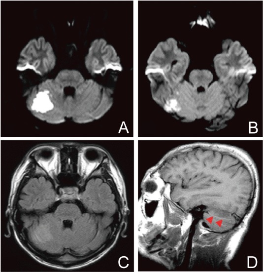
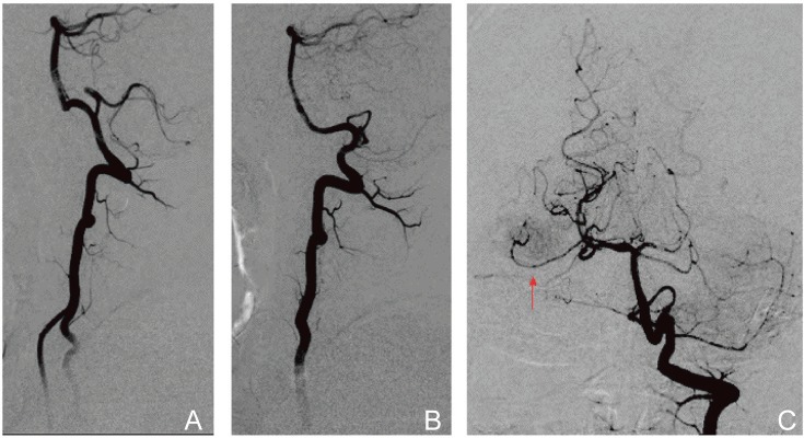
A healthy 23-year-old man suffered a helmet-to-helmet collision with an opponent during a game of American football. He immediately experienced transient blurred vision for a few minutes and mild nausea and dizziness, without amnesia. The symptoms improved within 15 min and the team director allowed him to return to play the next day. Three days later, he was again hit on the head while playing American football. He subsequently experienced continuous vomiting and dizziness. He visited our hospital 18 h after sustaining the second injury. On admission, he showed mild dizziness without cerebellar sign. Magnetic resonance imaging (MRI) revealed acute infarction in the right cerebellar hemisphere (Fig. 1A–D), and magnetic resonance angiography (MRA) revealed stenosis of the right superior cerebellar artery (SCA) without vertebral artery (VA) dissection (Fig. 2A). Although vasculitis, autoimmune diseases, encephalitis, and cardiogenic cerebral embolism were considered, laboratory and cardiac investigations yielded normal results. We therefore diagnosed cerebellar infarction due to traumatic arterial spasm or dissection of the SCA and started conservative treatment. Dizziness gradually improved, and MRA 3 days after the second injury revealed improved stenosis of the right SCA (Fig. 2B). Vertebral angiography 7 days after the second injury demonstrated no abnormalities (Fig. 3A–C). The patient was discharged home, after 2 weeks, without neurological deficits. Follow-up MRI and MRA 3 months after the second injury revealed old infarction in the right cerebellar hemisphere without vascular abnormality (Fig. 2C).
Fig. 1.

A, B: Diffusion weighted image on admission (18 h after the second injury) revealed acute infarction in the right cerebellar hemisphere. C: Fluid-attenuated inversion recovery image on admission demonstrated the infarction as a slightly high intensity lesion. D: Sagittal T1-weighted image on admission revealed a hypotense lesion just below the tentorium (arrow-heads), indicating that infarction occurred in the territory of the right superior cerebellar artery.
Fig. 2.

A: Magnetic resonance angiography (MRA) on admission revealed stenosis of the right superior cerebellar artery (SCA) (arrows) without vertebral artery dissection. B: Follow-up MRA 5 days after the second injury revealed improved stenosis of the right SCA (arrow). C: Follow-up MRA 3 months after the second injury revealed no abnormality.
Fig. 3.

A, B: Vertebral angiograms 7 days after the second injury, lateral view, demonstrated no abnormality such as vertebral artery dissection (A: right side, B: left side). C: Right vertebral angiogram, anteroposterior view, demonstrated no abnormality of the right superior cerebellar artery (arrow).
Discussion
Although posttraumatic cerebral infarction is a well-recognized complication in patients with severe head injury, cerebral infarction after minor head injury has been reported on rare occasions. Such stroke appears more common in children and possible mechanisms include traumatic vasospasm, endothelial lesion, or dissection.1–7) In body contact sports, head hits are common, and ischemic stroke after sport-related head injury has been reported only in children.19–22) In such cases, sports-related trauma leading to arterial dissection with resulting thromboembolism is suspected to represent an important mechanism leading to stroke.19–22) Generally speaking, ischemic stroke after minor head injury in adults is uncommon regardless of cause of injuries.
Cerebellar infarction after head injury is usually caused by VA injury such as dissection.23–28) In those cases, severe head or cervical injuries resulting from events such as traffic accidents or falls cause cerebellar infarction secondary to VA injury accompanying cervical hyperextension, head rotation, or cervical spine fracture.23–28) In children, even minor head injuries can cause cerebellar infarction secondary to VA injury.23,24) On the other hand, cerebellar infarction without VA injury has been reported.25,29) In such cases, severe local trauma with instantaneous deformity of bone may result in injury to the cerebellar cortical artery, leading to cerebellar infarction under the occipital bone.25,29)
In the present case, no VA dissection was detected and the injury was not severe; therefore, the mechanism of cerebellar infarction was neither secondary to VA injury nor involving cerebellar cortical artery injury, as previously reported. Due to the stenosis of the SCA on MRA, we initially diagnosed traumatic arterial spasm or dissection of the SCA causing cerebellar infarction. At present, we consider that arterial spasm of the SCA rather than dissection of the SCA may have caused infarction, because of the transient nature of SCA stenosis on MRA. Although there remains much contention over the existence of trauma-induced arterial spasm in the absence of arterial wall damage, several reports of traumatic arterial spasm have been reported.30,31) Previous reports of cerebral infarction after minor head injury have reported that rapid time course, recurrent and transient symptoms, and reversible imaging changes are consistent with intermittent arterial spasm.5–7) They would support our hypothesis.
In general, posttraumatic vasospasm is a delayed complication that involves the large basal intracranial arteries and causes ischemic damage following moderate to severe head injury.32) Intradural bleeding extends into the cerebrospinal fluid and plays a role in the pathogenesis of vasospasm.32) In the present case, the injury was mild and no intracranial hemorrhage occurred. The pathophysiology may thus differ from that of general posttraumatic vasospasm. Symon has reported an experimental evidence that trauma to the cerebral artery can lead to arterial spasm.33) Moreover, Haug has reported that external mechanical stimulation caused by neighboring tissues or transmitted shockwaves may cause arterial spasm.30) In American football, head impacts can cause linear or rotational acceleration.9,11) Such acceleration may have forced the SCA against neighboring tissues such as the tentorium or may have injured the SCA directly. The repeated impacts in the short term may have caused arterial spasm of the SCA.
In our case, after first injury, the patient experienced transient blurred vision, dizziness, and nausea. These symptoms indicated that the first injury caused concussion. Although the majority of concussions resolve within 7–10 days,15–17) dizziness at the time of injury was found to be the greatest predictor in football players for a recovery taking longer.15) In the present case, potential brain damage might not have resolved fully in spite of transient dizziness. Because the patient was again hit on the head 3 days after the first injury, there is a possibility that the second concussion before the first concussion has properly healed may cause arterial spasm. A second injury before the brain has recovered results in worsening cellular metabolic changes in animal laboratory models.15,18) Repeated mild head injuries may cause cumulative damage to the medial smooth muscle cells and result in arterial spasm.
In general, there are potential health risks of returning an athlete with persistent symptoms to play including the possibility of SIS or diffuse cerebral swelling, and increased susceptibility to a recurrent or more severe concussion and prolonged duration of symptoms.15) Hence, the cornerstone of concussion management is physical and cognitive rest until the acute symptoms resolve.15) Once concussion is diagnosed, an athlete should not return to play on the same day. An athlete must be asymptomatic prior to return to play, and should take a graduated return to play protocol lasting around 1 week.14–17) In the present case, symptoms improved within 15 min and the team director allowed him to return to play the next day. The team director should have referred to a clinician with experience in managing athletes with concussion and the patient should have been controlled in graduated return to play. Prevention of the concussion is probably the most important issue, and athletes themselves need to consider how they can prevent a head injury.10) Moreover, we keep in the possibility that a second head injury in the short term may cause a special TBI as in this case.
References
- 1). Ahn JY, Han IB, Chung YS, Yoon PH, Kim SH: Posttraumatic infarction in the territory supplied by the lateral lenticulostriate artery after minor head injury. Childs Nerv Syst 22: 1493– 1496, 2006 [DOI] [PubMed] [Google Scholar]
- 2). Dharker SR, Mittal RS, Bhargava N: Ischemic lesions in basal ganglia in children after minor head injury. Neurosurgery 33: 863– 865, 1993 [DOI] [PubMed] [Google Scholar]
- 3). Kieslich M, Fiedler A, Heller C, Kreuz W, Jacobi G: Minor head injury as cause and co-factor in the aetiology of stroke in childhood: a report of eight cases. J Neurol Neurosurg Psychiatr 73: 13– 16, 2002 [DOI] [PMC free article] [PubMed] [Google Scholar]
- 4). Matsumoto H, Kohno K: Posttraumatic cerebral infarction due to progressive occlusion of the internal carotid artery after minor head injury in childhood: a case report. Childs Nerv Syst 27: 1169– 1175, 2011 [DOI] [PubMed] [Google Scholar]
- 5). Nabika S, Kiya K, Satoh H, Mizoue T, Oshita J, Kondo H: Ischemia of the internal capsule due to mild head injury in a child. Pediatr Neurosurg 43: 312– 315, 2007 [DOI] [PubMed] [Google Scholar]
- 6). Muthukumar N: Basal ganglia-internal capsule low density lesions in children with mild head injury. Br J Neurosurg 10: 391– 393, 1996 [DOI] [PubMed] [Google Scholar]
- 7). Shaffer L, Rich PM, Pohl KR, Ganesan V: Can mild head injury cause ischaemic stroke? Arch Dis Child 88: 267– 269, 2003 [DOI] [PMC free article] [PubMed] [Google Scholar]
- 8). Bailes JE, Petraglia AL, Omalu BI, Nauman E, Talavage T: Role of subconcussion in repetitive mild traumatic brain injury. J Neurosurg 119: 1235– 1245, 2013 [DOI] [PubMed] [Google Scholar]
- 9). Bartsch A, Benzel E, Miele V, Prakash V: Impact test comparisons of 20th and 21st century American football helmets. J Neurosurg 116: 222– 233, 2012 [DOI] [PubMed] [Google Scholar]
- 10). Clark D: Effects of repeated mild head impacts in contact sports: adding it up. Neurology 78: e140– e142, 2012 [DOI] [PubMed] [Google Scholar]
- 11). Crisco JJ, Wilcox BJ, Beckwith JG, Chu JJ, Duhaime AC, Rowson S, Duma SM, Maerlender AC, McAllister TW, Greenwald RM: Head impact exposure in collegiate football players. J Biomech 44: 2673– 2678, 2011 [DOI] [PMC free article] [PubMed] [Google Scholar]
- 12). Rowson S, Duma SM: Brain injury prediction: assessing the combined probability of concussion using linear and rotational head acceleration. Ann Biomed Eng 41: 873– 882, 2013 [DOI] [PMC free article] [PubMed] [Google Scholar]
- 13). Weinstein E, Turner M, Kuzma BB, Feuer H: Second impact syndrome in football: new imaging and insights into a rare and devastating condition. J Neurosurg Pediatr 11: 331– 334, 2013 [DOI] [PubMed] [Google Scholar]
- 14). Giza CC, Kutcher JS, Ashwal S, Barth J, Getchius TS, Gioia GA, Gronseth GS, Guskiewicz K, Mandel S, Manley G, McKeag DB, Thurman DJ, Zafonte R: Summary of evidence-based guideline update: evaluation and management of concussion in sports: report of the Guideline Development Subcommittee of the American Academy of Neurology. Neurology 80: 2250– 2257, 2013 [DOI] [PMC free article] [PubMed] [Google Scholar]
- 15). Harmon KG, Drezner JA, Gammons M, Guskiewicz KM, Halstead M, Herring SA, Kutcher JS, Pana A, Putukian M, Roberts WO: American Medical Society for Sports Medicine position statement: concussion in sport. Br J Sports Med 47: 15– 26, 2013 [DOI] [PubMed] [Google Scholar]
- 16). McCrory P, Meeuwisse WH, Aubry M, Cantu B, Dvorák J, Echemendia RJ, Engebretsen L, Johnston K, Kutcher JS, Raftery M, Sills A, Benson BW, Davis GA, Ellenbogen RG, Guskiewicz K, Herring SA, Iverson GL, Jordan BD, Kissick J, McCrea M, McIntosh AS, Maddocks D, Makdissi M, Purcell L, Putukian M, Schneider K, Tator CH, Turner M: Consensus statement on concussion in sport: the 4th International Conference on Concussion in Sport held in Zurich, November 2012. Br J Sports Med 47: 250– 258, 2013 [DOI] [PubMed] [Google Scholar]
- 17). Nagahiro S, Tani S, Ogino M, Kawamata T, Maeda T, Noji M, Nariai T, Nakayama H, Fukuda O, Abe T, Suzuki M, Yamada K, Katayama Y: Neurosurgical management of sport-related head injuries—Interium consensus statment for guideline development. Neurotraumatology 36: 119– 128, 2013 [Google Scholar]
- 18). Slemmer JE, Matser EJ, De Zeeuw CI, Weber JT: Repeated mild injury causes cumulative damage to hippocampal cells. Brain 125: 2699– 2709, 2002 [DOI] [PubMed] [Google Scholar]
- 19). Brosch JR, Golomb MR: American childhood football as a possible risk factor for cerebral infarction. J Child Neurol 26: 1493– 1498, 2011 [DOI] [PubMed] [Google Scholar]
- 20). Podberesky DJ, Unsell BJ, Anton CG: Imaging of American football injuries in children. Pediatr Radiol 39: 1264– 1274; quiz 1385–1266, 2009 [DOI] [PubMed] [Google Scholar]
- 21). Sepelyak K, Gailloud P, Jordan LC: Athletics, minor trauma, and pediatric arterial ischemic stroke. Eur J Pediatr 169: 557– 562, 2010 [DOI] [PMC free article] [PubMed] [Google Scholar]
- 22). Tekin S, Aykut-Bingöl C, Aktan S: Case of intracranial vertebral artery dissection in young age. Pediatr Neurol 16: 67– 70, 1997 [DOI] [PubMed] [Google Scholar]
- 23). Byrd LR, Vogel HL: Ischemic cerebellar infarct in a 5-year-old boy: sequela to minor back trauma. J Am Osteopath Assoc 96: 245– 249, 1996 [DOI] [PubMed] [Google Scholar]
- 24). Duval EL, Van Coster R, Verstraeten K: Acute traumatic stroke: a case of bow hunter's stroke in a child. Eur J Emerg Med 5: 259– 263, 1998 [PubMed] [Google Scholar]
- 25). Agrawal A, Kakani A: Cerebellar infarction after head injury. J Emerg Trauma Shock 3: 207– 209, 2010 [DOI] [PMC free article] [PubMed] [Google Scholar]
- 26). Galtés I, Borondo JC, Cos M, Subirana M, Martin-Fumadó C, Martín C, Castellà J, Medallo J: Traumatic bilateral vertebral artery dissection. Forensic Sci Int 214: e12– e15, 2012 [DOI] [PubMed] [Google Scholar]
- 27). Muratsu H, Doita M, Yanagi T, Sekiguchi K, Nishida K, Tomioka M, Kurosaka M: Cerebellar infarction resulting from vertebral artery occlusion associated with a Jefferson fracture. J Spinal Disord Tech 18: 293– 296, 2005 [PubMed] [Google Scholar]
- 28). Tyagi AK, Kirollos RW, Marks PV: Posttraumatic cerebellar infarction. Br J Neurosurg 9: 683– 686, 1995 [DOI] [PubMed] [Google Scholar]
- 29). Taniura S, Okamoto H: Traumatic cerebellar infarction. J Trauma 64: 1674, 2008 [DOI] [PubMed] [Google Scholar]
- 30). Haug RH: Management of low-caliber, low-velocity gunshot wounds of the maxillofacial region. J Oral Maxillofac Surg 47: 1192– 1196, 1989 [DOI] [PubMed] [Google Scholar]
- 31). Peach G, Antoniou GA, El Sakka K, Hamady M: Traumatic arterial spasm causing transient limb ischaemia: a genuine clinical entity. Ann R Coll Surg Engl 92: W3– W4, 2013 [DOI] [PMC free article] [PubMed] [Google Scholar]
- 32). Shahlaie K, Keachie K, Hutchins IM, Rudisill N, Madden LK, Smith KA, Ko KA, Latchaw RE, Muizelaar JP: Risk factors for posttraumatic vasospasm. J Neurosurg 115: 602– 611, 2011 [DOI] [PubMed] [Google Scholar]
- 33). Symon L: An experimental study of traumatic cerebral vascular spasm. J Neurol Neurosurg Psychiatr 30: 497– 505, 1967 [DOI] [PMC free article] [PubMed] [Google Scholar]


