Abstract
Postoperative visual outcome is a major concern in transsphenoidal surgery (TSS). Intraoperative visual evoked potential (VEP) monitoring has been reported to have little usefulness in predicting postoperative visual outcome. To re-evaluate its usefulness, we adapted a high-power light-stimulating device with electroretinography (ERG) to ascertain retinal light stimulation. Intraoperative VEP monitoring was conducted in TSSs in 33 consecutive patients with sellar and parasellar tumors under total venous anesthesia. The detectability rates of N75, P100, and N135 were 94.0%, 85.0%, and 79.0%, respectively. The mean latencies and amplitudes of N75, P100, and N135 were 76.8 ± 6.4 msec and 4.6 ± 1.8 μV, 98.0 ± 8.6 msec and 5.0 ± 3.4 μV, and 122.1 ± 16.3 msec and 5.7 ± 2.8 μV, respectively. The amplitude was defined as the voltage difference from N75 to P100 or P100 to N135. The criterion for amplitude changes was defined as a > 50% increase or 50% decrease in amplitude compared to the control level. The surgeon was immediately alerted when the VEP changed beyond these thresholds, and the surgical manipulations were stopped until the VEP recovered. Among the 28 cases with evaluable VEP recordings, the VEP amplitudes were stable in 23 cases and transiently decreased in 4 cases. In these 4 cases, no postoperative vision deterioration was observed. One patient, whose VEP amplitude decreased without subsequent recovery, developed vision deterioration. Intraoperative VEP monitoring with ERG to ascertain retinal light stimulation by the new stimulus device was reliable and feasible in preserving visual function in patients undergoing TSS.
Keywords: visual evoked potential, transsphenoidal surgery, pituitary adenomas, visual outcome
Introduction
A deterioration in visual function has occasionally been noted after standard transsphenoidal surgery (TSS) for pituitary adenomas.1) However, when extended TSS has been applied to treat patients with suprasellar craniopharyngiomas or tuberculum sellar meningiomas, the complication rate of postoperative visual deterioration has dramatically increased, even in surgeries performed by the most experienced surgeons.2–4) In the expanding application of TSS in patients with sellar and parasellar lesions, the intraoperative monitoring of visual function is mandatory in ensuring the safety of the surgery. Wright et al. first applied visual evoked potential (VEP) monitoring during orbital tumor surgery.5) Since then, several researchers have demonstrated the importance of VEP monitoring during the removal of pituitary tumors.6–8) As opposed to somatosensory and auditory evoked potentials, intraoperative VEP has been regarded as unreliable because of its intra-individual variability and instability.8–10) Even recently, Chung et al. have reported that intraoperative VEP has no association with postoperative visual function in patients who were treated with pituitary TSS.8) However, progress in clinical science occurs in small steps most of the time, and a method once declared unsuitable for a given purpose may prove more useful under mildly changed basic conditions.11) In order to re-evaluate the usefulness of VEP monitoring during TSS, the authors adapted a high-power light-stimulating device and simultaneous electroretinography (ERG), which has been developed by Sasaki et al.,12) in order to ascertain retinal light stimulation. In this situation involving confirmed retinal stimulation, the reproducibility of VEPs during TSS and the relationship between intraoperative VEP amplitude changes and postoperative visual functions were examined in patients with sellar and parasellar tumors.
Materials and Methods
Between May 2012 and July 2013, we performed VEP monitoring during TSS in 33 consecutive patients with sellar and parasellar tumors at the University Hospital of Hamamatsu University School of Medicine. Among these patients, 25 had pituitary adenomas, 4 had craniopharingiomas, 3 had pouches of Rathke, and 1 had a choroid sarcoma. All TSSs were performed by experienced neurosurgeons by endoscopic-assisted microscopic surgery. Extended TSSs were performed on two cases of craniopharyngiomas. During surgeries, direct exposure and manipulation of the optic nerve nerves were performed in dissecting the tumors. Patient data, such as pre- and postoperative magnetic resonance (MR) images, histopathological diagnoses, the results of intraoperative VEP monitoring, and pre- and postoperative examinations of visual functions, were evaluated. In these patients, 22 patients had visual disturbances preoperatively, and the other 11 patients had no visual disturbances. Written informed consents for the surgery, intraoperative VEP monitoring, and general clinical research were obtained from all patients.
After induction with a bolus injection of propofol (1.5–2 mg/kg) and fentanyl (2 μg/kg), anesthesia was maintained by the continuous infusion of propofol (6–10 mg/kg) and an additional injection of fentanyl (2 μg/kg) which determined the depth of anesthesia by a bispectral index (BIS) sensor connected to a QE-910P BIS processor (Covidien, Aspect Medical System, Massachusetts, USA). Anesthesia was adjusted in order to maintain the BIS values between the recommended 40–60. For this study, we adapted a high-power light-stimulating devices consisting of 16 red high-luminosity (100 mCd) LEDs embedded in a 2 cm diameter soft silicone disk that had been developed by Sasaki et al.12) (Unique Medical Co., Ltd., Tokyo). The light-stimulating devices were applied to both closed eyelids, and plate electrodes for the ERG were placed at both canthus. The VEP recording plate electrodes were placed bilaterally at a point 4 cm above and 4 cm lateral from the external occipital protuberance (inion), and the reference electrodes were placed at both mastoid processes. The contact impedance of the plate electrodes was adjusted to below 10 Ω.
We used a high-end signal processor machine (Neuropack X1 MEB-2312, NIHON KOHDEN, Tokyo) in order to analyze the ERG and VEP waveforms. The configuration of the luminosity was changed from 1.000 lux to 13.000 lux by referring to the waveforms. The duration of each stimulus was 20 msec, and the frequency was 1 Hz. As we performed the summations of 100 responses, each recording session required 100 sec. The analysis time was 200 msec. We used low—(20 Hz) and high—(500 Hz) band pass filters. Before the start of TSS, a minimum of two recording sessions of light stimulation to both eyes and unilateral left and right light stimulations were obtained in order to confirm the reproducibility of the data. Light stimulation to both eyes was usually used during TSS. In the critical stage during TSS, left and right unilateral stimulation was used. We focused the large positive peak around 100 msec (P100) and the large negative peak before and after P100 around 75 msec (N75) and 135 msec (N135). As P100 has been reported to be mostly related to the primary visual cortex,13,14) we defined the amplitude as the voltage difference from P100 to the larger negative peak (N75 or N135) in this study. The criterion for amplitude changes was defined as a > 50% increase or 50% decrease in amplitude compared to the control level. The surgeon was immediately alerted when the VEP changed beyond these thresholds, and the surgical manipulations were stopped until the VEPs recovered. The VEP recordings were monitored continuously every 5 min. Postoperative visual function was evaluated within 4 weeks after surgery in all cases. We evaluated the VEP data (the latency, amplitude, and reproducibility ratios of N75, P100, and N135) and the changes in pre- and postoperative visual function.
Results
Tumor excisions were sufficiently performed to release the compression on the optic chasm in all cases. Stable and reproducible ERG data were obtained in all of the cases. As for the VEP data, the detectability rates of N75, P100, and N135 were 94.0% (31 of 33 patients), 85.0% (28 of 33 patients), and 79.0% (26 of 33 patients), respectively. The mean latencies and amplitudes of N75, P100, and N135 were 76.8 ± 6.4 msec and 4.6 ± 1.8 μV, 98.0 ± 8.6 msec, and 5.0 ± 3.4 μV, and 122.1 ± 16.3 msec, and 5.7 ± 2.8 μV, respectively. In the 22 patients with preoperative visual disturbances, visual acuity was improved in 13 cases (59%), and visual field was improved in 7 cases (32%) immediately after surgery. The other patients except one case (representative Case 1) gradually improved over a period of several months. Among these patients, the VEP amplitudes (N75-P100 or P100-N135) were detected in 19 patients (86.0%) and not detected in 3 patients (14.0%). In these 3 patients, 2 had severe visual impairments. The reason of a failure to detect VEP amplitude in a case with preoperative visual disturbance was due to failure of the electrode attachment. In the 11 patients without preoperative visual disturbances, the VEP amplitudes were detected in 9 patients (81.8%) and not detected in 2 patients (18.2%) due to detachment of the recording electrodes.
The relationship between intraoperative VEP changes and postoperative visual functions in 28 patients with evaluable VEP recordings are summarized in Table 1. The VEP amplitudes were stable in 23 cases including 2 cases of craniopharyngiomas dissected by extended TSSs, and transiently decreased in 4 cases. In these 27 cases, no postoperative vision deteriorations were observed. We experienced a case (representative Case 1) in which the patient exhibited decreased VEP amplitude without subsequent recovery and developed visual field deterioration. In this study, we did not experience any cases in which the VEP amplitudes improved in connection with a recovery from the visual disturbances.
Table 1.
Intraoperative VEP change and postoperative visual outcome in 28 patients with evaluable VEP recording
| VEP change | No. of cases | Visual acuity | No. of cases | Visual field | No. of cases |
|---|---|---|---|---|---|
| Stable | 23 | No change | 12 | No change | 18 |
| Improved | 11 | Improved | 5 | ||
| Worsened | 0 | Worsened | 0 | ||
| Improved | 0 | ||||
| Decreased | 1 | No change | Worsened | 1 | |
| Transient decreased | 4 | No change | 2 | No change | 2 |
| Improved | 2 | Improved | 2 | ||
| Worsened | 0 | Worsened | 0 |
VEP: visual evoked potential.
Representative Cases
I. Case 1
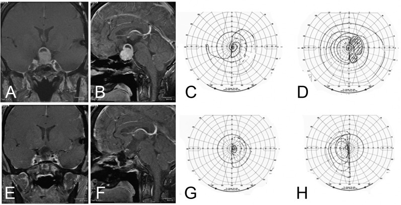
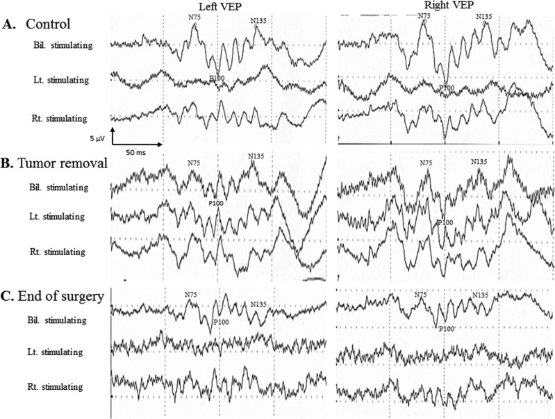
This 32-year-old woman with a repeated recurrence of nonfunctioning pituitary adenoma presented with a gradual deterioration of visual acuities in both eyes and bitemporal hemianopsia (Fig. 1). She had undergone TSS twice in our hospital and a transcranial surgery once in another hospital. At this time, she underwent TSS with intraoperative VEP monitoring for total resection. At the final stage of the TSS when we pulled out the final piece of tumor, bilateral stimulating VEP amplitudes (N75 to P100) on both sides were decreased to below 50% of the control level (Fig. 2). We directly observed the chiasm and noticed that the final piece of tumor was adhered to the chiasm without arachnoid membrane. The surgical manipulations were stopped, and 1000 mg of methylprednisolone was administered. However, the bilateral VEP waveforms did not recover to the control level. Although the tumor was resected sub totally, postoperative examination of visual functions revealed complete bitemporal hemianopsia (Fig. 1).
Fig. 1.
Preoperative Gd-enhanced MR images: coronal (A), sagittal (B), and visual fields: left (C), right (D) postoperative Gd-enhanced MR images: coronal (E), sagittal (F), and visual fields: left (G), right (H). The tumor was removed subtotally, but visual field demonstrated complete bitemporal hemoanopsia postoperatively. Gd: gadolinium, MR: magnetic resonance.
Fig. 2.
Intraoperative VEP findings at the beginning of surgery as control (A), at the stage of tumor removal (B), and at the end of surgery (C). Negatively is shown as an upward deflection. The VEP amplitude was defined as the voltage difference from P100 to N75. During tumor removal, the VEP amplitude decreased and did not recover to the control level. Bil: bilateral, Lt: left, Rt: right, VEP: visual evoked potential.
II. Case 2
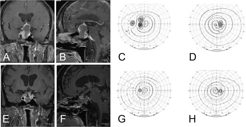
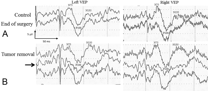
This 71-year old man with a nonfunctioning pituitary adenoma presented with loss of visual acuity on both eyes [right vision (RV) = (0.6), left vision (LV) = (0.6)] and bitemporal hemianopsia (Fig. 3). He underwent TSS with intraoperative VEP monitoring. At the final stage of the TSS when a relatively fibrous and firm tumor was curetted, bilateral stimulating VEP amplitudes on both sides were decreased to below 50% of the control level (Fig. 4). The surgical manipulation was stopped, and the VEP waveforms on both sides recovered to the control level in 5 min. Although the tumor resection was incomplete, a postoperative examination revealed improvements in the visual acuities on both eyes [RV = (1.0), LV = (1.0)] and a recovery of the visual field defects (Fig. 4).
Fig. 3.
Preoperative Gd-enhanced MR images: coronal (A), sagittal (B), and visual fields: left (C), right (D) postoperative Gd-enhanced MR images: coronal (E), sagittal (F), and visual fields: left (G), right (H). Although, residual tumor was observed under chiasm, visual field recovered postoperativeply. Gd: gadolinium, MR: magnetic resonance.
Fig. 4.
Intraoperative VEP findings at the beginning of surgery as control and the end of surgery (A) and the stage of tumor removal (B). Negatively is shown as an upward deflection. The VEP amplitude was defined as the voltage difference from P100 to N75. During tumor removal, the VEP amplitude decreased transiently (B: arrow) and recovered to the control level during suspended surgical manipulation for 5 min. VEP: visual evoked potential.
Discussion
The intraoperative monitoring of VEP has not prevailed for over 30 years because of its high intra-individual variability and instability.11) It has been concluded that VEPs are unstable and not regularly recordable and that they are not suited as a valid intraoperative indicator of visual function.9,10) As for pituitary surgeries, Chung et al. have reported that intraoperative VEP has no association with postoperative visual outcome in TSS. At present, the novelty value of any study on VEP monitoring depends on whether methodological improvements have been achieved.11) In order to ensure the feasibility and clinical validity of intraoperative VEP monitoring, Kodama et al. have suggested the importance of patient selection, total intravenous anesthesia, and a performance of light stimulation device.15) They obtained a stunning 97% rate of successful and stable VEP recordings, and found an excellent correlation between the VEP results and visual outcome.15) Sasaki et al. have developed a high-power light-stimulating device in a soft silicone disk and they have introduced ERG in order to ascertain retinal light stimulation.12) In their cases, all patients without an intraoperative decrease in the VEP amplitude were without severe postoperative deterioration in visual function.12)
In this study, we adapted the methods of Sasaki et al. in 33 consecutive patients with sellar and parasellar tumors.12) When retinal stimulation is confirmed by ERG recording, the inability to VEP recordings in patients with preoperative visual disturbances is thus attributed to the pre-existing visual disturbances rather than to light axis deviation. In the previous reports that have suggested unreliable intraoperative VEP monitoring, ERG was not recorded in order to ascertain adequate light stimulation to the patients.9–11) We obtained stable and reproducible ERG data in all 33 cases. Therefore, we confirmed that adequate light stimulations were delivered to all cases with our high-power light-stimulating device. We achieved stable VEP monitoring during TSS in 28 of the 33 patients (85%). In 5 cases with failed VEP monitoring, 2 cases had severe preoperative visual impairments. Although chiasmal compression due to pituitary adenomas has been reported to cause a reduction in amplitudes and the prolongation of latencies in the VEP responses,16) the degree of visual disturbance at which the VEP disappears has not been elucidated. In order to reveal the correlation between preoperative visual functions and the pattern of VEP waves, preoperative VEP recording on the patients with the same device might be helpful. In the other 3 cases in which VEP monitoring failed, all of the failures were caused by the detachment of the VEP recording electrodes from the occipital head during surgery. Simple but serious technical failures occurred in the beginning of this study, and they were resolved by the fixation of the electrode by a surgical stapler to the skin.
The anesthetic regimen, in particular halogenated agents, has a major influence on intraoperative VEP stability.17) Total venous anesthesia with propofol facilitates the detection of slight VEP changes during surgery.12,15) In our series, all cases were maintained by the continuous infusion of propofol which determined the depth of anesthesia with the BIS values. BIS has been shown to decrease linearly as propofol blood concentration increases.18) Although BIS does not reflect the changes in real electroencephalography, we observed that VEP amplitudes slightly changed with changes in the BIS values during TSS. Therefore, we recommend maintaining the BIS values between 40 and 60 in order to obtain more stable intraoperative VEP monitoring.
In this study, the intraoperative VEP amplitudes were stable in 23 cases. In these cases, no postoperative vision deteriorations were observed. In 4 cases with transient VEP decreases, VEP changes were observed when traction force was applied to the optic nerves or the chiasm in removal manipulations with ring curette or cup forceps in the final stage of tumor resection. In these 4 cases, no postoperative vision deteriorations were observed. We experienced a case with a decreased VEP without subsequent recovery that developed vision deterioration (Fig. 2). In nonfunctioning pituitary adenomas, vision preservation of visual acuity and the fields is more important than total tumor resection. Therefore, this was a case that showed us the importance of intraoperative VEP monitoring in TSS. However, some large nonfunctioning adenomas result in postoperative critical bleeding and increase mass effect with vision deterioration following incomplete resection. Therefore, operators are frequently faced with difficult decisions whether to continue the surgery or stop the surgery when VEP decreased. In such situations, direct observation by using the extended approach with meticulous microsurgical manipulation might be useful to continue with the further resection.
In this study, immediate postoperative recovery of visual field was observed in only 32% even though sufficient tumor excisions were performed to release the compression on the optic chasm in all cases. However, the remaining cases except one case (representative Case 1) gradually improved over a period of several months. Recovery of nerve conduction in the optic chiasm might take more time than in the optic nerve.
In our intraoperative VEP monitoring, each recording session required 100 sec. This time lag of VEP recording should be taken into account, and we should perform surgical manipulations more carefully and pay close attention to the VEP changes during the critical stage. The direct recording of the flash stimulation-responded optic nerve potential is an alternative method for real-time visual function monitoring. Optic nerve potentials after flash stimulation have been reported to consist of a positive peak with a latency around 40 msec.19) In extended TSS for suprasellar craniopharyngiomas or tuberculum sellar meningiomas, direct optic nerve potential recording with VEP monitoring might be a more sensitive method that is useful for preserving postoperative visual function.
In conclusion, in spite of its intra-individual variability and instability compared to somatosensory and auditory evoked potentials, intraoperative VEP monitoring should be re-evaluated as a routine method for ensuring vision preservation in TSS. In order to obtain reproducible and reliable VEP wave forms, an adequate high-power device with ERG recording in order to ascertain retinal light stimulation is necessary. We should pay attention to minimizing technical failure. In addition, the lag time of VEP recording should be taken into account when manipulating a critical region.
Acknowledgments
The authors are grateful to Mr. Masahiko Kamoshita and Mrs. Asuka Tanaka (Clinical engineers of the University Hospital of Hamamatsu University School of Medicine) for their technical support and meticulous observation of VEP changes during surgery. Their sincere cooperation has guaranteed the safety of the surgery.
References
- 1). Cohen AR, Cooper PR, Kupersmith MJ, Flamm ES, Ransohoff J: Visual recovery after transsphenoidal removal of pituitary adenomas. Neurosurgery 17: 446– 452, 1985. [DOI] [PubMed] [Google Scholar]
- 2). Kitano M, Taneda M, Nakao Y: Postoperative improvement in visual function in patients with tuberculum sellae meningiomas: results of the extended transsphenoidal and transcranial approaches. J Neurosurg 107: 337– 346, 2007. [DOI] [PubMed] [Google Scholar]
- 3). Kitano M, Taneda M: Extended transsphenoidal surgery for suprasellar craniopharyngiomas: infrachiasmatic radical resection combined with or without a suprachiasmatic trans-lamina terminalis approach. Surg Neurol 71: 290– 298; discussion 298, 2009. [DOI] [PubMed] [Google Scholar]
- 4). Yamada S, Fukuhara N, Oyama K, Takeshita A, Takeuchi Y, Ito J, Inoshita N: Surgical outcome in 90 patients with craniopharyngioma: an evaluation of transsphenoidal surgery. World Neurosurg 74: 320– 330, 2010. [DOI] [PubMed] [Google Scholar]
- 5). Wright JE, Arden G, Jones BR: Continuous monitoring of the visually evoked response during intra-orbital surgery. Trans Ophthalmol Soc UK 93: 311– 314, 1973. [PubMed] [Google Scholar]
- 6). Wilson WB, Kirsch WM, Neville H, Stears J, Feinsod M, Lehman RA: Monitoring of visual function during parasellar surgery. Surg Neurol 5: 323– 329, 1976. [PubMed] [Google Scholar]
- 7). Chacko AG, Babu KS, Chandy MJ: Value of visual evoked potential monitoring during trans-sphenoidal pituitary surgery. Br J Neurosurg 10: 275– 278, 1996. [DOI] [PubMed] [Google Scholar]
- 8). Chung SB, Park CW, Seo DW, Kong DS, Park SK: Intraoperative visual evoked potential has no association with postoperative visual outcomes in transsphenoidal surgery. Acta Neurochir (Wein) 154: 1505– 1510, 2012. [DOI] [PubMed] [Google Scholar]
- 9). Cedzich C, Schramm J, Fahlbusch R: Are flash-evoked visual potentials useful for intraoperative monitoring of visual pathway function? Neurosurgery 21: 709– 715, 1987. [DOI] [PubMed] [Google Scholar]
- 10). Cedzich C, Schramm J: Monitoring of flash visual evoked potentials during neurosurgical operations. Int Anesthesiol Clin 28: 165– 169, 1990. [DOI] [PubMed] [Google Scholar]
- 11). Neuloh G: Time to revisit VEP monitoring? Acta Neurochir (Wien) 152: 649– 650, 2010. [DOI] [PubMed] [Google Scholar]
- 12). Sasaki T, Itakura T, Suzuki K, Kasuya H, Munakata R, Muramatsu H, Ichikawa T, Sato T, Endo Y, Sakuma J, Matsumoto M: Intraoperative monitoring of visual evoked potential: introduction of a clinically useful method. J Neurosurg 112: 273– 284, 2010. [DOI] [PubMed] [Google Scholar]
- 13). Whittingstall K, Kevin W, Wilson D, Doug W, Schmidt M, Matthias S, Stroink G, Gerhard S: Correspondence of visual evoked potentials with FMRI signals in human visual cortex. Brain Topogr 21: 86– 92, 2008. [DOI] [PubMed] [Google Scholar]
- 14). Whittingstall K, Stroink G, Schmidt M: Evaluating the spatial relationship of event-related potential and functional MRI sources in the primary visual cortex. Hum Brain Mapp 28: 134– 142, 2007. [DOI] [PMC free article] [PubMed] [Google Scholar]
- 15). Kodama K, Goto T, Sato A, Sakai K, Tanaka Y, Hongo K: Standard and limitation of intraoperative monitoring of the visual evoked potential. Acta Neurochir (Wien) 152: 643– 648, 2010. [DOI] [PubMed] [Google Scholar]
- 16). Jayaraman M, Ambika S, Gandhi RA, Bassi SR, Ravi P, Sen P: Multifocal visual evoked potential recordings in compressive optic neuropathy secondary to pituitary adenoma. Doc Ophthalmol 121: 197– 204, 2010. [DOI] [PubMed] [Google Scholar]
- 17). Cedzich C, Schramm J, Mengedoht CF, Fahlbusch R: Factors that limit the use of flash visual evoked potentials for surgical monitoring. Electroencephalogr Clin Neurophysiol 71: 142– 145, 1988. [DOI] [PubMed] [Google Scholar]
- 18). Glass PS, Bloom M, Kearse L, Rosow C, Sebel P, Manberg P: Bispectral analysis measures sedation and memory effects of propofol, midazolam, isoflurane, and alfentanil in healthy volunteers. Anesthesiology 86: 836– 847, 1997. [DOI] [PubMed] [Google Scholar]
- 19). Benedicic M, Bosnjak R: Optic nerve potentials and cortical potentials after stimulation of the anterior visual pathway during neurosurgery. Doc Ophthalmol 122: 115– 125, 2011. [DOI] [PubMed] [Google Scholar]


