Significance
Metabolites of the mevalonate (MVA) pathway play essential roles in the regulation of growth and development in many organisms. In this study, we demonstrate that a key regulatory enzyme of the MVA pathway is directly involved in the signaling pathway that transduces endosymbiotic microbial signals in Medicago truncatula. Furthermore, we show that exogenous MVA application is sufficient to activate this transduction pathway. The use of mutants in the signaling pathway and a heterologous expression system provides evidence that the MVA pathway is a missing link between the initial perception of microbial signals at the host plasma membrane and the regulation of symbiotic gene expression in the nucleus.
Keywords: HMG-CoA reductase, mevalonate, calcium signaling, legume nodulation, arbuscular mycorrhization
Abstract
Rhizobia and arbuscular mycorrhizal fungi produce signals that are perceived by host legume receptors at the plasma membrane and trigger sustained oscillations of the nuclear and perinuclear Ca2+ concentration (Ca2+ spiking), which in turn leads to gene expression and downstream symbiotic responses. The activation of Ca2+ spiking requires the plasma membrane-localized receptor-like kinase Does not Make Infections 2 (DMI2) as well as the nuclear cation channel DMI1. A key enzyme regulating the mevalonate (MVA) pathway, 3-Hydroxy-3-Methylglutaryl CoA Reductase 1 (HMGR1), interacts with DMI2 and is required for the legume–rhizobium symbiosis. Here, we show that HMGR1 is required to initiate Ca2+ spiking and symbiotic gene expression in Medicago truncatula roots in response to rhizobial and arbuscular mycorrhizal fungal signals. Furthermore, MVA, the direct product of HMGR1 activity, is sufficient to induce nuclear-associated Ca2+ spiking and symbiotic gene expression in both wild-type plants and dmi2 mutants, but interestingly not in dmi1 mutants. Finally, MVA induced Ca2+ spiking in Human Embryonic Kidney 293 cells expressing DMI1. This demonstrates that the nuclear cation channel DMI1 is sufficient to support MVA-induced Ca2+ spiking in this heterologous system.
The mevalonate (MVA) pathway controls the biosynthesis of hundreds of isoprenoids (sterols, carotenoids, prenyl side chains, etc.) in eukaryotes. These isoprenoids contribute to membrane integrity and development, among many other functions (1). Here, we report that the MVA pathway is also necessary for the earliest responses of plants to symbiotic signals produced by nitrogen-fixing rhizobia and arbuscular mycorrhizal (AM) fungi. These two types of endosymbiotic associations require a common set of genes in host plants to allow successful bacterial and fungal colonization. The molecular mechanisms controlling the establishment of the legume–rhizobium symbiosis have been extensively studied in model legumes such as Medicago truncatula and Lotus japonicus (2, 3). Rhizobia secrete lipochitooligosaccharides (LCOs) known as nodulation (Nod) factors, which are perceived by LysM-type receptor kinases, such as Nod factor perception (NFP) and LYK3 in M. truncatula and are required for both rhizobial infection and nodule organogenesis (4, 5). Similarly, AM fungi release signal molecules, so-called Myc factors, which are likely perceived by other LysM-type receptor kinases in both legumes as well as nonleguminous plants (6–8). The perception of Nod and Myc factors initiates early symbiotic responses in host plants through the activation of the receptor-like kinase Does not Make Infections 2 (DMI2), which is believed to act as a coreceptor (9). Although these signaling components reside on the plasma membrane (10, 11), the perception of symbiotic signals triggers sustained oscillations in Ca2+ concentration both within the nucleus (nuclear Ca2+ spiking) and around the nucleus (perinuclear Ca2+ spiking) (12, 13). As a result, the generation of second messengers transducing the signals from the plasma membrane to the nuclear envelope has long been hypothesized (12–19).
Elegant mathematical models have been developed to explain the mechanism of nuclear Ca2+ spiking and the primary role of DMI1 (a nuclear envelope-localized cation channel) in its initiation and maintenance (13, 18, 20–22). Downstream decoding of Ca2+ spiking involves the nuclear Ca2+/calmodulin-dependent protein kinase DMI3 (23). In M. truncatula, DMI1, DMI2, and DMI3 are essential components of the common symbiosis pathway that is required for establishing both root nodulation and the AM symbiosis, as the respective mutants are defective for both symbioses (2). DMI1 is thus viewed as the first known target of the unidentified second messenger(s) transducing signals from the plasma membrane to the nucleus.
In a previous study, a yeast two-hybrid screen identified a 3-hydroxy 3-methylglutaryl CoA reductase 1 (HMGR1) as strongly interacting with DMI2 (24). HMGRs are well-known regulatory enzymes of the MVA pathway in plants and animals, catalyzing the conversion of HMG-CoA into MVA. Furthermore, the pharmacological inhibition of HMGRs by statin drugs led to decreased nodulation (24). More specifically, silencing HMGR1 by RNA interference (RNAi) resulted in a drastic reduction in root infection and nodule development (24). However, despite these findings, the precise role of HMGR1 and MVA in symbiosis remained unclear. We now demonstrate that HMGR1 plays a key role during the initial symbiotic signaling between the host plant and both rhizobia and AM fungi. Using pharmacological, biochemical, and genetic approaches, we show that HMGR1 and the products of the MVA pathway act upstream of DMI1 in the symbiotic signaling cascade, providing the missing link between the perception of symbiotic signals at the plasma membrane and the activation of Ca2+ spiking in the nucleus.
Results
M. truncatula HMGR1 Possesses HMGR Activity.
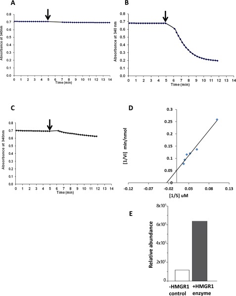
Because it was a considerable surprise to discover a metabolic enzyme as an interactor of the symbiotic receptor-like kinase DMI2 (24), we investigated whether MtHMGR1 is indeed a bona fide enzyme with HMG-CoA reductase activity. The catalytic domain of HMGR1 was tagged with a maltose-binding protein (MBP:HMGR1ΔN), expressed in Escherichia coli, and purified using amylose resin. Its kinetic properties were studied using spectrophotometry as described in Dale et al. (25). HMG-CoA was used to start the reaction, and the oxidation rate of NADPH was determined before (5 min) and after the addition of HMG-CoA using the absorbance at 340 nm and the initial rate of the reaction was calculated (Fig. S1 A and B). A Lineweaver–Burk plot was used to calculate the Vmax and Km of the reaction (Fig. S1D). The apparent Vmax and Km(HMG-CoA) were 3.62 ± 0.37 µmol/min/mg of protein and 38.88 ± 4.41 µM, respectively. The Km(HMG-CoA) of MBP:HMGR1∆N was thus within the same range as that of other known plant (e.g., rubber, 13 µM; maize, 10 µM; Arabidopsis, 8 µM) and animal (e.g., rat, 29–47 µM) HMGRs, indicating a similar affinity for HMG-CoA (25–28). We also evaluated the effect of lovastatin, a well-characterized competitive inhibitor of HMGRs, on the enzymatic activity of MtHMGR1. The addition of 50 µM lovastatin completely abolished MBP:HMGR1∆N activity (Fig. S1C), thus confirming that HMGR1 has typical HMG-CoA reductase activity. The production of MVA in the HMGR1 reaction mix was determined to be 5 times greater than the control based on the area under the curve corresponding to the extracted ion chromatogram of the MVA standard base peak (Fig. S1E).
Fig. S1.
Enzymatic analysis of MtHMGR1 activity. To evaluate the catalytic activity of M. truncatula HMGR1, assays were performed by measuring the oxidation of NADPH in the presence of the substrate HMG-CoA. (A) There is no detectible change in the oxidation of NADPH in the absence of MtHMGR1. (B) However, in the presence of HMGR1, addition of HMG-CoA results in a progressive oxidation of NADPH. (C) Addition of the competitive inhibitor lovastatin (50 µM) to the reaction mixture inhibits HMGR1 activity, confirming that MtHMGR1 has HMG-CoA reductase activity. Arrows indicate the addition of HMG-CoA to initiate the reaction, and the oxidation of NADPH was determined by measuring the absorbance at 340 nm. (D) The double reciprocal Lineweaver–Burk plot was used to calculate the Vmax as 3.62 ± 0.37 µmoles/min/mg of protein and Km as 38.88 ± 4.41 µM. (E) Confirmation of the production of MVA in HMGR1 ezymatic assay as determined by ion chromatogram obtained through ultra gas chromatograph coupled to a quadrupole/orbitrap mass spectrometer. MVA production in the HMGR1 enzymatic assay was 5 times greater than the control.
HMGR1 Silencing Affects Nod Factor-Induced ENOD11 Expression and Ca2+ Spiking in M. truncatula.
M. truncatula HMGR1 interacts with DMI2, and RNAi-based silencing of HMGR1 strongly reduces the ability of transgenic roots to develop nodules when inoculated with Sinorhizobium meliloti (24). Because a knockout mutant of HMGR1 was not available, we used the same RNAi-based silencing strategy to investigate the role of HMGR1 in early symbiotic signaling. We analyzed the induction of nuclear-associated Ca2+ spiking and the expression of the M. truncatula early nodulin 11 (ENOD11) gene, two early symbiotic responses that have been used extensively to study symbiotic signaling (29–31).
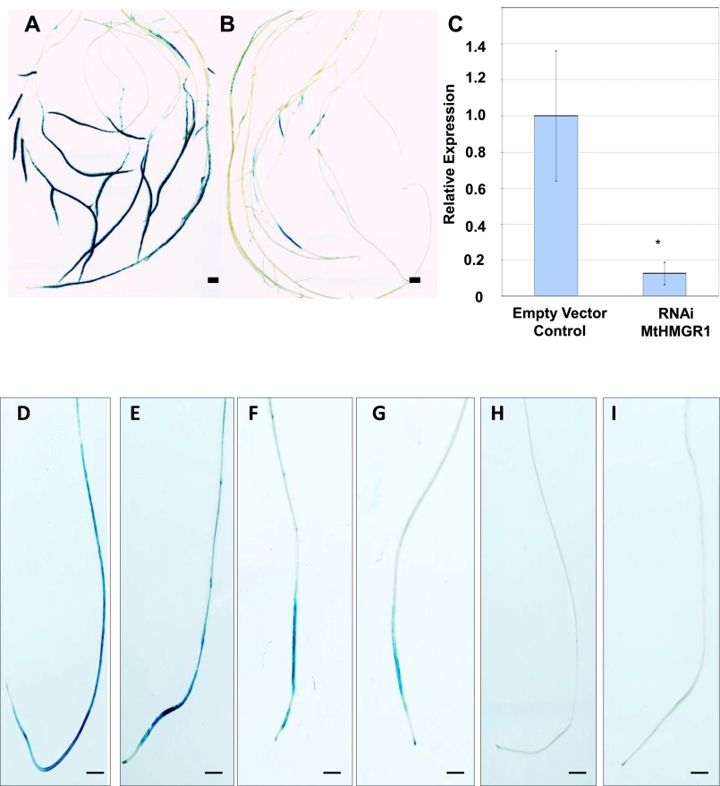
ENOD11 expression was analyzed qualitatively in Jemalong A17 plants expressing pENOD11-GUS and also quantitatively using RT-PCR. Control pENOD11-GUS–expressing roots transformed with the RNAi vector alone displayed the normal dark blue staining 12 h after 10−8 M Nod factor addition, whereas HMGR1-RNAi roots were only lightly stained (Fig. S2 A and B), thus indicating a clear reduction in reporter expression. This was confirmed by quantitative RT-PCR, which revealed that ENOD11 expression was approximately eightfold lower in HMGR1-RNAi transgenic roots compared with that of the control (Fig. S2C).
Fig. S2.
Nod factor- and MVA-induced ENOD11 expression in M. truncatula wild-type, HMGR1-RNAi transgenic roots, and symbiotic defective mutants. (A) Control M. truncatula roots expressing the cytoplasmic cameleon YC3.6 show intense Nod factor-induced ENOD11-GUS expression after 12 h of treatment. (B) Silencing HMGR1 significantly reduces Nod factor-induced ENOD11 expression. (A and B scale bar, 5 mm.) (C) Quantitative RT-PCR confirms a major reduction (approx. eightfold) in Nod factor-induced ENOD11 induction in roots expressing the HMGR1-RNAi cassette. (D and E) ENOD11-GUS expression induced in wild-type M. truncatula plants after 24 h of treatment with 10 nM Nod factors (D) and 100 µM MVA (E). (F–I) Similarly, 100 µM MVA elicits ENOD11-GUS expression in the nfp-2 mutant (F), the dmi2 mutant background (G), but not in dmi1 (H) or dmi3 (I) mutants, thus showing that MVA acts downstream of DMI2 and upstream of DMI1 during symbiotic signaling. (D–I scale bar, 2 mm.)
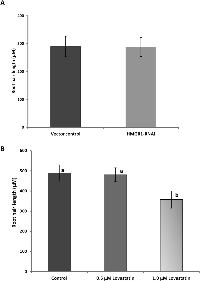
Because actively growing root hairs are used for Ca2+ measurements, we tested both the effect of silencing HMGR1 and the effect of inhibiting the MVA pathway on root hair growth. Silencing HMGR1 had no significant effect on root hair growth (Fig. S3A). Similarly, the application of lovastatin at 0.5 µM, the concentration that reduced nodulation (24), did not affect root hair growth (Fig. S3B). It should be noted that the negative effect of lovastatin on root hair growth at the higher 1 µM concentration (Fig. S3B) could be due to nonspecific effects. Taken together, these results indicate that the components of the MVA pathway (including HMGR1) are not essential for root hair growth.
Fig. S3.
Role of MVA pathway and HMGR1 in root hair growth in M. truncatula. (A) Silencing HMGR1 did not affect root hair growth. (B) The effect of lovastatin (0.5 µM and 1 µM) on root hair growth in M. truncatula. Application of lovastatin at 0.5 µM concentration, which inhibited nodulation (1), did not affect root hair growth.
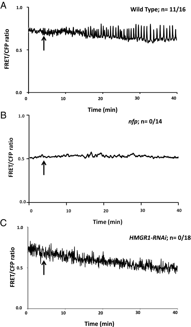
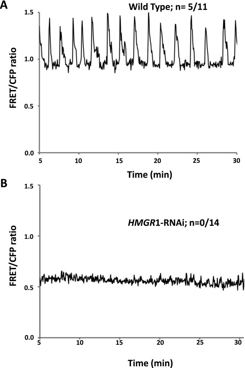
Nod factor-induced Ca2+ spiking was analyzed in M. truncatula seedlings expressing both the HMGR1-RNAi construct and the Ca2+ sensor Yellow Cameleon 3.6 (YC3.6). Transgenic roots expressing YC3.6 alone were used as controls. The application of Nod factors at 10−8 M triggered nucleus-associated Ca2+ spiking in wild-type control roots 10–15 min after Nod factor application (Fig. 1A). Nod factor-induced Ca2+ spiking was not observed in the nfp-1 mutant (Fig. 1B), which served as a negative control for this experiment. The application of Nod factors failed to trigger detectable Ca2+ spiking in epidermal cells of HMGR1-silenced roots (Fig. 1C). Ca2+ spiking was not detected either in these HMGR1-silenced roots in response to germinated spore exudates (GSEs) from the AM fungus Rhizophagus irregularis (Fig. S4 A and B). Altogether, these observations indicate that HMGR1 plays a role in early symbiotic signaling upstream of Ca2+ spiking and ENOD11 expression. We therefore examined whether products of HMGR1 activity such as MVA are able to elicit Ca2+ spiking in root epidermal cells.
Fig. 1.
Effect of silencing M. truncatula HMGR1 on nuclear-associated Ca2+ spiking in root epidermal cells. (A) Nod factor-induced Ca2+ spiking in M. truncatula root hair cells expressing YC3.6. (B) Absence of Nod factor-induced Ca2+ spiking in a nfp mutant expressing YC3.6. (C) Silencing HMGR1 abolishes Nod factor-induced Ca2+ spiking in M. truncatula root hair cells. Arrows indicate the addition of Nod factors.
Fig. S4.
Effect of silencing HMGR1 on AM GSE-induced nuclear-associated Ca2+ spiking. (A) R. irregularis GSE activates Ca2+ spiking in wild-type M. truncatula roots expressing the YC3.6 cameleon. (B) Absence of GSE-induced Ca2+ spiking in HMGR1-RNAi M. truncatula roots expressing YC3.6.
MVA Induces ENOD11 Expression in M. truncatula and Ca2+ Spiking in Root Epidermal Cells of Legumes and Nonlegumes.
To determine whether the product of HMGR1 activity, MVA, is able to elicit ENOD11 expression in the absence of Nod factors, M. truncatula roots stably transformed with pENOD11:GUS were treated with a 100 µM solution of MVA, the lowest concentration required to induce signaling events (SI Materials and Methods). Although the responses were weaker compared with Nod factor treatment (Fig. S2D), clear GUS staining was observed in wild-type roots 24 h after MVA treatment (Fig. S2E). However, we were unable to quantify the MVA-induced ENOD11 expression in M. truncatula roots through quantitative RT-PCR, most likely due to the weaker expression of ENOD11 during MVA treatment compared with Nod factor treatment.
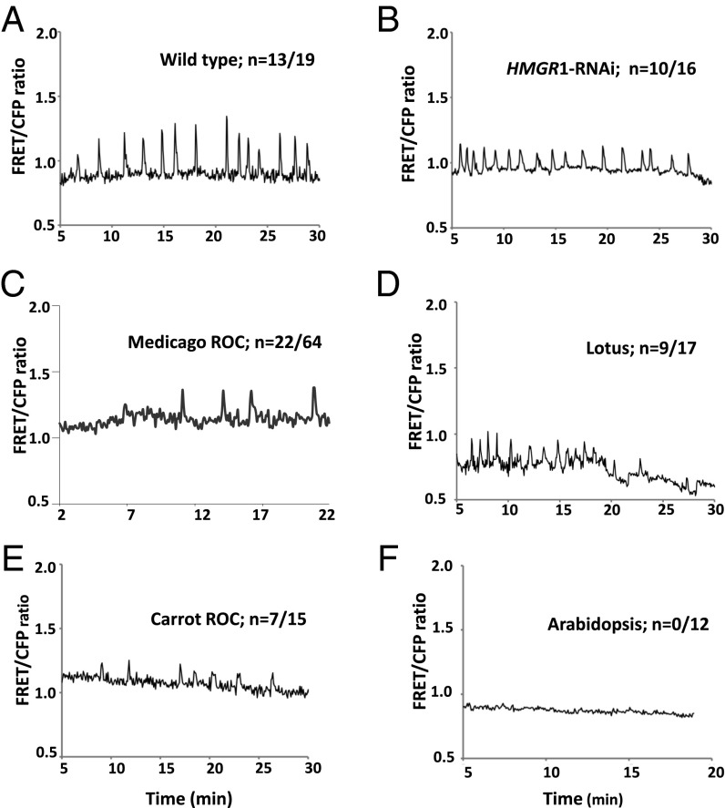
We then investigated whether MVA is also able to activate nuclear-associated Ca2+ spiking in the root epidermal cells of M. truncatula. The application of the same concentration of MVA triggered sustained Ca2+ spiking within 5–10 min in over 50% of cells examined (Fig. 2A). Such MVA-induced Ca2+ spiking was also observed in roots of the other model legume, L. japonicus (Fig. 2D). Significantly, 100 µM MVA elicited Ca2+ spiking (albeit of lower frequency) in epidermal cells of root organ cultures of M. truncatula (Fig. 2C). This is particularly interesting, as these root cultures do not respond to rhizobial Nod factors but can be colonized by AM fungi and respond to germinating AM fungal spore exudates (15). Finally, MVA induction of nuclear-associated Ca2+ spiking was observed in root organ cultures of carrot, a nonlegume AM host plant (Fig. 2E). In contrast, MVA failed to trigger nuclear-associated Ca2+ spiking in trichoblast cells of Arabidopsis thaliana, which is unable to develop AM associations or to form root nodules (Fig. 2F).
Fig. 2.
MVA-induced nuclear-associated Ca2+ spiking in legumes and nonlegumes. (A) Exogenous application of 100 µM MVA triggers Ca2+ spiking in M. truncatula root hair cells expressing YC3.6. (B) MVA restores Ca2+ spiking in roots silenced for HMGR1. (C) MVA induces nuclear Ca2+ spiking in atrichoblast cells of a M. truncatula root organ culture expressing nuclear-targeted YC2.1. (D) MVA-induced nuclear spiking in root hairs of L. japonicus expressing NLS-YC3.6. (E) Atrichoblasts of carrot root organ cultures expressing nuclear-targeted YC2.1 also respond to MVA. (F) In contrast, MVA does not elicit nuclear-associated Ca2+ spiking in trichoblast cells of Arabidopsis expressing YC3.6, which is unable to form endosymbiotic associations with either rhizobia or AM fungi.
Taken together, these results suggest that the agonist activity of MVA in relation to Ca2+ spiking is comparable for both the rhizobial and AM symbioses, thus potentially placing this metabolite within the common symbiosis pathway. This hypothesis is consistent with the fact that HMGR1 silencing abolishes Ca2+ spiking in root epidermal cells of M. truncatula treated with GSE of R. irregularis (Fig. S4B). Furthermore, 100 µM MVA restored Ca2+ spiking in the same HMGR1-silenced roots (Fig. 2B), indicating a direct link between HMGR1 expression and MVA in symbiotic signaling.
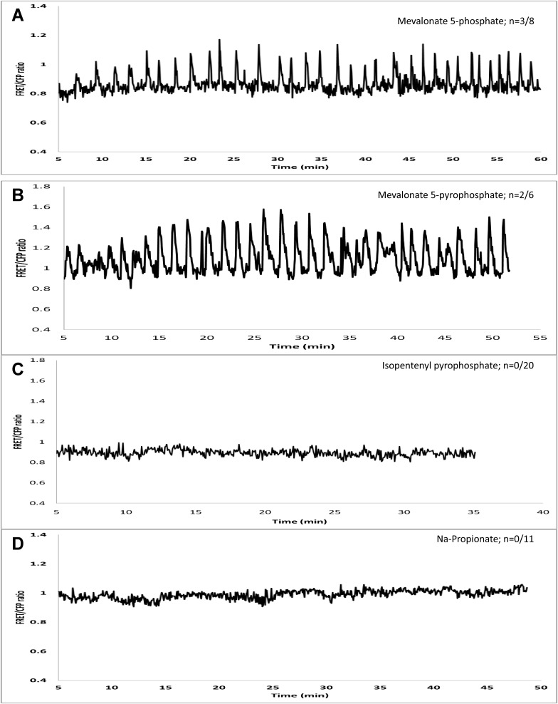
Because an array of additional metabolites are synthesized from MVA, it is also possible that some of these (isopentenyl pyrophosphate, geranylgeranyl pyrophosphate, etc.) may be responsible for triggering Ca2+ spiking in the root epidermis following the exogenous addition of MVA. The application of isopentenyl pyrophosphate (100 µM) did not induce nuclear Ca2+ spiking in root epidermal cells (Fig. S5C). Hence, we tested the effect of upstream components of the MVA pathway, such as MVA 5-phosphate and MVA 5-pyrophosphate. Both these phosphorylated versions of MVA triggered nuclear Ca2+ spiking in M. truncatula root hair cells (Fig. S5 A and B). To rule out the possibility that MVA and its phosphorylated versions might induce cytoplasmic acidification, which might account for the generation of nuclear-associated Ca2+ spiking, we mimicked such an effect by applying sodium propionate. As shown in Fig. S5D, sodium propionate did not induce Ca2+ spiking at concentrations ranging from 100 µM to 1 mM, thus consistent with a specific role for MVA or its immediate downstream products.
Fig. S5.
Investigating the effect of downstream products of the MVA pathway on nuclear-associated Ca2+ spiking that is not due to acidification of the cytoplasm. (A) MVA 5-phosphate (100 µM) and (B) MVA 5-pyrophosphate (100 µM) induce Ca2+ spiking in the root hairs of M. truncatula expressing YC3.6. (C) In contrast, isopentenyl pyrophosphate (100 µM) does not induce Ca2+ spiking. (D) Sodium propionate (100 µM), used here to mimic cytoplasmic acidification, does not trigger Ca2+ spiking in root hairs of YC3.6-expressing roots.
MVA-Induced ENOD11 Expression and Nuclear-Associated Ca2+ Spiking Are Dependent on Upstream Components of the Common Symbiosis Pathway.
To determine whether MVA-induced ENOD11 expression is dependent on the symbiotic signaling pathway, we treated M. truncatula nfp-2, dmi1, dmi2, and dmi3 mutants expressing pENOD11:GUS with 100 µM MVA and performed GUS staining 24 h after treatment. MVA induces detectable ENOD11 expression in nfp-2 and dmi2 mutants but not in dmi1 or dmi3 mutants, suggesting that HMGR1/MVA acts downstream of DMI2 but upstream of DMI1 and DMI3 (Fig. S2 F–I).
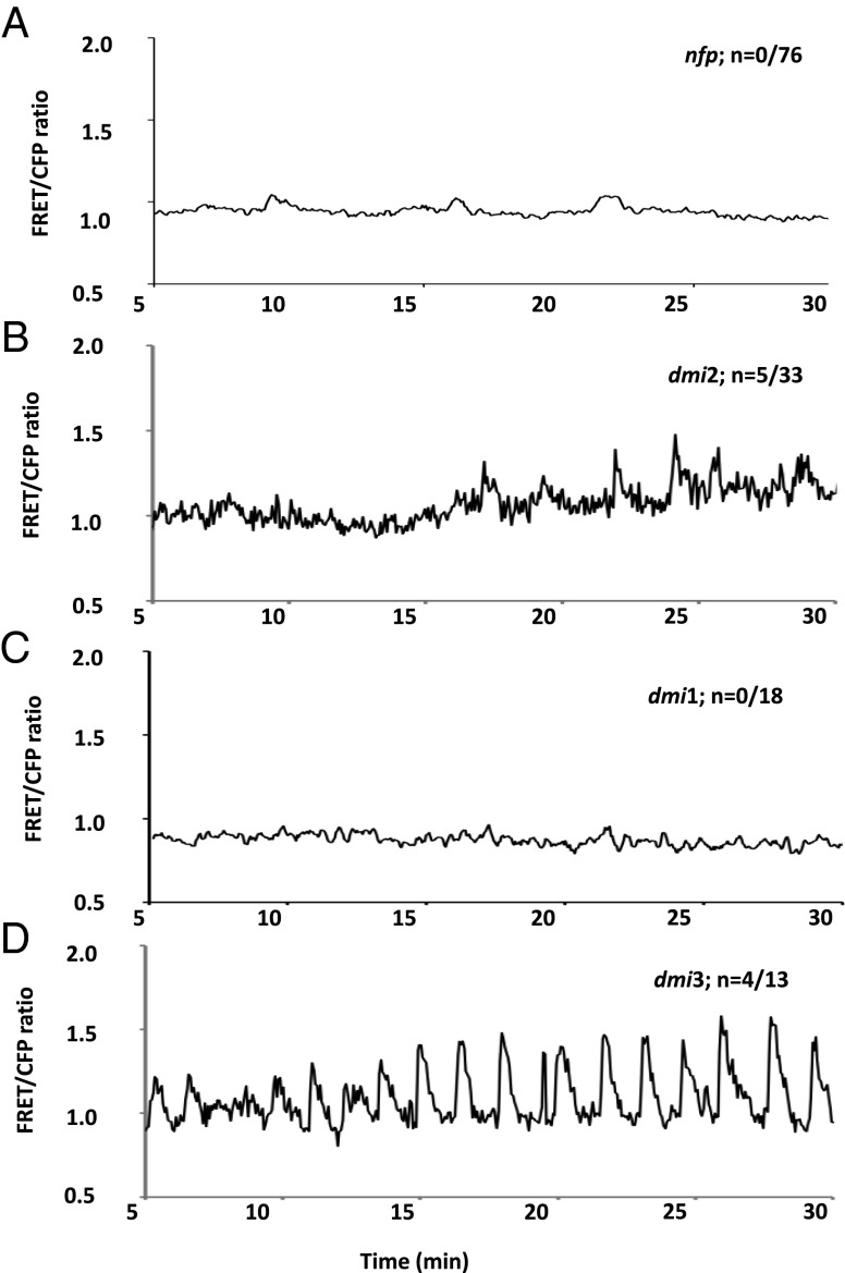
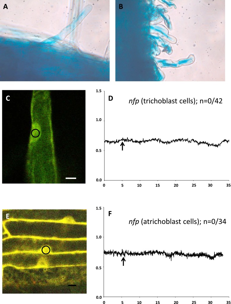
Parallel experiments were then performed on MVA-elicited Ca2+ spiking using nfp and various dmi mutants expressing the YC3.6 Ca2+ sensor. Consistent with the ENOD11 expression data, 100 µM MVA triggered nuclear-associated Ca2+ spiking in both dmi2 and dmi3 mutants but not in dmi1 mutants (Fig. 3 B–D). Nevertheless, it should be noted that the spiking profile for the dmi2 mutant had a noisy background as reported previously (32) and that the percentage of responding cells is significantly lower than in wild-type plants (Fig. 2A). On the other hand, MVA-elicited Ca2+ spiking was not observed in either trichoblast or atrichoblast cells of two different alleles of nfp (nfp-1 and nfp-2; Fig. 3A and Fig. S6). Despite this apparent contradiction with the ENOD11 expression data for nfp (see Discussion), we conclude that MVA itself or the products of the MVA pathway are likely to act downstream of DMI2 and upstream of DMI1 in triggering both Ca2+ spiking and regulating symbiotic gene expression. This hypothesis is consistent with HMGR1 acting downstream of its interacting protein partner DMI2. To our knowledge, this is the first phenotype that permits the uncoupling of dmi1 and dmi2 mutants.
Fig. 3.
Analyses of MVA-induced nuclear-associated Ca2+ spiking responses in symbiosis-defective mutants of M. truncatula. (A) MVA-induced Ca2+ spiking was not observed in an nfp mutant, which acts upstream of DMI2. (B) MVA activates Ca2+ spiking in the dmi2 mutant expressing YC3.6, (C) but not in the dmi1 mutant. (D) As expected, MVA-elicited Ca2+ spiking is not modified in the dmi3 mutant, as DMI3 acts downstream of the Ca2+ spiking machinery.
Fig. S6.
Analyses of MVA-induced ENOD11 expression and Ca2+ spiking in nfp mutants (nfp-1 and nfp-2). (A) MVA-induced ENOD11 expression in the trichoblast and atrichoblast cells of wild type. (B) MVA-induced ENOD11 expression in the trichoblast and atrichoblast cells of nfp-2. (C) Representative trichoblast cell expressing YC3.6 used for Ca2+ spiking analyses. (D) MVA failed to induce nuclear Ca2+ spiking in trichoblast cells of both nfp mutants. (E) Representative atrichoblast cells expressing YC3.6 used for Ca2+ spiking analyses. (F) MVA failed to induce Ca2+ spiking in atrichoblast cells of both nfp mutants. (Scale bar, 10 µm.) The small circle in panels C and E indicates the region of interest used for obtaining FRET and CFP channel intensities to analyze nuclear Ca2+ spiking.
MVA Elicits Ca2+ Spiking in Human Embryonic Kidney 293 Cells Expressing the M. truncatula DMI1 Protein.
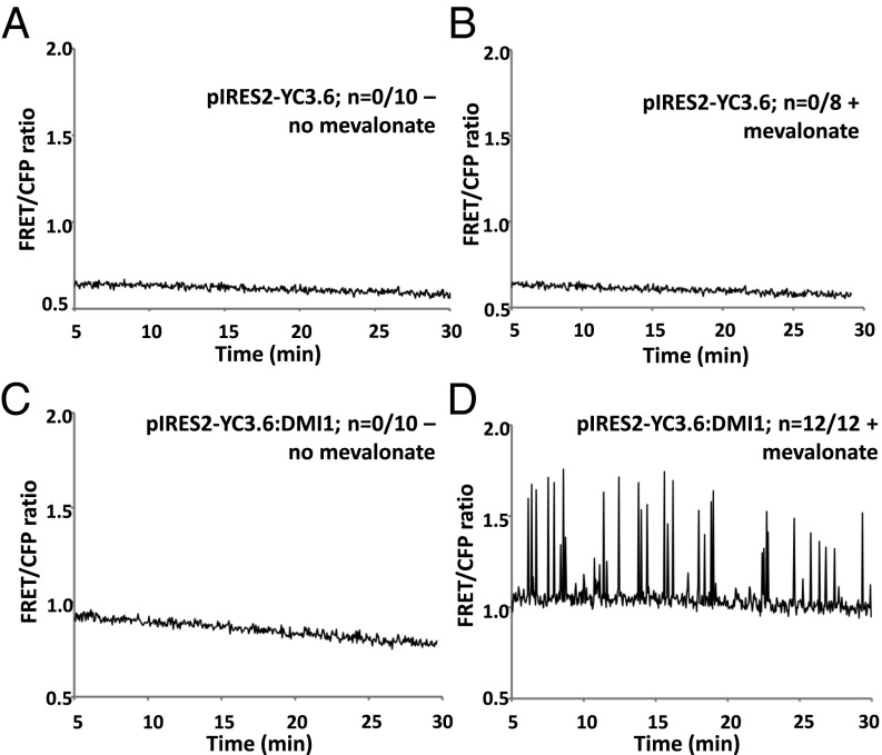
To determine whether MVA and DMI1 can be sufficient to trigger Ca2+ spiking, we used Human Embryonic Kidney 293 (HEK-293) cells as a heterologous expression system. We have shown in a previous study (18) that DMI1 maintains its nuclear envelope localization in HEK-293 cells when expressed under the control of the cytomegalovirus (CMV) promoter. Furthermore, following exogenous Ca2+ treatment, these cells display perinuclear Ca2+ oscillations (18). To determine whether MVA can trigger DMI1-mediated Ca2+ spiking in HEK-293, this cell line was transfected with either the Ca2+ sensor alone (pIRES2-YC3.6) or the Ca2+ sensor along with DMI1 (pIRES2-YC3.6::DMI1). For all experiments, the growth medium was replaced by bath solution, either with or without 100 µm MVA. In the absence of MVA, cells expressing pIRES2-YC3.6 or pIRES2-YC3.6::DMI1 did not exhibit de novo Ca2+ spiking (Fig. 4 A and C). Similarly, the exogenous application of MVA to cells expressing the Ca2+ sensor alone did not trigger Ca2+ spiking (Fig. 4B). By contrast, MVA application to HEK-293 cells expressing DMI1 triggered an intense Ca2+ spiking response (Fig. 4D). These observations, in which DMI1 is expressed as the sole M. truncatula protein in a heterologous system, provide further evidence that DMI1 expression is sufficient to support MVA-induced Ca2+ spiking.
Fig. 4.
MVA-induced Ca2+ spiking in HEK-293 cells expressing DMI1. (A and B) Absence of Ca2+ spiking in HEK-293 cells expressing the vector control pIRES2-YC3.6 in the absence (A) or presence of exogenous MVA (100 µM) (B). (C and D) Ca2+ spiking is only observed when MVA is added to HEK-293 cells expressing the pIRES2-YC3.6::DMI1 vector.
SI Materials and Methods
HMGR1 Enzymatic Assay.
The catalytic domain of HMGR1 along with the linker region (HMGR1ΔN) was cloned into pDONR221 (Invitrogen). This construct was verified by sequencing, and using the Gateway LR recombination reaction (Invitrogen), a maltose-binding protein MBP::HMGR1ΔN construct was assembled in the Gateway vector pVP16. A modified method from Horn and Walker (42) was adopted for the expression and purification of recombinant proteins. MBP::HMGR1ΔN was expressed in E. coli Rosetta pLaqI and purified at room temperature using an amylose resin (New England Biolabs). The protocol for the enzyme assay was adapted from Dale et al. (25). The assay was performed using a spectrophotometer with 1 mL of reaction mix containing 100 mM potassium phosphate, pH 7.0, 4 mM DTT, 50 mM NADPH, and varied concentrations of HMG-CoA. The reaction was started using HMG-CoA, and the absorbance at 340 nm was measured before and after the addition of HMG-CoA to calculate the oxidation of NADPH. The reaction was carried out at room temperature for 25 min, and the initial rate of reaction was used to calculate the Km.
Samples and buffer controls not containing an enzyme were evaporated and resuspended in dichloromethane before derivatization with N-Methyl-N-(trimethylsilyl) trifluoroacetamide (MSTFA) with 1% trimethylchlorosilane (Sigma Aldrich). Samples were incubated for 30 min before analysis by Thermo Trace Ultra Gas Chromatograph coupled to a Quadrupole/Orbitrap mass spectrometer. Metabolites were separated by thermal gradient and analyzed as previously described (43). MVA was identified by spectral and retention time comparison with an authentic standard (Sigma Aldrich). Relative quantitation was performed by comparison of peak areas generated from extracted ion chromatograms using Thermo Xcaliber Software.
RNAi of MtHMGR1.
The entry clone pENTR/D:HMGR1-RNAi carrying the 5′ untranslated region with a short fragment of the coding region of HMGR1 (226 bp), described in a previous article (24), was used in this study to clone a HMGR1-RNAi fragment into a modified pH7GWIWG2(II)-YC3.6 binary vector using LR Clonase (Invitrogen) as previously described (44). This RNAi–hairpin loop binary vector that coexpresses YC3.6 along with the HMGR1-RNAi cassette makes it possible to perform Ca2+ imaging in HMGR1-RNAi roots. A. rhizogenes-mediated hairy root transformation was used to introduce this construct and the vector control pH7GWIWG2(II)-YC3.6 into M. truncatula. Nod factor-induced, GSE-induced, and MVA-induced Ca2+ spiking were monitored as described below.
pENOD11:GUS Assays and qRT-PCR.
M. truncatula lines (Jemalong line A17, nfp-1, nfp-2, dmi2-1, dmi1-4, and dmi3-1) transformed with a pENOD11:GUS reporter were used for this study (41). Ten-day-old seedlings (n = 10) were treated with Nod factors (10−8 M) or MVA (100 µM, the lowest concentration that triggered nuclear Ca2+ spiking in M. truncatula root epidermal cells) for 12 or 24 h, respectively. The roots were then excised to evaluate ENOD11 expression using a GUS assay as previously described (41).
Similarly, 12 h after Nod factor treatment, the root tissues were flash-frozen in liquid nitrogen for total RNA isolation using the plant RNeasy Miniprep Kit (QIAGEN). The RNA preparations were treated with DNA-free (Ambion) to remove the residual genomic DNA contamination and quantified using NanoDrop. From each sample, 1 µg of total RNA was used for cDNA synthesis using the RevertAid First Strand cDNA Synthesis Kit (Fermentas). Quantitative RT-PCR was performed to monitor the expression level of MtENOD11 using MtActin and MtEF1a as reference genes using SYBR Advantage Real-Time qPCR Master Mix (Clontech). The primers that were used were as follows: ENOD11 (5′-CTCCATCCCACAATATGCCTCC and 5′-ATGGATGCTAGGTGGAGGCT), MtActin (5′-GCAGATGCTGAGGATATTAACCC and 5′-CGACCACTTGCATAGAGGGAGAGG), and MtEF1a (5′-GTCAAAACATGGTTGCTGCACAAGC and 5′-TTAGGTCACAAGGCAGATTGCAGG). The PCR cycling conditions consisted of an initial denaturation step at 95 °C for 3 min, followed by 40 cycles at 95 °C for 5 s for denaturation, 58.5 °C for 10 s for annealing, and 72 °C for 20 s for elongation.
Root Hair Growth Assays.
To test the effect of lovastatin on the growth of root hairs, Jemalong A17 plants were grown on Fahräeus agar medium that was supplemented with lovastatin as described in Kevei et al. (24). Briefly, a 12.5-mM lovastatin (Sigma Aldrich) stock solution was prepared in ethanolic NaOH, and the lactone ring was hydrolyzed by incubating at 60 °C for 1 h. Then, 0.5 µM and 1 µM lovastain plates were prepared using this stock solution, and 10 seedlings were planted on each plate. Plants that were 2 wk old were chosen for root hair measurement, and a total of 100 mature root hairs from at least 10 different plants for each concentration of lovastatin and control were measured for the analysis. To test the effect of silencing HMGR1 on root hair growth, roots transformed with HMGR1-RNAi or vector control were used for measuring the length of mature root hairs 3 wk after transformation.
Ca2+ Imaging in Root Epidermal Cells.
A Förster Resonance Energy Transfer (FRET)-based assay was used to analyze Ca2+ spiking. Two weeks after A. rhizogenes (MSU440) transformation of M. truncatula seedlings, transgenic roots were identified using the YC3.6 fluorescence. Plants with transgenic roots expressing YC3.6 were selected and mounted onto custom-built slide holders with a capacity of 10 mL of Fahräeus agar medium. The roots were mounted in such a way so that it was possible to focus on epidermal nuclei using an inverted confocal laser scanning microscope. After mounting, plants were incubated in a growth chamber for at least 24 h to establish normal root and root hair growth. Transformed L. japonicus and Arabidopsis plants were germinated and grown on either Fahräeus medium (L. japonicus) or Murashige–Skoog medium (Arabidopsis). M. truncatula and carrot root organ cultures expressing NUP:YC2.1 were grown on M-medium. After 2 wk, plants or root cultures were mounted in their respective medium using custom-built slide holders as described above for M. truncatula seedlings. (±)-Mevalonolactone (Sigma Aldrich, M4667) was dissolved (1 M) in Fahräeus medium and added to the mounted roots at various final concentrations (10, 50, 100, 250, 500, and 1,000 µM). Responses to MVA treatment, such as Ca2+ spiking and ENOD11 expression, were observed in plants starting from a 100 µM concentration. Hence, this lower concentration (100 µM) that triggers responses in plants was used for further experiments. The same concentration (100 µM) was used to test the effect of (±)-Mevalonic acid 5-phosphate trilithium salt (Sigma Aldrich, 79849), (±)-Mevalonic acid 5-pyrophosphate tetralithium salt (Sigma Aldrich, 94259), and isopentenyl pyrophosphate triammonium salt (Sigma Aldrich, I0503) on nuclear Ca2+ spiking. A Zeiss LSM 510 Meta confocal microscope (Carl Zeiss Inc.) was used to collect data on the intensity of the YFP and CFP fluorescent signals every 3 s for about 30 min. All of the experiments on Ca2+ measurements were made on actively growing root hair cells present on the primary roots of seedlings including Arabidopsis, with the exception of the root organ culture of M. truncatula and carrot, where Ca2+ measurements were made on atrichoblast cells. Because MVA induced ENOD11 expression but failed to activate Ca2+ spiking in root epidermal cells of nfp mutants (nfp-1 and nfp-2), we analyzed both trichoblast and atrichoblast cells for both ENOD11 expression and Ca2+ spiking (Fig. S6). Variations in the nuclear and perinuclear Ca2+ concentrations are indicated by FRET-based changes in the YFP/CFP ratio.
S. meliloti Nod factors (10−8 M) and GSEs of R. irregularis were suspended in Fahräeus medium broth and applied directly to roots to detect nuclear-associated Ca2+ spiking where appropriate.
Ca2+ Imaging Using YC3.6 in HEK-293 Cells and Data Analyses.
HEK-293 cells were cultured and transfected as previously described (18). At 36 h after transfection, the HEK-293 cells expressing the YC3.6 cytosolic Ca2+ reporter both with and without DMI1 were selected for confocal microscopy. The complete growth medium in the chamber was replaced with 1 mL of external bath solution (130 mM NaCl, 3 mM KCl, 0.6 mM MgCl2, 10 mM glucose, and 10 mM Hepes; pH 7.4). The transfected cells with high YC3.6 expression levels were selected for FRET analyses. The CFP emission (473–505 nm) and YFP emission (536–546 nm) were collected using a 458-nm primary dichroic mirror and the Metadetector of a Zeiss LSM 510 Meta confocal microscope (Carl Zeiss Inc.). MVA (100 µM) was applied to the external solution. The objective fields were scanned once every 3.5 s for a total of 500 cycles. The image analyses were performed using LSM Image Browser version 4.2 (Carl Zeiss Inc.). After correction for the background, the YFP/CFP ratio was calculated for each time point and plotted on the y axis in a scatter diagram over time (min) on the x axis.
Discussion
HMGR1 Is Required in the Early Symbiotic Signaling Cascade.
Genetic and genomic approaches have advanced our understanding of the molecular mechanisms of signal transduction during the initial stages of legume nodulation and AM symbioses. This research led to the identification of essential components of the common symbiosis pathway (2, 13). However, the secondary messengers that link the perception of microbial signals at the plasma membrane level to the regulation of ion channels and Ca2+ pumps on the nuclear envelope remain unknown (17, 33). With the identification of HMGR1 as both an interactor of the DMI2 coreceptor and a requirement for legume nodulation, we hypothesized that this MVA-producing enzyme might function in the common symbiosis signaling pathway. Metabolites from the MVA pathway play a wide variety of roles in many eukaryotes, including growth, development, and responses to environmental stimuli (34–36), by regulating cell-autonomous transcriptional and posttranscriptional processes (36). The observation that silencing HMGR1 affects Ca2+ spiking in response to Nod factors and GSEs of AM fungi supports the hypothesis that HMGR1 is indeed a component of the common symbiosis pathway (Fig. 5). In addition, the study of MVA-induced ENOD11 expression and Ca2+ spiking in various symbiosis-defective mutants indicated that addition of MVA can partly restore ENOD11 expression and Ca2+ spiking in dmi2 but not in dmi1 mutants. To our knowledge, this is the first report clearly uncoupling the phenotypes of dmi1 and dmi2 mutants. Because DMI1 and DMI2 belong to the common symbiosis pathway, these results place HMGR1 and its MVA-derived products downstream of DMI2 but upstream of DMI1 in the symbiotic cascade (3).
Fig. 5.
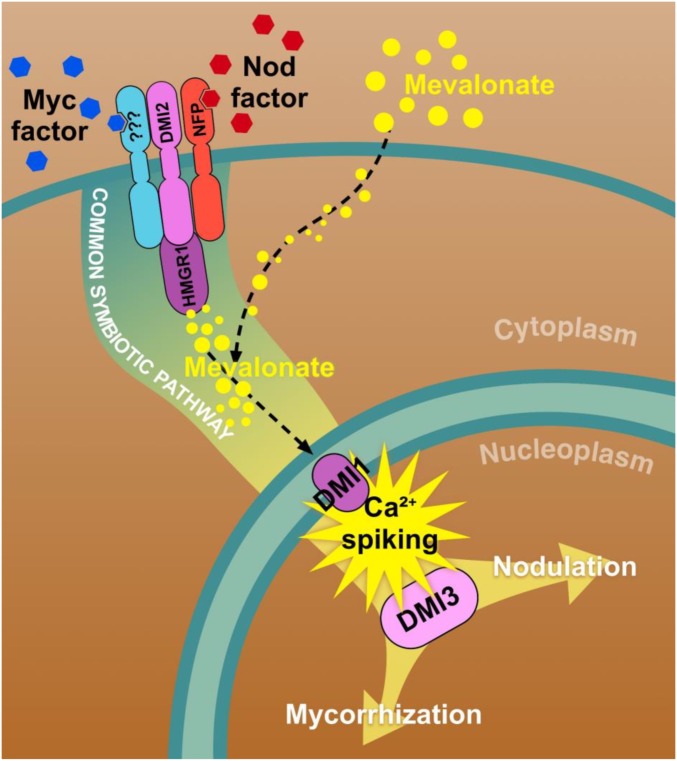
Model illustrating the proposed role of MVA within the common symbiosis pathway. Nod and Myc factors are perceived at the plasma membrane by a complex including the receptor-like kinase DMI2, interacting with either the Nod factor receptor component NFP or the so far unidentified Myc factor receptor. Based on our observations, we propose that HMGR1—which is known to interact with DMI2—generates MVA as a second messenger, transducing the signal from the plasma membrane to the nuclear compartment where DMI1, the nuclear envelope-localized cation channel, is required for the initiation of nuclear Ca2+ spiking. This Ca2+ response is then decoded by the Ca2+ and calmodulin-dependent kinase DMI3, which in turn leads to downstream endosymbiosis-related gene activation. In our experiments, the exogenous application of MVA is sufficient to activate the common symbiosis pathway and trigger nuclear Ca2+ spiking in the absence of receptor activation.
The conversion of HMG-CoA into MVA by HMGR is the first committed and rate-limiting step of the MVA biosynthetic pathway. The ability of MVA to trigger nuclear-associated Ca2+ spiking emphasizes the key role of HMGR1 as a link between signaling proteins residing on the plasma membrane and those located on the nuclear envelope. MVA-activated Ca2+ spiking is conserved across diverse AM host plants including legumes and nonlegumes. In contrast, the non-AM host Arabidopsis does not respond to either rhizobial or AM signals (2, 14), but does to MVA (Fig. 2F), suggesting that the targets of MVA (or its derived metabolites) are absent in Arabidopsis like many other signaling components required for AM associations.
Do MVA or Other Products of the MVA Pathway Act as Signaling Intermediates Linking the Plasma Membrane to the Nuclear Envelope?
Many classical secondary messengers, such as IP3, NAD+/NADH, cADP ribose, and Ca2+, have been considered as possible candidates for transducing symbiotic signal perception at the plasma membrane to the activation of Ca2+ spiking responses in the nucleus (33, 37, 38). Electrophysiological analyses have ruled out the possibility of IP3 or Ca2+ acting as modulators of Ca2+ spiking (33). However, MVA and its immediate phosphorylated derivatives (MVA 5-phosphate and MVA 5-pyrophosphate) elicited nuclear Ca2+ spiking, whereas isopentenyl pyrophosphate failed to elicit this response. This negative result for isopentenyl pyrophosphate could be due to restricted diffusion into root epidermal cells. Although of course we do not provide direct evidence that MVA is a second messenger in symbiotic signaling, we consider that the results presented in this article make MVA and its phosphorylated derivatives promising candidates for future studies.
Several isoforms of HMGR1 have been reported in M. truncatula (24), and MVA and its derivatives are abundant metabolites in plant cells, including root epidermal cells. Hence, it is interesting to discover that the exogenous application of MVA elicits Ca2+ spiking in root epidermal cells. Neither silencing HMGR1 nor the addition of lovastatin at a concentration that is inhibitory to nodulation (24) affected root hair growth, implying that the MVA pathway is not required for root hair development. We therefore hypothesize that the activation of DMI2 during symbiotic signaling may transiently activate HMGR1, leading to localized production of MVA in root epidermal cells (trichoblasts and atrichoblasts). The transient elevation in the MVA level inside the epidermal cells may in turn be responsible for activating nuclear Ca2+ spiking.
Differences clearly exist in the Ca2+ spiking patterns elicited by MVA, rhizobial Nod factors, and diffusible signals from AM fungi that were tested in different model plants and in different genetic backgrounds. If MVA activates the common symbiosis pathway by bypassing the receptor-ligand recognition step, it is therefore possible that downstream responses will not necessarily be specific to a particular microbial signal. In addition, it is likely that the exogenous application of MVA to root epidermal cells can only partially mimic an endogenous production of MVA in terms of intracellular concentration or subcellular localization. Thus, exogenous MVA is likely to activate the common symbiosis pathway both nonspecifically and suboptimally, probably explaining the high degree of variability in the observed Ca2+ spiking patterns.
If the model presented in Fig. 5 is correct, then it is surprising that ENOD11 expression was elicited by MVA in nfp mutants, whereas Ca2+ spiking was not detected in these mutants. The reason for this apparent discrepancy remains unclear. It is known that nfp mutants have more severe symbiotic phenotypes compared with dmi mutants, as for instance both root hair deformation and Ca2+ spiking are blocked in nfp, whereas only Ca2+ spiking is affected in dmi2 mutants in response to Nod factors (4, 10). Thus, it is possible that Ca2+ spiking may require not only MVA but also other signaling molecules that are produced in an NFP- but not DMI2-dependent manner. Alternatively, the apparent difference between pENOD11-GUS assays and Ca2+ spiking analyses in the nfp mutant may simply reflect different sensitivities of the respective techniques. The fact that ENOD11 expression in response to MVA addition cannot be detected by RT-PCR indicates a much lower level of gene induction with MVA than with Nod factors. This response may be even lower in some mutant backgrounds. Such a lower response was indeed observed for the Ca2+ spiking, as lower levels of spiking were observed in response to MVA in the dmi2 mutant compared with wild-type plants (compare Figs. 2A and 3B). If we are operating at the detection limit for the Ca2+ spiking assay, then this could explain the failure to observe Ca2+ spiking in the nfp background. However, at this stage, this question remains to be fully clarified.
Finally, the fact that MVA was able to trigger Ca2+ spiking in HEK-293 cells expressing M. truncatula DMI1 provides preliminary evidence that MVA may act directly on the cellular machinery that controls Ca2+ spiking in plants in response to symbiotic microbial signals (Fig. 5). We therefore hypothesize that the perception of symbiotic signals leads to the activation of HMGR1 bound to DMI2 and to the localized production of MVA, which then translocates to the nucleus activating in turn nuclear cation channels (DMI1/POLLUX, CASTOR, or Ca2+ channels), and thereby triggering nuclear-associated Ca2+ spiking. Although MVA is well known for its essential role in isoprenoid/sterol metabolism in eukaryotes, this study sheds light on a potential new role for this ubiquitous metabolite as a signaling intermediate in intracellular signaling pathways.
Materials and Methods
Ca2+ spiking analyses on M. truncatula were performed using the wild-type Jemalong A17 line and the symbiosis-defective mutants nfp-1, nfp-2, dmi2-1, dmi1-4, dmi1-2, and dmi3-1 (19, 39), after Agrobacterium rhizogenes-dependent transformation with the cytosolic YC3.6 yellow cameleon calcium sensor (40). For L. japonicus, the wild-type Gifu line was transformed via Agrobacterium tumefaciens with the nuclear-targeted Ca2+ sensor NES:YC3.6, and the Arabidopsis thaliana ecotype Col-0 was transformed with cytosolic YC3.6. GUS assays were performed on Jemalong A17 (pENOD11:GUS), nfp-1 (pENOD11:GUS), nfp-2 (pENOD11:GUS), dmi2-1 (pENOD11:GUS), dmi1-4 (pENOD11:GUS), and dmi3-1 (pENOD11:GUS) lines (38, 41). M. truncatula and carrot root organ cultures expressing the nuclear NUP-YC2.1 cameleon sensor were obtained via A. rhizogenes transformation (15).
For detailed description of methods pertaining to HMGR1 enzymatic assay, RNAi of MtHMGR1, pENOD11-GUS assays and RT-PCR, root hair growth assays, Ca2+ imaging in root epidermal cells, and Ca2+ imaging in HEK-293 cells, see SI Materials and Methods.
Acknowledgments
We thank Michael R. Sussman for technical assistance. We acknowledge the technical support of Steven Scholzen, Maxime Magne, Maegen Howes-Podoll, Pich Tea, and Gary Flores (University of Wisconsin–Madison) and Leanna Oltz and Kendell Welch (University of Wisconsin–Platteville). The confocal microscopy was primarily performed at the University of Wisconsin–Madison Newcomb Imaging Center and was supported by the National Science Foundation. This research was supported by National Science Foundation Grants IOS-0701846 and IOS-1021196 (to J.-M.A.) and United States Department of Agriculture-Agriculture and Food Research Initiative Grant 2015-67013-22899 (to J.-M.A. and M.V.). The work performed at the Laboratory of Plant-Microbe Interactions (Toulouse, France) is part of the TULIP Laboratory of Excellence (ANR6106LABX-41).
Footnotes
The authors declare no conflict of interest.
*This Direct Submission article had a prearranged editor.
This article contains supporting information online at www.pnas.org/lookup/suppl/doi:10.1073/pnas.1413762112/-/DCSupplemental.
References
- 1.Osbourn A. Saponins and plant defence—A soap story. Trends Plant Sci. 1996;1(1):4–9. [Google Scholar]
- 2.Venkateshwaran M, Volkening JD, Sussman MR, Ané JM. Symbiosis and the social network of higher plants. Curr Opin Plant Biol. 2013;16(1):118–127. doi: 10.1016/j.pbi.2012.11.007. [DOI] [PubMed] [Google Scholar]
- 3.Kistner C, et al. Seven Lotus japonicus genes required for transcriptional reprogramming of the root during fungal and bacterial symbiosis. Plant Cell. 2005;17(8):2217–2229. doi: 10.1105/tpc.105.032714. [DOI] [PMC free article] [PubMed] [Google Scholar]
- 4.Amor BB, et al. The NFP locus of Medicago truncatula controls an early step of Nod factor signal transduction upstream of a rapid calcium flux and root hair deformation. Plant J. 2003;34(4):495–506. doi: 10.1046/j.1365-313x.2003.01743.x. [DOI] [PubMed] [Google Scholar]
- 5.Fliegmann J, et al. Lipo-chitooligosaccharidic symbiotic signals are recognized by LysM receptor-like kinase LYR3 in the legume Medicago truncatula. ACS Chem Biol. 2013;8(9):1900–1906. doi: 10.1021/cb400369u. [DOI] [PubMed] [Google Scholar]
- 6.Maillet F, et al. Fungal lipochitooligosaccharide symbiotic signals in arbuscular mycorrhiza. Nature. 2011;469(7328):58–63. doi: 10.1038/nature09622. [DOI] [PubMed] [Google Scholar]
- 7.Mukherjee A, Ané JM. Germinating spore exudates from arbuscular mycorrhizal fungi: Molecular and developmental responses in plants and their regulation by ethylene. Mol Plant Microbe Interact. 2011;24(2):260–270. doi: 10.1094/MPMI-06-10-0146. [DOI] [PubMed] [Google Scholar]
- 8.Genre A, et al. Short-chain chitin oligomers from arbuscular mycorrhizal fungi trigger nuclear Ca2+ spiking in Medicago truncatula roots and their production is enhanced by strigolactone. New Phytol. 2013;198(1):190–202. doi: 10.1111/nph.12146. [DOI] [PubMed] [Google Scholar]
- 9.Antolín-Llovera M, Ried MK, Parniske M. Cleavage of the SYMBIOSIS RECEPTOR-LIKE KINASE ectodomain promotes complex formation with Nod factor receptor 5. Curr Biol. 2014;24(4):422–427. doi: 10.1016/j.cub.2013.12.053. [DOI] [PubMed] [Google Scholar]
- 10.Endre G, et al. A receptor kinase gene regulating symbiotic nodule development. Nature. 2002;417(6892):962–966. doi: 10.1038/nature00842. [DOI] [PubMed] [Google Scholar]
- 11.Limpens E, et al. Formation of organelle-like N2-fixing symbiosomes in legume root nodules is controlled by DMI2. Proc Natl Acad Sci USA. 2005;102(29):10375–10380. doi: 10.1073/pnas.0504284102. [DOI] [PMC free article] [PubMed] [Google Scholar]
- 12.Sieberer BJ, et al. A nuclear-targeted cameleon demonstrates intranuclear Ca2+ spiking in Medicago truncatula root hairs in response to rhizobial nodulation factors. Plant Physiol. 2009;151(3):1197–1206. doi: 10.1104/pp.109.142851. [DOI] [PMC free article] [PubMed] [Google Scholar]
- 13.Capoen W, et al. Nuclear membranes control symbiotic calcium signaling of legumes. Proc Natl Acad Sci USA. 2011;108(34):14348–14353. doi: 10.1073/pnas.1107912108. [DOI] [PMC free article] [PubMed] [Google Scholar]
- 14.Ané JM, et al. Medicago truncatula DMI1 required for bacterial and fungal symbioses in legumes. Science. 2004;303(5662):1364–1367. doi: 10.1126/science.1092986. [DOI] [PubMed] [Google Scholar]
- 15.Chabaud M, et al. Arbuscular mycorrhizal hyphopodia and germinated spore exudates trigger Ca2+ spiking in the legume and nonlegume root epidermis. New Phytol. 2011;189(1):347–355. doi: 10.1111/j.1469-8137.2010.03464.x. [DOI] [PubMed] [Google Scholar]
- 16.Ehrhardt DW, Wais R, Long SR. Calcium spiking in plant root hairs responding to Rhizobium nodulation signals. Cell. 1996;85(5):673–681. doi: 10.1016/s0092-8674(00)81234-9. [DOI] [PubMed] [Google Scholar]
- 17.Peiter E, et al. The Medicago truncatula DMI1 protein modulates cytosolic calcium signaling. Plant Physiol. 2007;145(1):192–203. doi: 10.1104/pp.107.097261. [DOI] [PMC free article] [PubMed] [Google Scholar]
- 18.Venkateshwaran M, et al. The recent evolution of a symbiotic ion channel in the legume family altered ion conductance and improved functionality in calcium signaling. Plant Cell. 2012;24(6):2528–2545. doi: 10.1105/tpc.112.098475. [DOI] [PMC free article] [PubMed] [Google Scholar]
- 19.Wais RJ, et al. Genetic analysis of calcium spiking responses in nodulation mutants of Medicago truncatula. Proc Natl Acad Sci USA. 2000;97(24):13407–13412. doi: 10.1073/pnas.230439797. [DOI] [PMC free article] [PubMed] [Google Scholar]
- 20.Granqvist E, et al. Buffering capacity explains signal variation in symbiotic calcium oscillations. Plant Physiol. 2012;160(4):2300–2310. doi: 10.1104/pp.112.205682. [DOI] [PMC free article] [PubMed] [Google Scholar]
- 21.Charpentier M, Vaz Martins T, Granqvist E, Oldroyd GE, Morris RJ. The role of DMI1 in establishing Ca2+ oscillations in legume symbioses. Plant Signal Behav. 2013;8(2):e22894. doi: 10.4161/psb.22894. [DOI] [PMC free article] [PubMed] [Google Scholar]
- 22.Miller JB, et al. Calcium/Calmodulin-dependent protein kinase is negatively and positively regulated by calcium, providing a mechanism for decoding calcium responses during symbiosis signaling. Plant Cell. 2013;25(12):5053–5066. doi: 10.1105/tpc.113.116921. [DOI] [PMC free article] [PubMed] [Google Scholar]
- 23.Lévy J, et al. A putative Ca2+ and calmodulin-dependent protein kinase required for bacterial and fungal symbioses. Science. 2004;303(5662):1361–1364. doi: 10.1126/science.1093038. [DOI] [PubMed] [Google Scholar]
- 24.Kevei Z, et al. 3-hydroxy-3-methylglutaryl coenzyme a reductase 1 interacts with NORK and is crucial for nodulation in Medicago truncatula. Plant Cell. 2007;19(12):3974–3989. doi: 10.1105/tpc.107.053975. [DOI] [PMC free article] [PubMed] [Google Scholar]
- 25.Dale S, et al. Bacterial expression of the catalytic domain of 3-hydroxy-3-methylglutaryl-CoA reductase (isoform HMGR1) from Arabidopsis thaliana, and its inactivation by phosphorylation at Ser577 by Brassica oleracea 3-hydroxy-3-methylglutaryl-CoA reductase kinase. Eur J Biochem. 1995;233(2):506–513. doi: 10.1111/j.1432-1033.1995.506_2.x. [DOI] [PubMed] [Google Scholar]
- 26.Bach TJ, Weber T, Motel A. 1990. Some properties of enzymes involved in the biosynthesis and metabolism of 3-hydroxy-3- methylglutaryl coenzyme A reductase in plants. Biochemistry of the Mevalonic Acid Pathway to Terpenoids, Recent Advances in Phytochemistry, eds Towers GHN, Stafford AHA (Plenum, New York.), pp 1–82.
- 27.Cenedella RJ, Kuszak JR, Al-Ghoul KJ, Qin S, Sexton PS. Discordant expression of the sterol pathway in lens underlies simvastatin-induced cataracts in Chbb: Thom rats. J Lipid Res. 2003;44(1):198–211. doi: 10.1194/jlr.m200002-jlr200. [DOI] [PubMed] [Google Scholar]
- 28.Wititsuwannakul R, Wititsuwannakul D, Suwanmanee P. 3-hydroxy-3-methylglutaryl co-enzyme a reductase from Hevea brasiliensis. Phytochemistry. 1990;29(5):1401–1403. [Google Scholar]
- 29.Horváth B, et al. Medicago truncatula IPD3 is a member of the common symbiotic signaling pathway required for rhizobial and mycorrhizal symbioses. Mol Plant Microbe Interact. 2011;24(11):1345–1358. doi: 10.1094/MPMI-01-11-0015. [DOI] [PubMed] [Google Scholar]
- 30.Gobbato E, et al. A GRAS-type transcription factor with a specific function in mycorrhizal signaling. Curr Biol. 2012;22(23):2236–2241. doi: 10.1016/j.cub.2012.09.044. [DOI] [PubMed] [Google Scholar]
- 31.Roberts NJ, et al. Rhizobial and mycorrhizal symbioses in Lotus japonicus require lectin nucleotide phosphohydrolase, which acts upstream of calcium signaling. Plant Physiol. 2013;161(1):556–567. doi: 10.1104/pp.112.206110. [DOI] [PMC free article] [PubMed] [Google Scholar]
- 32.Shaw SL, Long SR. Nod factor elicits two separable calcium responses in Medicago truncatula root hair cells. Plant Physiol. 2003;131(3):976–984. doi: 10.1104/pp.005546. [DOI] [PMC free article] [PubMed] [Google Scholar]
- 33.Charpentier M, et al. Lotus japonicus CASTOR and POLLUX are ion channels essential for perinuclear calcium spiking in legume root endosymbiosis. Plant Cell. 2008;20(12):3467–3479. doi: 10.1105/tpc.108.063255. [DOI] [PMC free article] [PubMed] [Google Scholar]
- 34.Stermer BA, Bianchini GM, Korth KL. Regulation of HMG-CoA reductase activity in plants. J Lipid Res. 1994;35(7):1133–1140. [PubMed] [Google Scholar]
- 35.Chappell J. The biochemistry and molecular biology of isoprenoid metabolism. Plant Physiol. 1995;107(1):1–6. doi: 10.1104/pp.107.1.1. [DOI] [PMC free article] [PubMed] [Google Scholar]
- 36.Edwards PA, Ericsson J. Sterols and isoprenoids: Signaling molecules derived from the cholesterol biosynthetic pathway. Annu Rev Biochem. 1999;68:157–185. doi: 10.1146/annurev.biochem.68.1.157. [DOI] [PubMed] [Google Scholar]
- 37.Engstrom EM, Ehrhardt DW, Mitra RM, Long SR. Pharmacological analysis of nod factor-induced calcium spiking in Medicago truncatula. Evidence for the requirement of type IIA calcium pumps and phosphoinositide signaling. Plant Physiol. 2002;128(4):1390–1401. doi: 10.1104/pp.010691. [DOI] [PMC free article] [PubMed] [Google Scholar]
- 38.Charron D, Pingret JL, Chabaud M, Journet EP, Barker DG. Pharmacological evidence that multiple phospholipid signaling pathways link Rhizobium nodulation factor perception in Medicago truncatula root hairs to intracellular responses, including Ca2+ spiking and specific ENOD gene expression. Plant Physiol. 2004;136(3):3582–3593. doi: 10.1104/pp.104.051110. [DOI] [PMC free article] [PubMed] [Google Scholar]
- 39.Catoira R, et al. Four genes of Medicago truncatula controlling components of a nod factor transduction pathway. Plant Cell. 2000;12(9):1647–1666. doi: 10.1105/tpc.12.9.1647. [DOI] [PMC free article] [PubMed] [Google Scholar]
- 40.Boisson-Dernier A, et al. Agrobacterium rhizogenes-transformed roots of Medicago truncatula for the study of nitrogen-fixing and endomycorrhizal symbiotic associations. Molecular Plant-Microbe Interactions. 2001;14(6):695–700. doi: 10.1094/MPMI.2001.14.6.695. [DOI] [PubMed] [Google Scholar]
- 41.Journet EP, et al. Medicago truncatula ENOD11: A novel RPRP-encoding early nodulin gene expressed during mycorrhization in arbuscule-containing cells. Mol Plant Microbe Interact. 2001;14(6):737–748. doi: 10.1094/MPMI.2001.14.6.737. [DOI] [PubMed] [Google Scholar]
- 42.Horn MA, Walker JC. Biochemical properties of the autophosphorylation of RLK5, a receptor-like protein kinase from Arabidopsis thaliana. Biochim Biophys Acta. 1994;1208(1):65–74. doi: 10.1016/0167-4838(94)90160-0. [DOI] [PubMed] [Google Scholar]
- 43.Benkeblia N, Shinano T, Osaki M. Metabolite profiling and assessment of metabolome compartmentation of soybean leaves using non-aqueous fractionation and GGMS analysis. Metabolomics. 2007;3(3):297–305. [Google Scholar]
- 44.Riely BK, et al. Identification of legume RopGEF gene families and characterization of a Medicago truncatula RopGEF mediating polar growth of root hairs. Plant J. 2011;65(2):230–243. doi: 10.1111/j.1365-313X.2010.04414.x. [DOI] [PubMed] [Google Scholar]


