



Abstract
Hypoxia-Inducible Gene Domain Family Member 1A (HIGD1A) is a survival factor induced by Hypoxia-inducible Factor-1 (HIF1). HIF1 regulates many responses to oxygen deprivation but viable cells within hypoxic perinecrotic solid tumor regions frequently lack HIF1α. HIGD1A is induced in these HIF-deficient extreme environments and interacts with the mitochondrial electron transport chain to repress oxygen consumption, enhance AMPK activity and lower cellular ROS levels. Importantly, HIGD1A decreases tumor growth but promotes tumor cell survival in vivo. The human Higd1a gene is located on chromosome 3p22.1, where many tumor suppressor genes reside. Consistent with this, the Higd1a gene promoter is differentially methylated in human cancers, preventing its hypoxic induction. When hypoxic tumor cells are confronted with glucose deprivation, however, DNA methyltransferase activity is inhibited, enabling HIGD1A expression, metabolic adaptation and possible dormancy induction. Our findings therefore reveal important new roles for this family of mitochondrial proteins in cancer biology.
Introduction
Heart disease, stroke and cancer are associated with hypoxia (Semenza, 2014) and nutrient deprivation (Hardie et al., 2012). Hypoxia inducible factor 1 (HIF1) is a widely expressed transcription factor that regulates the survival of cells during oxygen and glucose deprivation (Iyer et al., 1998; Maltepe et al., 1997; Ochiai et al., 2011; Ryan et al., 1998). HIF can also regulate tumor metabolism by repressing respiration (Kim et al., 2006; Papandreou et al., 2006) while promoting glycolysis, which enables rapid tumor cell proliferation (Vander Heiden et al., 2009). When severe, cancer cells can survive hypoxia and/or nutrient-deprivation by entering a dormant state, which suppresses their growth (Bragado et al., 2012; Sosa et al., 2013). Since most cancer therapies target proliferating cells, oxygen/nutrient-deprived tumor regions frequently become resistant and contribute to tumor recurrence. New agents are therefore being developed to target these regions (Harada et al., 2012; Zhang et al., 2014). Paradoxically, chronically oxygen starved tumor regions frequently lack HIF1α expression (Ameri et al., 2010; Sobhanifar et al., 2005), likely due to simultaneous glucose deprivation (Catrina et al., 2004; Osada-Oka et al., 2010). However, some HIF1 target genes such as CAIX remain either due to greater protein stability (Sobhanifar et al., 2005) or HIF1-independent pathways (van den Beucken et al., 2009).
Oxygen or glucose deprivation promotes reactive oxygen species (ROS) production, which can trigger adaptive responses such as HIF induction (Sena and Chandel, 2012) or can induce apoptosis (Malhotra et al., 2008). Therefore, cells need to modulate both oxygen consumption and ROS production in order to survive oxygen/glucose-deprivation. One pathway that cells utilize to achieve this relies on AMP-dependent protein kinase (AMPK) activation (Jeon et al., 2012). AMPK can activate multiple adaptive pathways, including antioxidant mechanisms. Interestingly, the effects of AMPK on tumor growth are complex, acting as oncogene or tumor suppressor depending on context (Hardie and Alessi, 2013).
Hypoxia-Inducible Gene Domain Family Member 1A (HIGD1A) is a survival factor regulated by Hypoxia-inducible Factor-1 (HIF1) (Wang et al., 2006). We previously demonstrated that HIGD1A is expressed in regions of severe ischemia in vivo (Ameri et al., 2013) that frequently lack detectable HIF1 activity. To investigate this phenomenon, we interrogated the function of HIGD1A in RAS-transformed HIF1-deficient MEFs (Ryan et al., 2000) as well as in human cancers in vitro and in vivo. Our studies identify novel functions for HIGD1A with implications for tumor cell survival and dormancy mechanisms.
Results
HIGD1A protects from oxygen/glucose-deprivation but suppresses growth
HIGD1A can protect cells from glucose and oxygen deprivation induced death (Wang et al., 2006). To confirm this, we generated HIGD1A “knockdown” mouse embryonic fibroblasts (MEFs) (Fig. 1Ai), which exhibited poor survival during oxygen/glucose deprivation (Fig. 1Aii, and Aiii). To isolate the function of this single HIF1α target from other HIF-dependent effects, we generated HIF1α deficient MEFs (Hif-1α−/− MEFs) that stably expressed HIGD1A to levels observed in wild-type MEFs exposed to hypoxia (Fig. 1Bi). Sustained HIGD1A expression in Hif-1α−/− MEFs had negligible effects on colony formation under normoxic or hypoxic conditions (Fig. 1 Bii). Glucose deprivation reduced colony size and number in Hif-1α−/− MEFs expressing either HIGD1A or GFP (Fig. 1Bii). However, Hif-1α−/− MEFs expressing HIGD1A produced even fewer numbers of colonies that were also smaller in size during glucose- or combined glucose/oxygen-deprivation (Fig. 1B ii). Interestingly, HIGD1A expression protected Hif-1α−/− MEFs from death during glucose deprivation (Fig. 1C), suggesting that increased cell death was not the cause for reduced colony number or size. Consistent with these results, tumors derived from Hif-1α−/− MEFs expressing HIGD1A were much smaller than control Hif-1α−/− MEF tumors (Fig. 1D) and did not contain any appreciable areas of necrosis, which was widely seen in control tumors (Fig. 1Ei). Furthermore, these tumors also exhibited significantly less apoptosis (Fig. 1Eii). These results indicate that HIGD1A can promote cell survival during nutrient deprivation while simultaneously suppressing growth in vitro and in vivo.
Figure 1. HIGD1A protects against glucose starvation and suppresses tumor growth with diminished apoptosis.

(Ai) Immunoblot analysis of HIGD1A levels following shRNA-mediated knockdown in wt MEFs (control shRNA=ctrl). (A ii and iii) Phase contrast microscopy as well as trypan blue exclusion count indicate that HIGD1A is necessary for survival of cells during glucose starvation/hypoxia. 20,000 cells were seeded in 6-well plates and counted after 4 days. (Bi) Immunoblot analysis comparing protein levels of HIGD1A in HIF-deficient (Hif-1α−/−) MEFs stably expressing HIGD1A versus wild-type MEFs (Hif-1α+/+) exposed to hypoxia. (Bii) Colony formation assays showing that HIGD1A expression in HIF-deficient (Hif-1α−/−) MEFs results in fewer as well as smaller colonies during combined hypoxia/glucose deprivation or glucose deprivation alone. (C) Viability assay of Hif-1α−/− MEFs expressing HIGD1A compared with control GFP cells following three days of glucose deprivation. (D) Hif-1α−/− MEFs stably expressing HIGD1A resulted in significantly smaller tumor xenografts when grown for 3 weeks subcutaneously in mice. (Ei) Histopathological analysis indicating lack of necrosis in Hif-1α−/− HIGD1A tumors, but profound necrosis in Hif-1α−/− GFP control tumors. Cleaved-caspase-3 immunohistochemical staining shows significantly more apoptosis in Hif-1α−/− GFP control tumors (Eii). Error bars represent ±SD. * p<0.05. Five mice per group were used for tumor growth and analysis.
HIGD1A interaction with the electron transport chain modulates mitochondrial ROS production and oxygen consumption
HIGD1A is an inner mitochondrial protein and recently orthologs of HIGD1A and the related HIGD2A were shown to interact with complex IV in yeast (Chen et al., 2012; Strogolova et al., 2012). Immunoprecipitation assays with extracts derived from MEFs expressing GFP-tagged HIGD1A detected an interaction between murine HIGD1A and Complex III subunit 2 of the mitochondrial electron transport chain (ETC), but not with Complex IV subunit I (Fig. 2A). Complex III is an important site for mitochondrial superoxide (O2−) production (Buetler et al., 2004; Chen and Gibson, 2008), which can be increased when the proton motive force increases, as occurs with decreased flow through the respiratory chain (Murphy, 2009). Therefore, we examined the level of mitochondrial O2− production via FACS-mediated analysis of Mitosox Red intensity. This was increased to a greater extent in HIGD1A expressing Hif-1α−/− MEFs during glucose starvation than their GFP expressing counterparts (Fig. 2B). Additionally, HIGD1A expression in Hif-1α−/− MEFs resulted in an approximately two-fold reduction in cellular oxygen consumption during glucose deprivation (Fig. 2Ci and ii). Interestingly, following re-introduction of glucose, oxygen consumption was reversed more rapidly in cells stably expressing HIGD1A (Fig. 2D i and ii). These results indicate that HIGD1A expression can modulate mitochondrial ROS production and oxygen consumption during conditions of glucose deprivation via interaction with the ETC.
Figure 2. HIGD1A can regulate mitochondrial superoxide and oxygen consumption during glucose starvation.

(A) Immunoprecipitation assay showing HIGD1A can interact with complex III subunit 2 of the respiratory chain. (B) FACS analysis showing that HIF-deficient cells overexpressing HIGD1A have increased mitochondrial ROS (superoxide) during glucose starvation compared to control cells overexpressing GFP. (Ci and Cii) Oxygen consumption is lower during glucose deprivation when HIGD1A is overexpressed in Hif-1α−/− cells. (Di and Dii) When glucose is re-introduced to glucose-starved cells, HIGD1A expressing cells increase their oxygen consumption at a faster rate than control GFP expressing cells. Error bars represent ±SD, * p<0.05.
HIGD1A induces AMPK activity and decreases cellular ROS to promote survival
ROS generation during glucose deprivation can result in cell death (Gao et al., 2012; Lenin et al., 2012; Malhotra et al., 2008). AMPK can be induced by glucose deprivation and mitochondrial O2− production (Wu and Wei, 2012) (Mackenzie et al., 2013) to reduce total ROS via the promotion of pentose phosphate shunt-mediated NADPH production (Jeon et al., 2012). Consistent with these studies, Hif-1α−/− MEFs that stably expressed HIGD1A increased pAMPK levels to a greater extent than control cells during glucose deprivation (Fig. S1 A) and this effect was reduced by the mitochondria-targeted antioxidant MitoQ (Kelso et al., 2001) (Fig. S1B). Furthermore, this diminished total cellular ROS in HIGD1A expressing cells during glucose starvation (Fig. S1C), and a cell permeable form of the antioxidant glutathione (Graham et al., 2012) improved the viability of control Hif-1α−/− MEFs during glucose deprivation (Fig. S1D). Thus, HIGD1A triggers increased mitochondrial O2− production to activate AMPK and decrease total cellular ROS levels to promote cell survival. To determine whether AMPK activity was necessary for the protective effect of HIGD1A during glucose deprivation, we stably transfected Ampk1/2−/− MEFs with HIGD1A or GFP as control, and examined their survival during glucose starvation. AMPK-deficient cells do not reduce total cellular ROS levels and fail to induce autophagy during glucose starvation, compromising their survival (Jeon et al., 2012; Kim et al., 2011). Interestingly, HIGD1A expression did not protect Ampk1/2−/− MEFs from glucose starvation (Fig. S1E), indicating that AMPK activation is necessary for the protective effect of HIGD1A during glucose deprivation. We also examined AMPK activation in vivo utilizing tumors derived from Hif-1α−/− MEFs expressing HIGD1A previously described in Fig. 1D and found that they demonstrated pAMPK immunoreactivity that was more intense as well as more diffusely distributed than control tumors (Fig. S1F), consistent with the reduction of cell death. Immunoreactivity of pAMPK was ablated when treated with pAMPK blocking peptide (Fig. S1G). Finally, we questioned whether the effects of HIGD1A-dependent pAMPK induction during glucose deprivation could be due to autophagy induction, since it can also be regulated by AMPK to increase cell survival during glucose starvation (Kim et al., 2011). As seen in figure S2, autophagy induction was dispensable for the protective effects of HIGD1A.
HIGD1A is not induced by HIF1α in hypoxic human cancers but is triggered by additional metabolic stressors
We next examined the mode of regulation of HIGD1A in a variety of human cancer cell lines. Human HT1080 fibrosarcoma and HeLa cervical cancer cell lines exhibited basal levels of HIGD1A that were surprisingly not further induced by hypoxia, despite inducing HIF-1α (Fig. 3A). BNIP3, another HIF1 target mitochondrial protein (Sowter et al., 2001), was induced, however. The gene encoding HIGD1A is located on human chromosome 3p22.1, where many tumor suppressor genes reside, and these are often inactivated via epigenetic mechanisms (Bhat Singh and Amare Kadam, 2013; Buchhagen et al., 1994). From genome-wide analyses of aberrant DNA methylation in glioblastoma multiforme (GBM) (Nagarajan et al., 2014), we identified two candidate regions near the Higd1a promoter that exhibited GBM-specific hypermethylation. As indicated in figure 3B, one of these regions, located upstream of the 5′ CpG island promoter, in a CpG island “shore,” contains consensus core hypoxia response elements (HRE) (blue underlined). Two CpG sites within this region are interrogated on the Illumina HumanMethylation450 methylation array, and exhibit high methylation in several cancer cells, and only partial methylation in normal human NH-A astrocytes (ENCODE data), indicating that it may be a differentially methylated region (DMR, red and green CG). ChIP-seq profiles from the Roadmap Epigenomics Project confirmed the presence of histone modifications associated with enhancers in both brain and breast, consistent with it harboring potential HREs. ChIP analysis confirmed that HIF1α was able to bind the HRE within this DMR in HeLa cells during hypoxia (Fig. 3C), despite its expression not being induced. Treating HeLa cells with the DNA methylation inhibitor 5-aza-2′-deoxycytidine, however, enhanced HIGD1A expression during hypoxia (Fig. 3D). Reduced expression of DNA methyl transferase 1 (DNMT1) can reactivate tumor suppressors (Xiang et al., 2014; Yao et al., 2014), and glucose starvation can reduce expression of the gene encoding DNMT1 (Lin et al., 2012). Glucose starvation reduced DNMT1 expression in hypoxic HeLa cells suggesting that DNA methylation pathways are inhibited during combined oxygen/glucose deprivation (Fig. 3E). Reduction of DNMT1 correlated with enhanced HIGD1A expression during glucose starvation (Fig. 3F). Unlike canonical tumor suppressor genes that are sometimes strongly and permanently silenced by dense methylation across their promoter CpG islands, the Higd1a gene locus shows more nuanced epigenetic regulation in human cancer. DNA methylation at upstream HREs might prevent transactivation of HIGD1A via HIF1 and thereby suppress enhanced HIGD1A expression during growth permissive hypoxic conditions, but allow epigenetic activation when environmental conditions favoring HIGD1A expression are encountered.
Figure 3. Expression and regulation of HIGD1A in cancer.
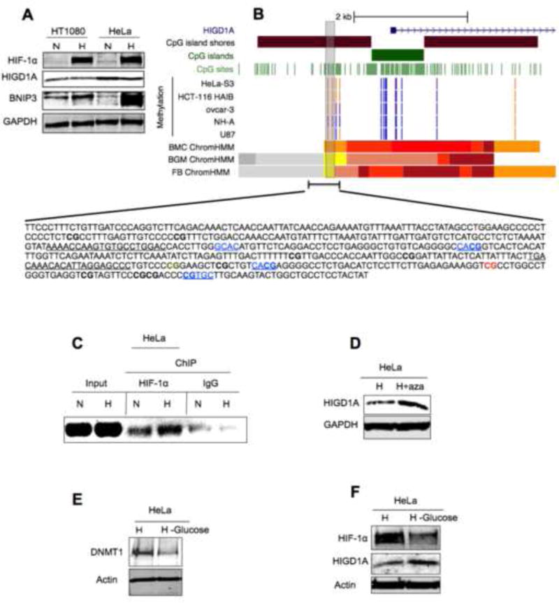
(A) Immunoblot analysis of HIGD1A, HIF1α and BNIP3 expression in human HT1080 and HeLa cancer cell lines during normoxia or hypoxia (B) Data from Illumina HumanMethylation450 methylation array, and the ENCODE consortium showing high methylation level (vertical orange lines) upstream of the 5′ CpG island promoter, in a CpG island “shore.” The HIGD1A CpG island itself is generally unmethylated (vertical blue and violet lines) in both cancer cell lines (U87, ovcar-3, HCT-116, HeLa) as well as in normal human astrocytes (NH-A). Two of the CpGs (within vertical grey rectangle) in the 5′ shore of that CpG island have high methylation levels in HeLa, ovcar-3, HCT-116, and U87 cell lines, and partial methylation in normal human astrocytes, indicating a potential differential methylated region (DMR, red and green CG in the sequence given). The sequence of the entire 5′ region (region chr3: 42846997–42847502) including the two specific CpGs (highlighted as green and red in the sequence) of this putative DMR neighbors several HRE-core sequences (blue underlined). ChIP-seq data from the Roadmap Epigenomics Project indicate that this region is marked by histone modifications associated with enhancers (yellow and orange bars) in both brain (FB ChromHMM, BGM ChromHMM) and breast (BMC ChromHMM). Primers used for CHIP analysis in black underlined. (C) ChIP analysis performed on normoxic (N) or hypoxic (H) HeLa cells using primers (black underlined in sequence) within the 5′ region that contains the two specific CpGs (highlighted as green and red in the sequence) of this putative DMR. (D) Immunoblot analysis demonstrating expression of HIGD1A protein in the human cervical cancer cell line HeLa in hypoxia (H) versus hypoxia combined with the DNA methylation inhibitor (DNMT-inhibitor) 5-aza-2′-deoxycytidine (H+aza). (E) Immunoblot analysis showing that glucose starvation (-glucose) during hypoxia (H) reduces expression of DNMT1. (F) Glucose starvation induces HIGD1A in hypoxic HeLa cells. H=hypoxia (1% O2)
HIGD1A expression is enhanced in severely ischemic tumor regions in vivo
We next examined whether HIGD1A expression is induced in a similar fashion in human cancers in vivo, particularly since reduced methyl cytosine levels have been reported in ischemic tumor regions due to reduced DNMT activity (Shahrzad et al., 2007; Skowronski et al., 2010). MDA-MB 231 breast cancer xenografts demonstrated severely ischemic perinecrotic regions as evidenced by strong staining with the hypoxia marker pimodinazole, along with diminished HIF1α immunoreactivity, indicating likely glucose starvation (Fig. 4A). As shown in Figure S3, lack of HIF1α was also observed in severely ischemic myocardial regions following experimental myocardial infarction in mice, suggesting that this may be a common indicator of starvation severity. As indicated in Fig. 4B and S3, these areas of ischemia demonstrated enhanced HIGD1A expression. Similar to our observations with HeLa cells, DNMT1 levels were also reduced in ischemic MDA-MB 231 cells (Fig. 4C). In addition, HIGD1A expression was enhanced in vitro in ischemic MDA-MB 231 cells when compared with hypoxia (Fig. 4D). Circulating tumor cells (CTCs) derived from MDA-MB 231 xenografts were previously reported to be more resistant to anoxia (Ameri et al., 2010). Both MDA-MB231 cells and their CTCs induced HIF1α but not HIGD1A during hypoxia (Fig. 4E). Interestingly, basal levels of HIGD1A were higher in CTCs. These results further confirm that hypoxic HIF1α induction is not sufficient to enhance HIGD1A expression in human cancers in vitro or in vivo, but that additional pathways triggered by severe metabolic stressors are necessary. Finally, we analyzed HIGD1A expression patterns in glioblastoma multiforme (GBM) biopsies from patients before and after treatment with the anti-angiogenesis agent bevacizumab, which is known to induce severe tumor ischemia. Primary GBM biopsies exhibited hypoxic areas as evident by increased CA9 expression (Fig. 4F). These areas did not demonstrate significant HIGD1A expression. However, after treatment with bevacizumab, HIGD1A was strongly induced (Fig. 4F). This indicates that HIGD1A expression is prevented during physiological hypoxia and that additional metabolic stressors are needed to induce HIGD1A in human cancers in vivo.
Figure 4. Expression of HIGD1A in vivo and in circulating tumor cells.

(A) Pimonidazole and HIF1α staining of MDA-MB 231 xenografts showing diminished expression of HIF1α within perinecrotic regions where pimonidazole staining is strongest. (B) MDA-MB 231 xenografts showing enhanced expression of HIGD1A at perinecrotic regions where HIF1α expression is diminished. (C) Immunoblot analysis showing that expression of DNMT1 during hypoxia (H) versus hypoxia and glucose starvation (H -Glucose). (D) Glucose starvation during hypoxia enhances HIGD1A protein level in MDA-MB 231 cells. (E) Immunoblot analysis of HIGD1A expression in MDA-MB231 cells from which the xenografts where made, and in CTCs derived from the xenografts via blood extraction, as a function of oxygen. (F) Human primary glioblastoma biopsies demonstrate lack of HIGD1A induction in hypoxic regions where Ca9 induction is evident. Induction of HIGD1A is evident only after treatment with the anti-angiogenesis agent bevacizumab. N=normoxia (21% oxygen), H=hypoxia (1% oxygen), HBS=HIF binding site, HRE=hypoxia response element, CTC=circulating tumor cell.
Discussion
We and others previously documented (Ameri et al., 2010; Sobhanifar et al., 2005), and confirmed once again here, that some of the most metabolically compromised tumor regions found around their necrotic cores fail to induce HIF activity, potentially due to glucose starvation (Catrina et al., 2004; Osada-Oka et al., 2010). Interestingly, these regions still express the HIF1 target HIGD1A. One way that cells can survive metabolic stress is via lowering cellular ROS and oxygen consumption, which are parameters associated with quiescence and dormancy-mediated survival (Endo et al., 2014; Lagadinou et al., 2013; Lopes et al., 2010). Consistent with this, we found that HIGD1A interacts with the mitochondrial electron transport chain, modulates oxygen consumption, ROS production and AMPK activity to promote cell survival during glucose starvation, while simultaneously suppressing tumor growth in vivo. Furthermore, anti-VEGF therapy has previously been shown to induce AMPK activity to promote tumor cell survival in vivo (Nardo et al., 2011), which we also confirm to be associated with enhanced HIGD1A activity in human GBM. Multiple studies have previously linked ROS suppression and cell survival with AMPK activation, likely via phosphatase inhibition (Davies et al., 1995; Faubert et al., 2013; Han et al., 2010; Hofstetter et al., 2012; Indraccolo, 2013; Ingebritsen et al., 1983; Jeon et al., 2012; Klaus et al., 2012; Kwan et al., 2013; Rotte et al., 2010; Wu and Wei, 2012). Our studies confirm these observations and identify HIGD1A as an important upstream component of this signaling cascade. Furthermore, HIGD1A repression is associated with tumor recurrence in breast cancers following therapy (Chanrion et al., 2008), consistent with our observations that HIGD1A expression helps repress tumor growth. These findings therefore provide novel insights into tumor cell adaptation mechanisms to extreme environments and suggest that HIGD1A may play an important role in tumor dormancy or recurrence mechanisms (Giancotti, 2013).
The ability of HIGD1A expression to be regulated epigenetically provides an attractive model whereby environmental factors can regulate HIGD1A expression independent of HIF activity to modulate tumor growth. The gene encoding HIGD1A is located on human chromosome 3p22.1, where many tumor suppressors reside, and many of which are inactivated via epigenetic mechanisms (Bhat Singh and Amare Kadam, 2013; Buchhagen et al., 1994). Our analysis of the human Higd1a locus indicated hypermethylation of the upstream promoter region in various cancer cell lines that was able to bind HIF1α but not drive its hypoxic expression. This suggested that additional pathways are required to induce HIGD1A in human cancers in vivo and we found that reducing the expression or activity of DNA methyl transferases (DNMTs) increased HIGD1A expression in vitro. This result is consistent with previous reports linking DNMT1 inhibition with tumor suppressor reactivation in response to environmental stressors in vivo (Xiang et al., 2014; Yao et al., 2014) (Lin et al., 2012). Constitutive basal expression of HIGD1A might be beneficial during growth permissive hypoxic conditions without glucose deprivation. When glucose deprivation becomes severe, enhanced HIGD1A expression modulated by epigenetic mechanisms may help trigger a state of dormancy and tumor growth inhibition (LaRue et al., 2004; Sutherland, 1988). Such dormant cells are typically resistant to many therapies, enabling tumor cell survival and cancer recurrence (Indraccolo, 2013; Lin et al., 2012). Novel molecules are therefore being developed to target these dormant cells (Zhang et al., 2014). Small molecule modulators of the HIG family of mitochondrial proteins (Lindert et al., 2014) may therefore prove useful in the fight against cancer.
Experimental Procedures
Cell Culture
MEFs, HT1080 and HeLa cells were cultured in RPMI-1640, 10%FBS, and 110 μg/ml Sodium Pyruvate. Glucose starvation was achieved by using glucose-free RPMI 1640. Normoxic cells were incubated at 5% CO2 and 21% O2 while hypoxia experiments were performed at 1% O2 with 5% CO2.
Oxygen consumption and ROS measurements
O2 consumption measured via use of the SeaHorse Extracellular flux Analyzer according to the manufacturer’s protocol. Mitochondrial ROS measured via FACS-mediated analysis of Mito-Sox Red, and total ROS assayed by measuring Cell-Rox Deep Red fluorescence via manufacturer’s instructions.
Immunohistochemistry
Formalin-fixed paraffin-embedded (FFPE) MEF and human tumor sections were cut at 5 μm, subjected to antigen retrieval, treated with M.O.M. kit, incubated for 1 hour at 37°C with primary antibody. Glioblastoma FFPE biopsies were cut at 16 μm sections. Sections were incubated overnight at 4°C in primary antibody. List of antibodies and suppliers available in supplement. Imaging performed with a Zeiss Imager Z.2 fluorescence microscope equipped with an Apotome and axiovision-ZEN software for optical sectioning and analysis.
Immunoprecipitation and immunoblotting
GFP-fusion proteins were immunoprecipitated with Chromotek-GFP-Trap bead according to manufacturer’s recommendations. Pulldowns, as well as all other immunoblotting, performed via SDS-PAGE and blotted onto Immobilon-FL membranes using semi-dry transfer. Membranes were blocked in blocking buffer from LI-COR Biosciences and probed with primary antibodies in LI-COR blocking buffer.
ChIP Assays
The ExactaCHIP kit was used for chromatin immunoprecipitation assays according to the manufacturer’s protocol.
Tumor models and human glioblastoma samples
Described in supplementary experimental procedures.
Supplementary Material
Acknowledgments
This research was made possible by a grant from the NIH R01 HD072455 and from the California Institute for Regeneration Medicine (Grant Number TB1-01194). Hif-1α−/−MEFs were provided by R. Johnson (University of Cambridge); Ampk1/2−/− MEFs were provided by B. Viollet (INSERM); Atg5−/− MEFs were provided by N. Mizushima (University of Tokyo).
Footnotes
Publisher's Disclaimer: This is a PDF file of an unedited manuscript that has been accepted for publication. As a service to our customers we are providing this early version of the manuscript. The manuscript will undergo copyediting, typesetting, and review of the resulting proof before it is published in its final citable form. Please note that during the production process errors may be discovered which could affect the content, and all legal disclaimers that apply to the journal pertain.
References
- Ameri K, Luong R, Zhang H, Powell AA, Montgomery KD, Espinosa I, Bouley DM, Harris AL, Jeffrey SS. Circulating tumour cells demonstrate an altered response to hypoxia and an aggressive phenotype. Br J Cancer. 2010;102:561–569. doi: 10.1038/sj.bjc.6605491. [DOI] [PMC free article] [PubMed] [Google Scholar]
- Ameri K, Rajah AM, Nguyen V, Sanders TA, Jahangiri A, Delay M, Donne M, Choi HJ, Tormos KV, Yeghiazarians Y, et al. Nuclear localization of the mitochondrial factor HIGD1A during metabolic stress. PLoS One. 2013;8:e62758. doi: 10.1371/journal.pone.0062758. [DOI] [PMC free article] [PubMed] [Google Scholar]
- Arai R, Ueda H, Kitayama A, Kamiya N, Nagamune T. Design of the linkers which effectively separate domains of a bifunctional fusion protein. Protein Eng. 2001;14:529–532. doi: 10.1093/protein/14.8.529. [DOI] [PubMed] [Google Scholar]
- Bhat Singh R, Amare Kadam PS. Investigation of tumor suppressor genes apart from VHL on 3p by deletion mapping in sporadic clear cell renal cell carcinoma (cRCC) Urol Oncol. 2013 doi: 10.1016/j.urolonc.2011.08.012. [DOI] [PubMed] [Google Scholar]
- Bragado P, Sosa MS, Keely P, Condeelis J, Aguirre-Ghiso JA. Microenvironments dictating tumor cell dormancy. Recent Results Cancer Res. 2012;195:25–39. doi: 10.1007/978-3-642-28160-0_3. [DOI] [PMC free article] [PubMed] [Google Scholar]
- Buchhagen DL, Qiu L, Etkind P. Homozygous deletion, rearrangement and hypermethylation implicate chromosome region 3p14.3–3p21.3 in sporadic breast-cancer development. Int J Cancer. 1994;57:473–479. doi: 10.1002/ijc.2910570406. [DOI] [PubMed] [Google Scholar]
- Buetler TM, Krauskopf A, Ruegg UT. Role of superoxide as a signaling molecule. News Physiol Sci. 2004;19:120–123. doi: 10.1152/nips.01514.2003. [DOI] [PubMed] [Google Scholar]
- Catrina SB, Okamoto K, Pereira T, Brismar K, Poellinger L. Hyperglycemia regulates hypoxia-inducible factor-1alpha protein stability and function. Diabetes. 2004;53:3226–3232. doi: 10.2337/diabetes.53.12.3226. [DOI] [PubMed] [Google Scholar]
- Chanrion M, Negre V, Fontaine H, Salvetat N, Bibeau F, Mac Grogan G, Mauriac L, Katsaros D, Molina F, Theillet C, et al. A gene expression signature that can predict the recurrence of tamoxifen-treated primary breast cancer. Clin Cancer Res. 2008;14:1744–1752. doi: 10.1158/1078-0432.CCR-07-1833. [DOI] [PMC free article] [PubMed] [Google Scholar]
- Chen Y, Gibson SB. Is mitochondrial generation of reactive oxygen species a trigger for autophagy? Autophagy. 2008;4:246–248. doi: 10.4161/auto.5432. [DOI] [PubMed] [Google Scholar]
- Chen YC, Taylor EB, Dephoure N, Heo JM, Tonhato A, Papandreou I, Nath N, Denko NC, Gygi SP, Rutter J. Identification of a protein mediating respiratory supercomplex stability. Cell Metab. 2012;15:348–360. doi: 10.1016/j.cmet.2012.02.006. [DOI] [PMC free article] [PubMed] [Google Scholar]
- Davies SP, Helps NR, Cohen PT, Hardie DG. 5′-AMP inhibits dephosphorylation, as well as promoting phosphorylation, of the AMP-activated protein kinase. Studies using bacterially expressed human protein phosphatase-2C alpha and native bovine protein phosphatase-2AC. FEBS Lett. 1995;377:421–425. doi: 10.1016/0014-5793(95)01368-7. [DOI] [PubMed] [Google Scholar]
- DeLay M, Jahangiri A, Carbonell WS, Hu YL, Tsao S, Tom MW, Paquette J, Tokuyasu TA, Aghi MK. Microarray analysis verifies two distinct phenotypes of glioblastomas resistant to antiangiogenic therapy. Clin Cancer Res. 2012;18:2930–2942. doi: 10.1158/1078-0432.CCR-11-2390. [DOI] [PMC free article] [PubMed] [Google Scholar]
- Endo H, Okuyama H, Ohue M, Inoue M. Dormancy of Cancer Cells with Suppression of AKT Activity Contributes to Survival in Chronic Hypoxia. PLoS One. 2014;9:e98858. doi: 10.1371/journal.pone.0098858. [DOI] [PMC free article] [PubMed] [Google Scholar]
- Faubert B, Boily G, Izreig S, Griss T, Samborska B, Dong Z, Dupuy F, Chambers C, Fuerth BJ, Viollet B, et al. AMPK is a negative regulator of the Warburg effect and suppresses tumor growth in vivo. Cell Metab. 2013;17:113–124. doi: 10.1016/j.cmet.2012.12.001. [DOI] [PMC free article] [PubMed] [Google Scholar]
- Gao HJ, Zhu YM, He WH, Liu AX, Dong MY, Jin M, Sheng JZ, Huang HF. Endoplasmic reticulum stress induced by oxidative stress in decidual cells: a possible mechanism of early pregnancy loss. Mol Biol Rep. 2012;39:9179–9186. doi: 10.1007/s11033-012-1790-x. [DOI] [PubMed] [Google Scholar]
- Giancotti FG. Mechanisms governing metastatic dormancy and reactivation. Cell. 2013;155:750–764. doi: 10.1016/j.cell.2013.10.029. [DOI] [PMC free article] [PubMed] [Google Scholar]
- Graham NA, Tahmasian M, Kohli B, Komisopoulou E, Zhu M, Vivanco I, Teitell MA, Wu H, Ribas A, Lo RS, et al. Glucose deprivation activates a metabolic and signaling amplification loop leading to cell death. Mol Syst Biol. 2012;8:589. doi: 10.1038/msb.2012.20. [DOI] [PMC free article] [PubMed] [Google Scholar]
- Han Y, Wang Q, Song P, Zhu Y, Zou MH. Redox regulation of the AMP-activated protein kinase. PLoS One. 2010;5:e15420. doi: 10.1371/journal.pone.0015420. [DOI] [PMC free article] [PubMed] [Google Scholar] [Retracted]
- Harada H, Inoue M, Itasaka S, Hirota K, Morinibu A, Shinomiya K, Zeng L, Ou G, Zhu Y, Yoshimura M, et al. Cancer cells that survive radiation therapy acquire HIF-1 activity and translocate towards tumour blood vessels. Nat Commun. 2012;3:783. doi: 10.1038/ncomms1786. [DOI] [PMC free article] [PubMed] [Google Scholar]
- Hardie DG, Alessi DR. LKB1 and AMPK and the cancer-metabolism link – ten years after. BMC Biol. 2013;11:36. doi: 10.1186/1741-7007-11-36. [DOI] [PMC free article] [PubMed] [Google Scholar]
- Hardie DG, Ross FA, Hawley SA. AMPK: a nutrient and energy sensor that maintains energy homeostasis. Nat Rev Mol Cell Biol. 2012;13:251–262. doi: 10.1038/nrm3311. [DOI] [PMC free article] [PubMed] [Google Scholar]
- Hofstetter CP, Burkhardt JK, Shin BJ, Gursel DB, Mubita L, Gorrepati R, Brennan C, Holland EC, Boockvar JA. Protein phosphatase 2A mediates dormancy of glioblastoma multiforme-derived tumor stem-like cells during hypoxia. PLoS One. 2012;7:e30059. doi: 10.1371/journal.pone.0030059. [DOI] [PMC free article] [PubMed] [Google Scholar]
- Hu YL, DeLay M, Jahangiri A, Molinaro AM, Rose SD, Carbonell WS, Aghi MK. Hypoxia-induced autophagy promotes tumor cell survival and adaptation to antiangiogenic treatment in glioblastoma. Cancer Res. 2012;72:1773–1783. doi: 10.1158/0008-5472.CAN-11-3831. [DOI] [PMC free article] [PubMed] [Google Scholar]
- Indraccolo S. Insights into the regulation of tumor dormancy by angiogenesis in experimental tumors. Adv Exp Med Biol. 2013;734:37–52. doi: 10.1007/978-1-4614-1445-2_3. [DOI] [PubMed] [Google Scholar]
- Ingebritsen TS, Stewart AA, Cohen P. The protein phosphatases involved in cellular regulation. 6. Measurement of type-1 and type-2 protein phosphatases in extracts of mammalian tissues; an assessment of their physiological roles. Eur J Biochem. 1983;132:297–307. doi: 10.1111/j.1432-1033.1983.tb07362.x. [DOI] [PubMed] [Google Scholar]
- Iyer NV, Kotch LE, Agani F, Leung SW, Laughner E, Wenger RH, Gassmann M, Gearhart JD, Lawler AM, Yu AY, et al. Cellular and developmental control of O2 homeostasis by hypoxia-inducible factor 1 alpha. Genes Dev. 1998;12:149–162. doi: 10.1101/gad.12.2.149. [DOI] [PMC free article] [PubMed] [Google Scholar]
- Jeon SM, Chandel NS, Hay N. AMPK regulates NADPH homeostasis to promote tumour cell survival during energy stress. Nature. 2012;485:661–665. doi: 10.1038/nature11066. [DOI] [PMC free article] [PubMed] [Google Scholar]
- Kelso GF, Porteous CM, Coulter CV, Hughes G, Porteous WK, Ledgerwood EC, Smith RA, Murphy MP. Selective targeting of a redox-active ubiquinone to mitochondria within cells: antioxidant and antiapoptotic properties. J Biol Chem. 2001;276:4588–4596. doi: 10.1074/jbc.M009093200. [DOI] [PubMed] [Google Scholar]
- Kim J, Kundu M, Viollet B, Guan KL. AMPK and mTOR regulate autophagy through direct phosphorylation of Ulk1. Nat Cell Biol. 2011;13:132–141. doi: 10.1038/ncb2152. [DOI] [PMC free article] [PubMed] [Google Scholar]
- Kim JW, Tchernyshyov I, Semenza GL, Dang CV. HIF-1-mediated expression of pyruvate dehydrogenase kinase: a metabolic switch required for cellular adaptation to hypoxia. Cell Metab. 2006;3:177–185. doi: 10.1016/j.cmet.2006.02.002. [DOI] [PubMed] [Google Scholar]
- Klaus A, Polge C, Zorman S, Auchli Y, Brunisholz R, Schlattner U. A two-dimensional screen for AMPK substrates identifies tumor suppressor fumarate hydratase as a preferential AMPKalpha2 substrate. J Proteomics. 2012;75:3304–3313. doi: 10.1016/j.jprot.2012.03.040. [DOI] [PubMed] [Google Scholar]
- Koslowski M, Luxemburger U, Tureci O, Sahin U. Tumor-associated CpG demethylation augments hypoxia-induced effects by positive autoregulation of HIF-1alpha. Oncogene. 2011;30:876–882. doi: 10.1038/onc.2010.481. [DOI] [PubMed] [Google Scholar]
- Kuma A, Hatano M, Matsui M, Yamamoto A, Nakaya H, Yoshimori T, Ohsumi Y, Tokuhisa T, Mizushima N. The role of autophagy during the early neonatal starvation period. Nature. 2004;432:1032–1036. doi: 10.1038/nature03029. [DOI] [PubMed] [Google Scholar]
- Kwan HT, Chan DW, Cai PC, Mak CS, Yung MM, Leung TH, Wong OG, Cheung AN, Ngan HY. AMPK activators suppress cervical cancer cell growth through inhibition of DVL3 mediated Wnt/beta-catenin signaling activity. PLoS One. 2013;8:e53597. doi: 10.1371/journal.pone.0053597. [DOI] [PMC free article] [PubMed] [Google Scholar]
- Lagadinou ED, Sach A, Callahan K, Rossi RM, Neering SJ, Minhajuddin M, Ashton JM, Pei S, Grose V, O’Dwyer KM, et al. BCL-2 inhibition targets oxidative phosphorylation and selectively eradicates quiescent human leukemia stem cells. Cell Stem Cell. 2013;12:329–341. doi: 10.1016/j.stem.2012.12.013. [DOI] [PMC free article] [PubMed] [Google Scholar]
- LaRue KE, Khalil M, Freyer JP. Microenvironmental regulation of proliferation in multicellular spheroids is mediated through differential expression of cyclin-dependent kinase inhibitors. Cancer Res. 2004;64:1621–1631. doi: 10.1158/0008-5472.can-2902-2. [DOI] [PubMed] [Google Scholar]
- Lenin R, Maria MS, Agrawal M, Balasubramanyam J, Mohan V, Balasubramanyam M. Amelioration of glucolipotoxicity-induced endoplasmic reticulum stress by a “chemical chaperone” in human THP-1 monocytes. Exp Diabetes Res. 2012;2012:356487. doi: 10.1155/2012/356487. [DOI] [PMC free article] [PubMed] [Google Scholar]
- Lin HY, Kuo YC, Weng YI, Lai IL, Huang TH, Lin SP, Niu DM, Chen CS. Activation of silenced tumor suppressor genes in prostate cancer cells by a novel energy restriction-mimetic agent. Prostate. 2012;72:1767–1778. doi: 10.1002/pros.22530. [DOI] [PMC free article] [PubMed] [Google Scholar]
- Lindert S, Maslennikov I, Chiu EJ, Pierce LC, McCammon JA, Choe S. Drug screening strategy for human membrane proteins: from NMR protein backbone structure to in silica- and NMR-screened hits. Biochem Biophys Res Commun. 2014;445:724–733. doi: 10.1016/j.bbrc.2014.01.179. [DOI] [PMC free article] [PubMed] [Google Scholar]
- Lopes AS, Lane M, Thompson JG. Oxygen consumption and ROS production are increased at the time of fertilization and cell cleavage in bovine zygotes. Hum Reprod. 2010;25:2762–2773. doi: 10.1093/humrep/deq221. [DOI] [PubMed] [Google Scholar]
- Mackenzie RM, Salt IP, Miller WH, Logan A, Ibrahim HA, Degasperi A, Dymott JA, Hamilton CA, Murphy MP, Delles C, et al. Mitochondrial reactive oxygen species enhance AMP-activated protein kinase activation in the endothelium of patients with coronary artery disease and diabetes. Clin Sci (Lond) 2013;124:403–411. doi: 10.1042/CS20120239. [DOI] [PMC free article] [PubMed] [Google Scholar]
- Malhotra JD, Miao H, Zhang K, Wolfson A, Pennathur S, Pipe SW, Kaufman RJ. Antioxidants reduce endoplasmic reticulum stress and improve protein secretion. Proc Natl Acad Sci U S A. 2008;105:18525–18530. doi: 10.1073/pnas.0809677105. [DOI] [PMC free article] [PubMed] [Google Scholar]
- Maltepe E, Schmidt JV, Baunoch D, Bradfield CA, Simon MC. Abnormal angiogenesis and responses to glucose and oxygen deprivation in mice lacking the protein ARNT. Nature. 1997;386:403–407. doi: 10.1038/386403a0. [DOI] [PubMed] [Google Scholar]
- Murphy MP. How mitochondria produce reactive oxygen species. The Biochemical journal. 2009;417:1–13. doi: 10.1042/BJ20081386. [DOI] [PMC free article] [PubMed] [Google Scholar]
- Nagarajan RP, Zhang B, Bell RJ, Johnson BE, Olshen AB, Sundaram V, Li D, Graham AE, Diaz A, Fouse SD, et al. Recurrent epimutations activate gene body promoters in primary glioblastoma. Genome Res. 2014;24:761–774. doi: 10.1101/gr.164707.113. [DOI] [PMC free article] [PubMed] [Google Scholar]
- Nardo G, Favaro E, Curtarello M, Moserle L, Zulato E, Persano L, Rossi E, Esposito G, Crescenzi M, Casanovas O, et al. Glycolytic phenotype and AMP kinase modify the pathologic response of tumor xenografts to VEGF neutralization. Cancer Res. 2011;71:4214–4225. doi: 10.1158/0008-5472.CAN-11-0242. [DOI] [PubMed] [Google Scholar]
- Ochiai D, Goda N, Hishiki T, Kanai M, Senoo-Matsuda N, Soga T, Johnson RS, Yoshimura Y, Suematsu M. Disruption of HIF-1alpha in hepatocytes impairs glucose metabolism in diet-induced obesity mice. Biochem Biophys Res Commun. 2011;415:445–449. doi: 10.1016/j.bbrc.2011.10.089. [DOI] [PMC free article] [PubMed] [Google Scholar]
- Osada-Oka M, Hashiba Y, Akiba S, Imaoka S, Sato T. Glucose is necessary for stabilization of hypoxia-inducible factor-1alpha under hypoxia: contribution of the pentose phosphate pathway to this stabilization. FEBS Lett. 2010;584:3073–3079. doi: 10.1016/j.febslet.2010.05.046. [DOI] [PubMed] [Google Scholar]
- Papandreou I, Cairns RA, Fontana L, Lim AL, Denko NC. HIF-1 mediates adaptation to hypoxia by actively downregulating mitochondrial oxygen consumption. Cell Metab. 2006;3:187–197. doi: 10.1016/j.cmet.2006.01.012. [DOI] [PubMed] [Google Scholar]
- Rotte A, Pasham V, Eichenmuller M, Bhandaru M, Foller M, Lang F. Upregulation of Na+/H+ exchanger by the AMP-activated protein kinase. Biochem Biophys Res Commun. 2010;398:677–682. doi: 10.1016/j.bbrc.2010.06.135. [DOI] [PubMed] [Google Scholar]
- Ryan HE, Lo J, Johnson RS. HIF-1 alpha is required for solid tumor formation and embryonic vascularization. Embo J. 1998;17:3005–3015. doi: 10.1093/emboj/17.11.3005. [DOI] [PMC free article] [PubMed] [Google Scholar]
- Ryan HE, Poloni M, McNulty W, Elson D, Gassmann M, Arbeit JM, Johnson RS. Hypoxia-inducible factor-1alpha is a positive factor in solid tumor growth. Cancer Res. 2000;60:4010–4015. [PubMed] [Google Scholar]
- Semenza GL. Oxygen sensing, hypoxia-inducible factors, and disease pathophysiology. Annu Rev Pathol. 2014;9:47–71. doi: 10.1146/annurev-pathol-012513-104720. [DOI] [PubMed] [Google Scholar]
- Sena LA, Chandel NS. Physiological roles of mitochondrial reactive oxygen species. Mol Cell. 2012;48:158–167. doi: 10.1016/j.molcel.2012.09.025. [DOI] [PMC free article] [PubMed] [Google Scholar]
- Shahrzad S, Bertrand K, Minhas K, Coomber BL. Induction of DNA hypomethylation by tumor hypoxia. Epigenetics. 2007;2:119–125. doi: 10.4161/epi.2.2.4613. [DOI] [PubMed] [Google Scholar]
- Skowronski K, Dubey S, Rodenhiser D, Coomber B. Ischemia dysregulates DNA methyltransferases and p16INK4a methylation in human colorectal cancer cells. Epigenetics. 2010;5:547–556. doi: 10.4161/epi.5.6.12400. [DOI] [PMC free article] [PubMed] [Google Scholar]
- Sobhanifar S, Aquino-Parsons C, Stanbridge EJ, Olive P. Reduced expression of hypoxia-inducible factor-1alpha in perinecrotic regions of solid tumors. Cancer Res. 2005;65:7259–7266. doi: 10.1158/0008-5472.CAN-04-4480. [DOI] [PubMed] [Google Scholar]
- Sosa MS, Bragado P, Debnath J, Aguirre-Ghiso JA. Regulation of tumor cell dormancy by tissue microenvironments and autophagy. Adv Exp Med Biol. 2013;734:73–89. doi: 10.1007/978-1-4614-1445-2_5. [DOI] [PMC free article] [PubMed] [Google Scholar]
- Sowter HM, Ratcliffe PJ, Watson P, Greenberg AH, Harris AL. HIF-1-dependent regulation of hypoxic induction of the cell death factors BNIP3 and NIX in human tumors. Cancer Res. 2001;61:6669–6673. [PubMed] [Google Scholar]
- Strogolova V, Furness A, Robb-McGrath M, Garlich J, Stuart RA. Rcf1 and Rcf2, members of the hypoxia-induced gene 1 protein family, are critical components of the mitochondrial cytochrome bc1-cytochrome c oxidase supercomplex. Mol Cell Biol. 2012;32:1363–1373. doi: 10.1128/MCB.06369-11. [DOI] [PMC free article] [PubMed] [Google Scholar]
- Sutherland RM. Cell and environment interactions in tumor microregions: the multicell spheroid model. Science. 1988;240:177–184. doi: 10.1126/science.2451290. [DOI] [PubMed] [Google Scholar]
- Vallet VS, Henrion AA, Bucchini D, Casado M, Raymondjean M, Kahn A, Vaulont S. Glucose-dependent liver gene expression in upstream stimulatory factor 2 −/− mice. J Biol Chem. 1997;272:21944–21949. doi: 10.1074/jbc.272.35.21944. [DOI] [PubMed] [Google Scholar]
- van den Beucken T, Koritzinsky M, Niessen H, Dubois L, Savelkouls K, Mujcic H, Jutten B, Kopacek J, Pastorekova S, van der Kogel AJ, et al. Hypoxia-induced expression of carbonic anhydrase 9 is dependent on the unfolded protein response. J Biol Chem. 2009;284:24204–24212. doi: 10.1074/jbc.M109.006510. [DOI] [PMC free article] [PubMed] [Google Scholar]
- Vander Heiden MG, Cantley LC, Thompson CB. Understanding the Warburg effect: the metabolic requirements of cell proliferation. Science. 2009;324:1029–1033. doi: 10.1126/science.1160809. [DOI] [PMC free article] [PubMed] [Google Scholar]
- Viollet B, Andreelli F, Jorgensen SB, Perrin C, Geloen A, Flamez D, Mu J, Lenzner C, Baud O, Bennoun M, et al. The AMP-activated protein kinase alpha2 catalytic subunit controls whole-body insulin sensitivity. J Clin Invest. 2003;111:91–98. doi: 10.1172/JCI16567. [DOI] [PMC free article] [PubMed] [Google Scholar]
- Wang J, Cao Y, Chen Y, Chen Y, Gardner P, Steiner DF. Pancreatic beta cells lack a low glucose and O2-inducible mitochondrial protein that augments cell survival. Proc Natl Acad Sci U S A. 2006;103:10636–10641. doi: 10.1073/pnas.0604194103. [DOI] [PMC free article] [PubMed] [Google Scholar]
- Wang L, Darling J, Zhang JS, Liu W, Qian J, Bostwick D, Hartmann L, Jenkins R, Bardenhauer W, Schutte J, et al. Loss of expression of the DRR 1 gene at chromosomal segment 3p21.1 in renal cell carcinoma. Genes Chromosomes Cancer. 2000;27:1–10. doi: 10.1002/(sici)1098-2264(200001)27:1<1::aid-gcc1>3.0.co;2-6. [DOI] [PubMed] [Google Scholar]
- Wu SB, Wei YH. AMPK-mediated increase of glycolysis as an adaptive response to oxidative stress in human cells: implication of the cell survival in mitochondrial diseases. Biochim Biophys Acta. 2012;1822:233–247. doi: 10.1016/j.bbadis.2011.09.014. [DOI] [PubMed] [Google Scholar]
- Xiang J, Luo F, Chen Y, Zhu F, Wang J. si-DNMT1 restore tumor suppressor genes expression through the reversal of DNA hypermethylation in cholangiocarcinoma. Clin Res Hepatol Gastroenterol. 2014;38:181–189. doi: 10.1016/j.clinre.2013.11.004. [DOI] [PubMed] [Google Scholar]
- Yao J, Zhou B, Zhang J, Geng P, Liu K, Zhu Y, Zhu W. A new tumor suppressor LncRNA ADAMTS9-AS2 is regulated by DNMT1 and inhibits migration of glioma cells. Tumour Biol. 2014;35:7935–7944. doi: 10.1007/s13277-014-1949-2. [DOI] [PubMed] [Google Scholar]
- Yeghiazarians Y, Gaur M, Zhang Y, Sievers RE, Ritner C, Prasad M, Boyle A, Bernstein HS. Myocardial improvement with human embryonic stem cell-derived cardiomyocytes enriched by p38MAPK inhibition. Cytotherapy. 2012;14:223–231. doi: 10.3109/14653249.2011.623690. [DOI] [PMC free article] [PubMed] [Google Scholar]
- Yusa K, Rad R, Takeda J, Bradley A. Generation of transgene-free induced pluripotent mouse stem cells by the piggyBac transposon. Nat Methods. 2009;6:363–369. doi: 10.1038/nmeth.1323. [DOI] [PMC free article] [PubMed] [Google Scholar]
- Zhang X, Fryknas M, Hernlund E, Fayad W, De Milito A, Olofsson MH, Gogvadze V, Dang L, Pahlman S, Schughart LA, et al. Induction of mitochondrial dysfunction as a strategy for targeting tumour cells in metabolically compromised microenvironments. Nat Commun. 2014;5:3295. doi: 10.1038/ncomms4295. [DOI] [PMC free article] [PubMed] [Google Scholar]
Associated Data
This section collects any data citations, data availability statements, or supplementary materials included in this article.