Abstract
Objective
To present the incidence and management of dural tears and cerebrospinal fluid leakage during corrective osteotomy [Pedicle Subtraction Osteotomy (PSO) or Smith-Petersen Osteotomy (SPO)] for ankylosing spondylitis with kyphotic deformity.
Methods
A retrospective study was performed for ankylosing spondylitis patients with fixed sagittal imbalance, who had undergone corrective osteotomy (PSO or SPO) at lumbar level. 87 patients were included in this study. 55 patients underwent PSO, 32 patients underwent SPO. The mean age of the patients at the time of surgery was 41.7 years (21-70 years). Of the 87 patients, 15 patients had intraoperative dural tears.
Results
The overall incidence of dural tears was 17.2%. The incidence of dural tears during PSO was 20.0%, SPO was 12.5%. There was significant difference in the incidence of dural tears based on surgical procedures (PSO vs. SPO) (p<0.05). The dural tears ranged in size from 12 to 221 mm2. A nine of 15 patients had the relatively small dural tears, underwent direct repair via watertight closure. The remaining 6 patients had the large dural tears, consequently direct repair was impossible. The large dural tears were repaired with an on-lay graft of muscle, fascia or fat harvested from the adjacent operation site. All patients had a successful repair with no patient requiring reoperation for the cerebrospinal fluid leak.
Conclusion
The overall incidence of dural tears during PSO or SPO for ankylosing spondylitis with kyphotic deformity was 17.2%. The risk factor of dural tears was complexity of surgery. All dural tears were repaired primarily using direct suture, muscle, fascia or fat graft.
Keywords: Dural tears, Ankylosing spondylitis, Corrective osteotomy
INTRODUCTION
Dural tears and cerebrospinal fluid (CSF) leakage are common intraoperative complications of spinal surgery2,7,9,11,12,14,16,18,19,26). The incidence of dural tear and CSF leakage during lumbar spinal surgery has been reported in the range of 1.8-17.4%2,9,24,25,26,27). The reported incidence of dural tear varies depending on the indications, type of procedure, and study2,9,11,25,26). However, no studies have been reported concerning the incidence and risk factors of dural tears and managing CSF leak during corrective osteotomy for ankylosing spondylitis with kyphotic deformity. Then, this study was designed to present single-institution incidence and management of dural tears and cerebrospinal fluid leakage that occurred during corrective osteotomy for ankylosing spondylitis with kyphotic deformity at lumbar level.
MATERIALS AND METHODS
A retrospective study was performed for ankylosing spondylitis patients with fixed sagittal imbalance, who had undergone corrective osteotomy at lumbar level between June 2006 and April 2009. We collected information on demographics, details of the incidental durotomy, treatment, postoperative evaluation. Thus, a total of 87 patients (male 80 and female 7) underwent corrective osteotomy at lumbar level. Of these, 55 patients underwent Pedicle Subtraction Osteotomy (PSO) (L1 PSO in 16 cases, L2 PSO in 33 cases, L3 in 5 cases, L4 in 1 case), 32 patients underwent Smith-Petersen Osteotomy (SPO) (L1-2 SPO in 29 cases, L2-3 SPO in 2 cases, L3-4 SPO in 1 case). The mean age at the time of surgery was 41.7 years (21-70 years) and the mean follow-up periods was 17 months (3-37 months).
All recognized dural tears were diagnosed and repaired at the time of surgery. The dural tear size was calculated through the longest tear distance multiplied by the shortest tear distance.
In the relatively small dural tears, direct repair using 4-0 silk sutures without intraoperative placement of a lumbar drain were performed. However, in the large dural tears in which direct repair was impossible, we used muscle, fascia or fat grafts to repair dural tears and subsequent intraoperative placement of lumbar drains which were removed 5 to 7 days postoperatively. Postoperatively, bed rest for 5 to 7 days was recommended.
Our institutional ethics committee has approved the human protocol for this investigation that all investigations were conducted in conformity with ethical principles of research, and that informed consent was obtained. For statistical analysis, the SPSS 17.0 was used and the p-value less than 0.05 were considered significant.
RESULTS
Of the 87 patients, 15 (12 men and 3 women) had intraoperative dural tears, yielding an overall incidence of 17.2%. Their mean age at the time of surgery was 42.2 years (32-55 years). The mean age of the patients who did not have intraoperative dural tears was 41.6 years (21-70 years). There was no significant difference in age (p>0.05). Dural tears occurred in 11 (20.0%) of 55 patients who underwent PSO, 4 (12.5%) of 32 patients who underwent SPO. There was significant difference in the incidence of dural tears based on surgical procedures (PSO vs. SPO) (p<0.05).
The number of dural tears according to PSO level was as follows : 3 (18.75%) in 16 patients who underwent L1 PSO, 6 (18.18%) in 33 patients who underwent L2 PSO, 1 (20.0%) in 5 patients who underwent L3 PSO, 1 (100.0%) in 1 patients who underwent L4 PSO. There was no significant difference in the incidence of dural tears based on PSO level (p>0.05).
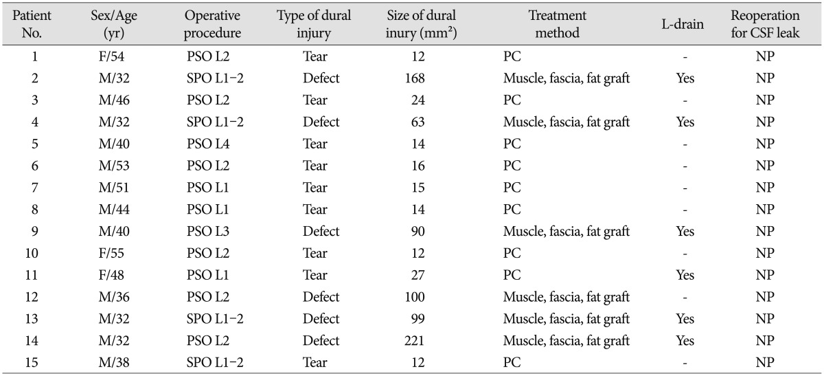
The dural tears ranged in size from a few mm to about 17 mm (12-221 mm2). All patients had a successful repair, with no patient requiring reoperation for the CSF leak (Table 1).
Table 1. Pateient demographics and treatment result.

CSF : cerebrospinal fluid, SPO : Smith-Petersen Osteotomy, PSO : Pedicle Subtraction Osteotomy, NP : not performed, PC : primary closure
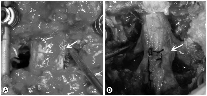
A nine of 15 patients had the relatively small dural tears, thus underwent direct repair via watertight closure without any difficulty or requiring intraoperative placement of lumbar drainage (Fig. 1A). The remaining 6 patients had the large dural tears, consequently direct repair was impossible. The size of the dural tears ranged from 63 to 221 mm2. The large dural tears were characterized by dural defect rather than dural tear. The dural defect was repaired with an on-lay graft of muscle, fascia or fat harvested from the adjacent operation site (Fig. 1B). The muscle, fascia or fat was flattened with a mallet and placed over the dural tear to completely cover the defect. In case where the margin of dural defect to be repaired with an on-lay graft was too thin or too close to the osteotomy site, a high-speed drill with a 2-mm round burr was used to create a small burr hole of the adjacent lamina for dural tenting. Although in such an instance, an instrument such as bone punch could be used for adequate exposure of the adjacent normal dura, it was avoided for the possibility of destroying the posterior fusion bed, subsequently giving rise to complications such as pseudoarthrosis. The muscle, fascia or fat piece was large enough to fill the defect between the stitches. A semicircular round needle on 4-0 black silk was used for the stitch; the needle passed through the muscle, fascia or fat piece and either both sides of the dura mater or one side of dura mater and the other side of the drilled burr hole site. If the defects were extending to the ventral side, a large muscle, fascia or fat graft was loosely packed to fill the ventral defect to serve as filler. Such muscle, fascia or fat patches were employed to 'cover' or 'plug' the dural defect1,22). Then closed lumbar drain was inserted as cranial as possible into the dural defect.
Fig. 1. Intraoperative photographs show that (A) the relatively small dural tear was directly repaired (arrow) and (B) the large dural tear (defect) was repaired with an onlay fat graft (arrow).
The six patients who had large dural tears received a successful dural defect repair and were followed by intraoperative placement of a lumbar drain, thus eliminating not only the need for reoperation for CSF leak but also attendant risks such as meningitis, CSF leak from the wound, or formation of pseudomeningocele and failure of wound healing. One patient, however, due to malfunction of the intraoperatively placed lumbar drain, suffered from persistent CSF leak through subfascial drain. Although attempts had been made to place the tip of the catheter cranial to the osteotomy site intraoperatively, postoperative radiograph revealed that it had supposedly bent on the way to be only located at the site of osteotomy, resulting in malfunction. Upon the discovery, negative pressure gradient of CSF drainage was avoided, and 20 cc of epidural blood patch was injected with fluoroscope guidance on postoperative day 6. On postoperative day 8, another 20 cc of epidural blood patch was injected with reinforcement with autologous fibrin glue patch. Subfascial drain was then removed on postoperative day 10 once cessation of CSF leak was confirmed. All patients received successful dural repairs without complications such as meningitis, wound abscess or formation of pseudomeningocele developed from the persistent CSF leak.
DISCUSSION
Although dural tears are a well known potential intraoperative complication of spine surgery, there is a relative lack of information about the true incidence of this common occurrence. Most of the studies in the literature are based on experience with relatively small numbers of patients2,5,9,10,17,24,26,27). The largest study to determine the incidence of dural tears was published by Khan et al.19). In their study, the incidence of dural tear in primary lumbar surgeries was found to be 7.6%, and their incidence during revision lumbar surgeries was 15.9%19). Dural tears are commonly associated with complex surgical procedures3,5,11,25,27). In our series, the incidence of dural tears during corrective osteotomy for ankylosing spondylitis was 17.2% (15 of 87 cases). To our knowledge, our study is the first series to be reported in the literature. Our incidence of 17.2% CSF leak after corrective osteotomy for ankylosing spondylitis with kyphotic deformity is relatively high compared with those (from 1.8% to 17.4%) reported in the literatures. Specially, dural tears occurred in 11 (20.0%) of 55 patients who underwent PSO, 4 (12.5%) of 32 patients who underwent SPO. There was significant difference in the incidence of dural tears based on surgical procedures (PSO vs. SPO) (p<0.05). These results revealed that risk factor for dural tears were complex surgical procedures. We assume this is from inflammatory characteristics of ankylosing spondylitis and complexity of surgery (corrective osteotomy). Ankylosing spondylitis is a disease characterized by prominent inflammatory changes of ligaments and capsules of facet joints, which subsequently leads to destruction and ankylosis of the joints4,6,13). Severe adhesion between dura and facet joint capsule may also result in dural erosion or adhesion that is impractical for adhesiolysis, thus giving rise to inevitable dural tears or defects. Therefore, the following considerations should be taken in the effort to prevent dural tears and CSF leakage that occurred during corrective osteotomy for ankylosing spondylitis with kyphotic deformity. First, before performing corrective osteotomy, preoperative CT assessment is necessary to identify the spinal canal encroachment of hypertrophied facet joints and ossified ligamentum flavum at the level of osteotomy. Second, meticulous surgical technique is necessary because the dura often is adherent to overlying bone, hypertrophied facet joints and ossified ligamentum flavum at the level of osteotomy (Fig. 2).
Fig. 2. A 33-year-old man with kyphotic deformity. A : Anteroposterior and lateral preoperative radiograph showing severe kyphotic deformity and coronal imbalance. B : Preoperative CT showing hypertrophied facet joints and severe ossification of ligamentum flavum (arrows). C : Anteroposterior and lateral postoperative radiograph showing improvement of the kyphotic deformity and coronal imbalance.
One of the main goals of dural repair is to attain a watertight seal, yet the optimal method to achieve this remains unknown. Investigators have used a variety of techniques including suturing, dural closure with the addition of a muscle patch, fascia patch, or artificial dura, and repair with gelatin sponge, fibrin glue, lumbar drain, or lumboperitoneal shunt1,8,15,20,21,22,23).
In general, a watertight closure is recommended in repair of a dural tear and midline dural tears is repaired by applying direct sutures for it is easily approachable and amenable to watertight closure without a technical difficulty. However, in the 6 cases mentioned on results, dural tears were characterized by dural defect due to spinal canal encroachment of hypertrophied facet joint and ossified ligamentum flavum, which was very closely adherent to dura as to even erode it. Direct repair of such defects were inadequate owing not only to its size, shape, and the friability of dura matter surrounding the defect, but also to the inherent inflammatory characteristics of ankylosing spondylitis. Then, Eismont et al.10) recommended muscle, fascia or fat graft secured by interrupted sutures in the treatment of larger dural tears (defects). We also used muscle, fascia or fat graft to repair the larger dural tears (defects) in six patients.
In the early cases, the authors attempted to proceed deformity correction after the treatment of larger dural tear (defect) with dural substitute (Neuropatch; B. Braun Melsungen AG) under EP monitoring. EP monitoring, however, revealed abnormal patterns after deformity correction, which was subjected to kinking of dural substitute in the process of deformity correction. EP monitoring returned to normal patterns once we tented the kinking of the dural substitute. Upon reaching the conclusion that the harder texture of dural substitute (Neuropatch: B. Braun Melsungen AG), in comparison to a normal dura mater, had caused such kinking and the consequent compression of encased neural elements. we removed the dural substitute, underwent correction with the large dural tear (defect) exposed, and performed dural repair by filling the large dural tear (defect) between the stitches with either a piece of muscle, fascia or fat harvested from adjacent muscles. Such a procedure (dural repair after deformity correction) has considerable merits of reducing size of the large dural tear (defect) from dural kinking along the process of correction, resultantly facilitating repair.
CONCLUSION
The incidence of dural tears during corrective osteotomy for ankylosing spondylitis with kyphotic deformity was 17.2% (15 of 87 cases) in our series.
In patients undergoing corrective osteotomy for ankylosing spondylitis with kyphotic deformity, the risk factor of dural tears was complexity of surgery and spinal canal encroachment of hypertrophied facet joint and ossified ligamentum flavum. Then, preoperative CT evaluation of severely hypertrophied facet joints or ossified ligamentum flavum at the level of osteotomy is imperative to avoid inadvertent dural tears.
Dural repair after deformity correction has considerable merits of reducing size of the large dural tear (defect) from dural kinking along the process of correction, resultantly facilitating repair.
In case of CSF leak, rather than revision through reoperation, injections of epidural blood and application of autologous fibrin glue patch with fluoroscope guidance prior to the formation of CSF fistula, could be an easier approach to managing the leak.
References
- 1.Black P. Cerebrospinal fluid leaks following spinal or posterior fossa surgery : use of fat grafts for prevention and repair. Neurosurg Focus. 2000;9:e4. doi: 10.3171/foc.2000.9.1.4. [DOI] [PubMed] [Google Scholar]
- 2.Blecher R, Anekstein Y, Mirovsky Y. Incidental dural tears during lumbar spine surgery : a retrospective case study of 84 degenerative lumbar spine patients. Asian Spine J. 2014;8:639–645. doi: 10.4184/asj.2014.8.5.639. [DOI] [PMC free article] [PubMed] [Google Scholar]
- 3.Bosacco SJ, Gardner MJ, Guille JT. Evaluation and treatment of dural tears in lumbar spine surgery : a review. Clin Orthop Relat Res. 2001;(389):238–247. doi: 10.1097/00003086-200108000-00033. [DOI] [PubMed] [Google Scholar]
- 4.Braun J, Bollow M, Remlinger G, Eggens U, Rudwaleit M, Distler A, et al. Prevalence of spondylarthropathies in HLA-B27 positive and negative blood donors. Arthritis Rheum. 1998;41:58–67. doi: 10.1002/1529-0131(199801)41:1<58::AID-ART8>3.0.CO;2-G. [DOI] [PubMed] [Google Scholar]
- 5.Cammisa FP, Jr, Girardi FP, Sangani PK, Parvataneni HK, Cadag S, Sandhu HS. Incidental durotomy in spine surgery. Spine (Phila Pa 1976) 2000;25:2663–2667. doi: 10.1097/00007632-200010150-00019. [DOI] [PubMed] [Google Scholar]
- 6.Carette S, Graham D, Little H, Rubenstein J, Rosen P. The natural disease course of ankylosing spondylitis. Arthritis Rheum. 1983;26:186–190. doi: 10.1002/art.1780260210. [DOI] [PubMed] [Google Scholar]
- 7.Choi JH, Kim JS, Jang JS, Lee DY. Transdural nerve rootlet entrapment in the intervertebral disc space through minimal dural tear : report of 4 cases. J Korean Neurosurg Soc. 2013;53:52–56. doi: 10.3340/jkns.2013.53.1.52. [DOI] [PMC free article] [PubMed] [Google Scholar]
- 8.Dalgic A, Okay HO, Gezici AR, Daglioglu E, Akdag R, Ergungor MF. An effective and less invasive treatment of post-traumatic cerebrospinal fluid fistula : closed lumbar drainage system. Minim Invasive Neurosurg. 2008;51:154–157. doi: 10.1055/s-2008-1042437. [DOI] [PubMed] [Google Scholar]
- 9.Du JY, Aichmair A, Kueper J, Lam C, Nguyen JT, Cammisa FP, et al. Incidental durotomy during spinal surgery : a multivariate analysis for risk factors. Spine (Phila Pa 1976) 2014;39:E1339–E1345. doi: 10.1097/BRS.0000000000000559. [DOI] [PubMed] [Google Scholar]
- 10.Eismont FJ, Wiesel SW, Rothman RH. Treatment of dural tears associated with spinal surgery. J Bone Joint Surg Am. 1981;63:1132–1136. [PubMed] [Google Scholar]
- 11.Espiritu MT, Rhyne A, Darden BV., 2nd Dural tears in spine surgery. J Am Acad Orthop Surg. 2010;18:537–545. doi: 10.5435/00124635-201009000-00005. [DOI] [PubMed] [Google Scholar]
- 12.Gautschi OP, Stienen MN, Smoll NR, Corniola MV, Schaller K. Incidental durotomy in lumbar spine surgery - is there still a role for flat bed rest? Spine J. 2014;14:2522–2523. doi: 10.1016/j.spinee.2014.06.014. [DOI] [PubMed] [Google Scholar]
- 13.Geusens P, Vosse D, van der Linden S. Osteoporosis and vertebral fractures in ankylosing spondylitis. Curr Opin Rheumatol. 2007;19:335–339. doi: 10.1097/BOR.0b013e328133f5b3. [DOI] [PubMed] [Google Scholar]
- 14.Hawk MW, Kim KD. Review of spinal pseudomeningoceles and cerebrospinal fluid fistulas. Neurosurg Focus. 2000;9:e5. doi: 10.3171/foc.2000.9.1.5. [DOI] [PubMed] [Google Scholar]
- 15.Hida K, Yamaguchi S, Seki T, Yano S, Akino M, Terasaka S, et al. Nonsuture dural repair using polyglycolic acid mesh and fibrin glue : clinical application to spinal surgery. Surg Neurol. 2006;65:136–142. discussion 142-143. doi: 10.1016/j.surneu.2005.07.059. [DOI] [PubMed] [Google Scholar]
- 16.Hodges SD, Humphreys SC, Eck JC, Covington LA. Management of incidental durotomy without mandatory bed rest. A retrospective review of 20 cases. Spine (Phila Pa 1976) 1999;24:2062–2064. doi: 10.1097/00007632-199910010-00017. [DOI] [PubMed] [Google Scholar]
- 17.Jones AA, Stambough JL, Balderston RA, Rothman RH, Booth RE., Jr Long-term results of lumbar spine surgery complicated by unintended incidental durotomy. Spine (Phila Pa 1976) 1989;14:443–446. doi: 10.1097/00007632-198904000-00021. [DOI] [PubMed] [Google Scholar]
- 18.Jung YY, Ju CI, Kim SW. Bilateral subdural hematoma due to an unnoticed dural tear during spine surgery. J Korean Neurosurg Soc. 2010;47:316–318. doi: 10.3340/jkns.2010.47.4.316. [DOI] [PMC free article] [PubMed] [Google Scholar]
- 19.Khan MH, Rihn J, Steele G, Davis R, Donaldson WF, 3rd, Kang JD, et al. Postoperative management protocol for incidental dural tears during degenerative lumbar spine surgery : a review of 3,183 consecutive degenerative lumbar cases. Spine (Phila Pa 1976) 2006;31:2609–2613. doi: 10.1097/01.brs.0000241066.55849.41. [DOI] [PubMed] [Google Scholar]
- 20.Kitchel SH, Eismont FJ, Green BA. Closed subarachnoid drainage for management of cerebrospinal fluid leakage after an operation on the spine. J Bone Joint Surg Am. 1989;71:984–987. [PubMed] [Google Scholar]
- 21.Narotam PK, José S, Nathoo N, Taylon C, Vora Y. Collagen matrix (DuraGen) in dural repair : analysis of a new modified technique. Spine (Phila Pa 1976) 2004;29:2861–2867. discussion 2868-2869. doi: 10.1097/01.brs.0000148049.69541.ad. [DOI] [PubMed] [Google Scholar]
- 22.Park JS, Kong DS, Lee JA, Park K. Intraoperative management to prevent cerebrospinal fluid leakage after microvascular decompression : dural closure with a "plugging muscle" method. Neurosurg Rev. 2007;30:139–142. discussion 142. doi: 10.1007/s10143-006-0060-6. [DOI] [PubMed] [Google Scholar]
- 23.Stendel R, Danne M, Fiss I, Klein I, Schilling A, Hammersen S, et al. Efficacy and safety of a collagen matrix for cranial and spinal dural reconstruction using different fixation techniques. J Neurosurg. 2008;109:215–221. doi: 10.3171/JNS/2008/109/8/0215. [DOI] [PubMed] [Google Scholar]
- 24.Stolke D, Sollmann WP, Seifert V. Intra- and postoperative complications in lumbar disc surgery. Spine (Phila Pa 1976) 1989;14:56–59. doi: 10.1097/00007632-198901000-00011. [DOI] [PubMed] [Google Scholar]
- 25.Tafazal SI, Sell PJ. Incidental durotomy in lumbar spine surgery : incidence and management. Eur Spine J. 2005;14:287–290. doi: 10.1007/s00586-004-0821-2. [DOI] [PMC free article] [PubMed] [Google Scholar]
- 26.Takahashi Y, Sato T, Hyodo H, Kawamata T, Takahashi E, Miyatake N, et al. Incidental durotomy during lumbar spine surgery : risk factors and anatomic locations : clinical article. J Neurosurg Spine. 2013;18:165–169. doi: 10.3171/2012.10.SPINE12271. [DOI] [PubMed] [Google Scholar]
- 27.Wang JC, Bohlman HH, Riew KD. Dural tears secondary to operations on the lumbar spine. Management and results after a two-year-minimum follow-up of eighty-eight patients. J Bone Joint Surg Am. 1998;80:1728–1732. doi: 10.2106/00004623-199812000-00002. [DOI] [PubMed] [Google Scholar]


