Abstract
Although clinical congestive heart failure (CHF) is associated with significant left ventricular (LV) systolic dysfunction, recently it has been recognized that LV diastolic dysfunction also may occur in the absence of abnormal systolic performance. A retrospective study evaluated 23 patients with myocardial infarction and CHF who had undergone multigated blood pool scintigraphy and were found to have normal LV ejection fraction (≥ 50%). Average rapid filling velocity (RFV) and slow filling velocity (SFV) were both significantly reduced in CHF patients (5.1 ± 1.3 unit/s, 2.2 ± 1.4 unit/s respectively) compared with normal control group (3.9 ± 1.2 unit/s, 1.3 ± 0.8 unit/s respectively). Rapid filling time and total diastolic time were also significantly prolonged in CHF patients (p<0.01, p<0.05 respectively). There were no significant changes in heart rate and blood pressure between two groups. Thus, normal systolic LV function is encountered in patients with CHF and it appears to be prudent to evaluate diastolic performance as well for optimal therapeutic strategies for CHF patients.
Keywords: Acute myocardial infarction, Congestive heart failure, Diastolic function
INTRODUCTION
Congestive heart failure (CHF) is associated with significant ventricular systolic dysfunction. Current medical therapy is traditionally directed toward improving contractile function or minimizing ventricular work load. However, recently it has been encountered that left ventricular (LV) diastolic dysfunction also may occur in the absence of overt systolic dysfunction1–7) and patients with CHF and systolic impairment do not benefit significantly from inotropic agents8,9).
The objective of the present study was to identify patients with myocardial infarction and CHF who have normal global ejection fraction (EF) and to evaluate whether pulmonary congestive symptoms may in part account for impaired diastolic performance.
MATERIALS AND METHODS
1. Patient Selection
The records of all patients with acute myocardial infarction (AMI) and CHF who had multigated radionuclide angiography (RNA) between February 1983 and April 1985 are reviewed.
Diagnosis of AMI was made on clinical manifestations, cardiac enzymes, ECG, Tc-99 m PYP scan and coronary angiography. Diagnosis of CHF was based upon a history of dyspnea, cardiomegaly and pulmonary venous congestion on chest radiography or physical findings such as jugular venous distention, rales, S3 gallop, displacement of cardiac apex or peripheral edema.
Those patients with AMI and CHF were selected for the present study who had an intact systolic function, defined as a LVEF of 50% or more by RNA.
Nineteen patients (83%) were receiving digitalis or diuretics or both. Five patients (22%) were receiving calcium channel blocker or nitroglycerin in addition to digitalis or diuretics. There were 7 patients (30%) who were receiving vasodilators.
A control group of 11 patients (5 men, 6 women) without evidence of heart diseases was matched with respect to LVEF and diastolic variables.
2. Protocol
Multigated radionuclide angiography was performed in the supine position using red blood cells tagged with 20 mCi of Technetium-99 m pyrophosphate10). Counts were acquired in the LAO 45° view showing best separation of right and left ventricles, using a Technicare Sigma 120 portable gamma camera, equipped with a GAP collimator and interfaced to a Technicare MCS 560 portable computer. ECG-gated images were used to collect data into 16 frames, each frame having a duration of 1/16th of the R-R interval and an average left ventricular count value of 125 counts per pixel. After completion of imaging, time activity curves and LV ejection fraction were computed using Technicare software. LV ejection fraction was calculated by subtracting the LV end-systolic counts from LV end-diastolic counts, and dividing the result by LV end-diastolic counts.
3. Diastolic Parameters
Diastolic function curves were examined in the same manner as Lee et al.11). Several measurements were calculated from the time-activity curve (Fig. 1). Four diastolic points were marked by the operator; (1) start of rapid filling (SRF), (2) end of rapid filling (ERF), (3) start of slow filling (SSF) and (4) end of slow filling (ESF).
Fig. 1.
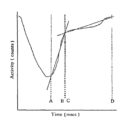
Left ventricular time-activity curve.
A = start of rapid filling (SRF)
B = end of rapid filling (ERF)
C = start of slow filling (SSF) and
D = end of slow filling (ESF)
Rapid filling time = A to B, slow filling time = C to D and total diastolic time = A to D
Rapid and slow filling velocity: A regression line was plotted for counts versus time between SRF and ERF and between SSF and ESF. Average rapid and slow filling velocities were taken as the slopes of these regression lines in counts/second divided by the end-diastolic counts and expressed as units/second.
Filling volume ratio: The ratio of rapid filling volume to rapid plus slow filling volume was defined as: filling volume ratio = counts at ERF-SRF/counts at ESF-SRF.
Rapid filling time, total diastolic time and diastolic time ratio: The duration of rapid and slow filling times and the ratio of rapid filling time to total diastolic time (diastolic time ratio) were calculated from the points marked on the time-activity curve.
RESULTS
1. Clinical Characteristics
In CHF patients group, jugular venous distention was present in 13 patients (57%). Sixteen patients (70%) had pulmonary rales. An S3 gallop was heard in 12 patients (52%). Peripheral edema was observed in 11 patients (48%) (Table 1).
Table 1.
Objective Findings in Congestive Heart Failure Patients
| Findings | No. of patients |
|---|---|
| Physical findings | |
| JVD* | 13 (57%) |
| Rales | 16 (70%) |
| S3 gallop | 12 (52%) |
| Edema | 11 (48%) |
| Chest radiograph | |
| Cardiomegaly | 16 (70%) |
| Pulmonary edema | 14 (61%) |
JVD : Jugular venous distention
2. Radionuclide Angiography
There were no significant differences between the control and CHF group in heart rate and blood pressure. The LVEF was 63 ± 6% in control and 60 ± 6% in CHF group, representing no differences between groups (Table 2).
Table 2.
Variables in Control and Congestive Heart Failure Group
| Control | Patients | P-value | |
|---|---|---|---|
| Heart rate (beats/min) | 81 ± 25 | 77 ± 17 | NS |
| Blood pressure (mmHg) | |||
| Systolic | 125 ± 22 | 126 ± 14 | NS |
| Diastolic | 77 ± 3 | 73 ± 9 | NS |
| Systolic function | |||
| EF (%) | 63 ± 6 | 60 ± 6 | NS |
| Diastolic function | |||
| RFV (units/sec) | 5.1 ± 1.3 | 3.9 ± 1.2 | P < 0.01 |
| SFV (units/sec) | 2.2 ± 1.4 | 1.3 ± 0.8 | P < 0.05 |
| FVR | 0.6 ± 0.2 | 0.7 ± 0.2 | NS |
| RFT (ms) | 121.7 ± 39.3 | 160.7 ± 37.2 | P < 0.01 |
| TDT (ms) | 258.20 | 349.2 ± 134.5 | P < 0.05 |
| DTR | 0.5 ± 0.1 | 0.5 ± 0.1 | NS |
Abbreviations : NS = Not significant, EF = Ejection fraction, RFV = Rapid filling velocity, SFV = Slow filling velocity, FVR = Filling volume ratio, RFT = Rapid filling time, TDT = Total diastolic time, DTR = Diastolic time ratio
Average rapid filling velocity was 5.1 ± 1.3 unit/s in control group and 3.9 ± 1.2 unit/s in CHF group (p<0.01) (Table 2, Fig. 2). Average slow filling-velocity was 2.2 ± 1.4 unit/s in control group and 1.3 ± 0.8 unit/s in CHF group (p<0.05) (Table 2, Fig. 3). There was no significant difference between groups in filling volume ratio. Rapid filling time (Fig. 4) and total diastolic time (Fig. 5) were significantly prolonged in CHF group compared with control group (p<0.01, p<0.05 respectively) (Table 2). No significant difference was found between groups in diastolic time ratio (Table 2).
Fig. 2.
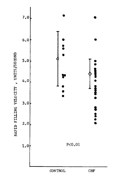
Rapid filling velocity in control and congestive heart failure group.
Fig. 3.
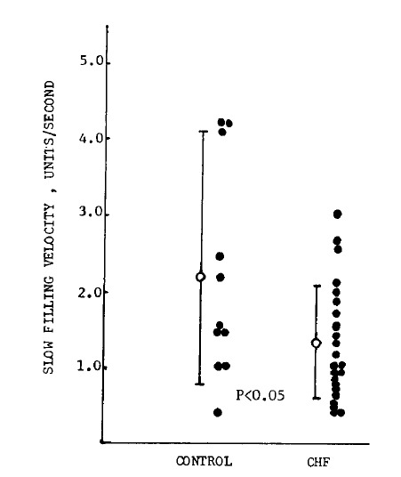
Slow filling velocity in control and congestive heart failure group.
Fig. 4.
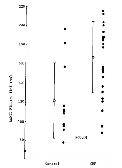
Rapid filling time in control and congestive heart failure group.
Fig. 5.
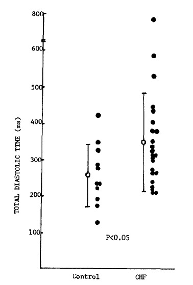
Total diastolic time in control and congestive heart failure group.
There were no significant differences in rapid and slow filling velocity according to the number of vessels involved in CHF group, even though rapid filling velocity was slightly decreased in 3 vessel disease compared with 1 vessel and 2 vessel diseases (Table 3). Also no significant differences were found in diastolic variables according to the location of myocardial infarction in CHF group.
Table 3.
Rapid and Slow Filling Velocity According to the Number of Vessels Involved
| Indices | Number of vessels involved
|
P value between groups | ||||
|---|---|---|---|---|---|---|
| 1 VD (11) | 2 VD (5) | 3 VD (3) | 1 + 2 VD (16) | 2 + 3 VD (8) | ||
| RFV | 4.2 ± 1.3 | 3.6 ± 1.0 | 3.2 ± 1.0 | 4.0 ± 1.4 | 3.3 ± 1.2 | NS |
| SFV | 1.3 ± 0.6 | 1.3 ± 0.9 | 1.6 ± 1.1 | 1.3 ± 0.5 | 1.4 ± 0.8 | NS |
VD : Vessels disease
Number of patients in parentheses
DISCUSSION
The clinical syndrome of congestive heart failure (CHF) is traditionally associated with inadequate myocardial contraction, volume overload and impaired ventricular filling. Accordingly, CHF therapy has been directed toward augmentation of contractile function with positive inotropic agents or alteration in preload and afterload with vasodilators and diuretics12,13).
Recently it has been recognized that left ventricular (LV) diastolic impairment may occur without systolic dysfunction and may develop coincident with or before abnormalities of systolic function. This has been reported in patients with coronary artery disease, pressure2–14) and volume-overload states15) and cardiomyopathy1,16,17). Several studies5,18–21) reported normal systolic LV function in patients with CHF. Souffer et al.22) noted intact systolic LV function in 42% of patients with clinical diagnosis of CHF referred to the nuclear cardiology laboratory during a 3 month sampling period. Echeverria et al.23), who used echocardiography noted normal systolic function in 40% of 50 consecutive patients with CHF, and Dougherty et al.24), who used radionuclide technique also noted a normal ejection fraction (>45%) in 36% of CHF. Warnowicz et al.25) reported 16 of 39 patients with acute myocardial infarction (AMI) and pulmonary edema having intact LVEF. These studies suggest that the frequency of normal systolic function in patients with CHF are more closely associated with diastolic than systolic performance. Moreover, Firth et al.9) have failed to show digitalis-induced augmentation of LVEF and Lee et al.8) noted that only 56% among the CHF patients improved clinically with digoxin.
Myocardial contraction and relaxation are loosely coupled processes that may be separately altered by certain stimuli26). Thus myocardial relaxation is not directly dependent upon contractile events and several studies17–19) have revealed the clinical relevance of diastolic impairment by its association with CHF. Pulmonary vascular congestion in the absence of abnormal systolic performance may be due to impaired ventricular filling21).
The mechanism for abnormal ventricular diastolic filling is not well known but it may be due to defects in calcium uptake shown in myocardial microsome and depletion of myocardial catecholamine in AMI and CHF21). Factors that may result in abnormal diastolic function in patients with CHF include volume-overload states, systemic hypertension, valvular heart disease, pericardial restriction, restrictive cardiomyopathy and increased heart rate, which reduces ventricular filling time5,17–20). In most of our patients with AMI and CHF, the authors could not identify such causes. Two patients in the present study had hypertension, but there was no evidence of LV hypertrophy by echocardiography.
LV performance was evaluated using radionuclide measurement. LVEF, used as useful parameter for detecting impaired myocardial function in the present study has been found of value in identifying patients with systolic dysfunction in several heart diseases. Diastolic function was assessed by 6 parameters, which obtained from time-activity curve. Although Bonow et al.1,3) and others2,4,17,22) have shown that peak filling rate can be used to evaluate diastolic function, the authors employed average filling velocity as measures of diastolic function. Average filling velocity are less sensitive to noise and independent of temporal resolution. Our methods for measuring average filling velocities differ slightly from those reported by Smith et al.27) in that only the relatively constant portions of rapid and slow filling periods were used. By not taking the minimum count point of the time-activity curve as the onset of rapid filling, our method revealed more rapid normal velocities in control group than the average rates reported by Smith et al.27).
In addition, average slow filling velocity and rapid filling time as well as total diastolic time were significantly different in CHF patients compared with normal control group.
Bonow et al.3) reported that diastolic indices in 1, 2, 3 vessel and left main diseases altered significantly in patients with CAD and normal ejection fraction compared with normal control group. But no difference between groups according to the number of vessels involved was noted. Reduto et al.4) noted that there was little change in EF and filling fraction at rest or during exercise between anterior and inferior MI patients. Similarly, the authors found no differences in diastolic variables between groups according to the number of vessels involved and between groups according to the localization of AMI.
In conclusion, evidence of impaired LV diastolic function without abnormal systolic function was identified in selected patients who had AMI and CHF. Radionuclide assessment of diastolic performance may be relevant for evaluation of LV performance and optimal therapy in patients with CHF.
REFERENCES
- 1.Bonow RO, Rosing DR, Bacharach SL, Green MV, Kent KM, Lipson LC, Maron BJ, Leon MB, Epstein SE. Effects of verapamil on left ventricular systolic function and diastolic filling in patients with hypertrophic cardiomyopathy. Circulation. 1981;64:787. doi: 10.1161/01.cir.64.4.787. [DOI] [PubMed] [Google Scholar]
- 2.Inouye I, Massie B, Loge D, Topic N, Silverstein D, Simpson P, Tubau J. Abnormal left ventricular filling: an early finding in mild to moderate systemic hypertension. Am J Cardiol. 1984;53:120. doi: 10.1016/0002-9149(84)90695-7. [DOI] [PubMed] [Google Scholar]
- 3.Bonow RO, Bacharach SL, Green MV, Kent KM, Rosing DR, Lipson LC, Leon MB, Epstein SE. Impaired left ventricular diastolic filling in patients with coronary artery disease: assessment with radionuclide angiography. Circulation. 1981;64:315. doi: 10.1161/01.cir.64.2.315. [DOI] [PubMed] [Google Scholar]
- 4.Reduto LA, Wickemeyer WJ, Young JB, Del Ventrura LA, Reid JW, Glaesor DH, Quinones MA, Miller RR. Left ventricular diastolic performance at rest and during exercise in patients with coronary artery disease. Circulation. 1981;63:1228. doi: 10.1161/01.cir.63.6.1228. [DOI] [PubMed] [Google Scholar]
- 5.Levine HJ, Gaasch WH. Diastolic compliance of the left ventricle. Mod Conc Cardiovasc Dis. 1978;47:95. [PubMed] [Google Scholar]
- 6.Bourdillon PD, Lorell BH, Mirsky I, Paulua WJ, Wynne J, Grossman W. Increased regional myocardial stiffness of the left ventricle during pacing induced angina in man. Circulation. 1983;67:316. doi: 10.1161/01.cir.67.2.316. [DOI] [PubMed] [Google Scholar]
- 7.Barry WH, Brooker JZ, Alderman EL, Harrison DC. Changes in diastolic stiffness and tone of the left ventricle during angina pectoris. Circulation. 1974;49:255. doi: 10.1161/01.cir.49.2.255. [DOI] [PubMed] [Google Scholar]
- 8.Lee DC, Johnson RA, Bingham JB, Leahy M, Dinsmore RE, Gorall AH, Newell JB, Strauss HW, Habor E. Heart failure in outpatients: a randomized trial of digoxin versus placebo. N Engl J Med. 1982;306:699. doi: 10.1056/NEJM198203253061202. [DOI] [PubMed] [Google Scholar]
- 9.Firth BG, Dehmer GJ, Corbett JR, Lewis SE, Parkey RW, Willerson JT. Effect of chronic oral digoxin therapy on ventricular function at rest and peak exercise in patients with ischemic heart disease. Am J Cardiol. 1980;46:481. doi: 10.1016/0002-9149(80)90019-3. [DOI] [PubMed] [Google Scholar]
- 10.Thrall JH, Freitas JE, Swanson D. Clinical comparison of cardiac blood pool visualization with technetium-99 m, red blood cells labelled in vivo and with technetium-99 m human serum albumin. J Nucl Med. 1978;19:796. [PubMed] [Google Scholar]
- 11.Lee BH, Goodenday LS, Muswick GJ, Yasnoff WA, Leighton RF, Skeel RT. Alterations in left ventricular diastolic function with doxorubicin therapy. J Am Coll Cardiol. 1987;9:184. doi: 10.1016/s0735-1097(87)80099-2. [DOI] [PubMed] [Google Scholar]
- 12.Liang C, Sherman LG, Doherty JU, Wellington K, Lee VW, Hood WB. Substained improvement of cardiac function in patients with congestive heart failure after short-term infusion of dobutamine. Circulation. 1984;69:113. doi: 10.1161/01.cir.69.1.113. [DOI] [PubMed] [Google Scholar]
- 13.Chatterjee K, Parmley WW. Vasodilator therapy in chronic heart failure. Annu Rev Pharmacol Toxicol. 1980;22:475. doi: 10.1146/annurev.pa.20.040180.002355. [DOI] [PubMed] [Google Scholar]
- 14.Dreslinski GR, Frohlich ED, Dunn FG, Messerli FH, Suarez DH, Reisin E. Echocardiographic diastolic ventricular abnormality in hypertensive heart disease: atrial emptying index. Am J Cardiol. 1981;47:1087. doi: 10.1016/0002-9149(81)90217-4. [DOI] [PubMed] [Google Scholar]
- 15.Lavine SJ, Follansbee WP, Shreiner DP. Pattern of left ventricular diastolic filling in chronic aortic regurgitation: A gated blood pool asessment. Am J Cardiol. 1985;55:127. doi: 10.1016/0002-9149(85)90313-3. [DOI] [PubMed] [Google Scholar]
- 16.Lorell BH, Paulus WJ, Grossman W, Wynne J, Conn PF. Modifications of abnormal left ventricular diastolic properties by nifedipine in patients with hypertrophic cardiomyopathy. Circulation. 1982;65:499. doi: 10.1161/01.cir.65.3.499. [DOI] [PubMed] [Google Scholar]
- 17.Alexander J, Dainiak N, Berger HJ, Goldman L, Honstone D, Reduto L, Duffy T, Schwartz P, Gottschalk A, Zaret BL. Serial assessment of doxorubicin cardiotoxicity with quantitative radionuclide angiocardiography. N Engl J Med. 1979;300:278. doi: 10.1056/NEJM197902083000603. [DOI] [PubMed] [Google Scholar]
- 18.Lewis BS, Gotsman MS. Current concepts of left ventricular relaxation and compliance. Am Heart J. 1980;99:101. doi: 10.1016/0002-8703(80)90320-8. [DOI] [PubMed] [Google Scholar]
- 19.Levine HJ. Compliance of the left ventricle. Circulation. 1972;45:423. doi: 10.1161/01.cir.46.3.423. [DOI] [PubMed] [Google Scholar]
- 20.Grossman W, Barry WH. Diastolic pressure-volume relations in the diseased heart. Fed Proc. 1980;39:148. [PubMed] [Google Scholar]
- 21.Grossman W, McLaurin LP. Diastolic properties of the left ventricle. Ann Intern Med. 1976;84:316. doi: 10.7326/0003-4819-84-3-316. [DOI] [PubMed] [Google Scholar]
- 22.Soufer R, Wohlgelernter D, Vita NA, Amuchestegui M, Sostmen D, Berger HJ, Zaret BL. Intact systolic left ventricular function in clinical congestive heart failure. Am J Cardiol. 1985;55:1032. doi: 10.1016/0002-9149(85)90741-6. [DOI] [PubMed] [Google Scholar]
- 23.Echeverria HH, Bilsker MS, Myerburg RJ, Kessler KM. Congestive heart failure: Echocardiographic insights. Am J Med. 1983;75:750. doi: 10.1016/0002-9343(83)90403-5. [DOI] [PubMed] [Google Scholar]
- 24.Dougherty AH, Maccarelli GV, Gray EI, Hicks CH, Goldstein RA. Congestive heart failure with normal systolic fuction. Am J Cardiol. 1984;54:778. doi: 10.1016/s0002-9149(84)80207-6. [DOI] [PubMed] [Google Scholar]
- 25.Warnowicz MA, Parker H, Cheltlin MD. Prognosis of patients with acute pulmonary edema and normal ejection fraction after acute myocardial infarction. Circulation. 1983;67:330. doi: 10.1161/01.cir.67.2.330. [DOI] [PubMed] [Google Scholar]
- 26.Parmley WW, Sonnenblick EH. Relation between mechanics of contraction and relaxation in mammalian cardiac muscle. Am J Physiol. 1969;216:1084. doi: 10.1152/ajplegacy.1969.216.5.1084. [DOI] [PubMed] [Google Scholar]
- 27.Smith VE, Schulman P, Karimeddini M. Rapid ventricular filling in left ventricular hypertrophy: II. Pathologic hypetrophy. J Am Coll Cardiol. 1985;5:869. doi: 10.1016/s0735-1097(85)80425-3. [DOI] [PubMed] [Google Scholar]


