

Abstract
We report a case of methimazole-induced acute hepatic failure, which occurred 17 weeks after initiation of the drug in a 43-year-old man with hyperthyroidism and hepatitis B surface antigenemia. Postmortem needle autopsy of the liver revealed an established micronodular cirrhosis secondary to hepatitis B with moderate septal/portal inflammation, marked cholestasis and scattered acidophilic bodies. The serum hepatitis B surface antigen (HBsAg) was positive, but reactivation of hepatitis B was unlikely in view of the absence of a serum hepatitis B e antigen (HBeAg) and hepatitis B virus deoxyribonucleic acid (HBV-DNA) and negative stain for HBsAg and hepatitis B core antigen (HBcAg) in the liver tissue.
Keywords: Methimazole, Acute hepatic failure, HBsAg carrier
INTRODUCTION
After the introduction of methimazole by Stanley and Astwood in 19491), this chemical has been a widely-used, safe and highly-effective drug for patients with hyperthyroidism. However, in 5–7% of the patients side effects occur2), including fever, skin rashes, gastrointestinal symptoms, loss of taste, neuropahty and often fatal agranulocytosis. Drug-induced hepatitis seems to be a very rare toxic reaction by this drug. Only 15 documented cases have been reported3–17). None of these reports mentioned acute hepatic failure induced by methimazole.
CASE REPORT
A 43-year-old man was admitted to the Endocinology Section of this hospital because of palpitations, weight loss of two years’ duration and recent onset of exertional dyspnea. The physical examination, laboratory and histological findings, including serum thyroxine, TSH level and needle biopsy of the thyroid, were compatible with hyperthyroidism and congestive heart failure. He was initially treated with propylthiouracil (300 mg daily), and the heart failure symptoms improved with appropriate care. On admission, serum HBsAg was tested as a part of routine clinical tests and was found to be positive. The patient denied any history of hepatitis exposure or any family history of hepatitis B or exposure to blood products. Serum aminotransferase activities were within normal limits. HBeAg and HBV–DNA were not detected in the serum. The patient was discharged on the 21st hospital day with markedly improved general condition. Two months later, propylthiouracil was substituted with methimazole (30 mg daily) for better compliance. Seven months later, the patient was readmitted to the hospital because of nausea, general malaise and jaundice of two weeks’ duration and transferred to the Hepatology Section. He denied any use of illicit drugs, herb medication or alcohol during the period. On examination the patient appeared acutely ill and icteric, although he was clinically euthyroid. No Spider angiomata, skin rash or palmar erythema was noted. The pulse was irregular with a rate of about 80 due to atrial fibrillation. The edge of the liver was palpable 5 cm below the right costal margin with a vertical span of 13 cm. The liver was diffusely tender.
No evidence of ascites was found. The hematocrit was 42 percent; the white cell count was 6400; with a normal differential count, the platelet count was 81,900/mm3. Agranulocytosis or eosinophilia had not developed during the illness. The serum protein was 7.1 g/dl (albumin 4.6 g and the globulin 2.5 g) and the bilirubin was 5.0 mg/dl (direct 3.5 mg and indirect 1.5 mg). The serum alanine aminotransferase (ALT) was 180 U/L (normal <40), the serum aspartate aminotransferase (AST), 848 U/L (normal <40), and the alkaline phosphatase, 848 IU/L (normal: 64–238). The prothrombin time was 13.7 seconds with a control 11.3 seconds. The T3 RIA was 109.85 ng per 100 ml (normal: 90–210), the thyroxine 3.01 ug per 100 ml (normal: 5–13) and the TSH 19.85 uU per ml (normal: 0.3–5). Serum HBsAg and antibody to hepatitis B core antigen (anti-HBc) were positive, but HBeAg, IgM anti-HBc, and HBV-DNA by dot and slot blot hybridization were negative. Antibody to hepatitis C virus (Anti-HCV) by Elisa method was negative. An ultrasonographic examination of the abdomen revealed a diffusely enlarged liver, although the spleen was not enlarged.
The methimazole was discontinued and symptomatic care was given. During the next four weeks the patient’s condition deteriorated rapidly (Fig. 1). From the 21st hospital day, dexamethasone was administered at a dose of 10 mg daily. On the 27th hospital day, hepatic encephalopathy developed and the patient died four days later. A postmortem needle autopsy of the liver was performed (Fig. 2, 3). The histologic findings revealed a micronodular cirrhosis secondary to a hepatitis B virus infection with moderate septal/portal inflammation, marked cholestasis and scattered acidophilic bodies. No HBsAg or HBcAg was detected in the liver by peroxidase-antiperoxidase method.
Fig. 1.
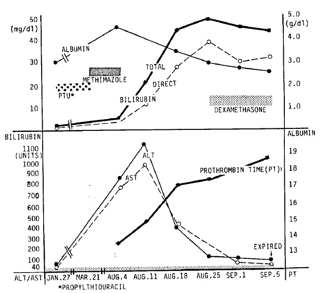
Schematic diagram of clinical and biochemical changes in the patient.
Fig. 2.
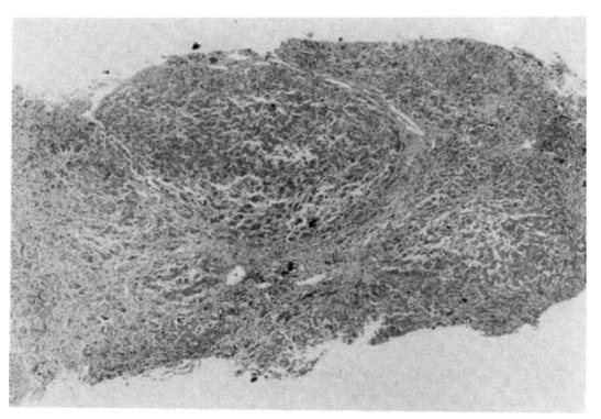
Micronodular cirrhosis with inflammatory cell infiltration and fibrosis of the portal tracts and septa. Hematoxylin and eosin ×40.
Fig. 3.
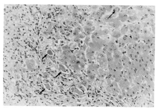
Portal inflammatory cell infiltration and bile plugs (thick arrows) in bile ducts and acidophilic body (thin arrow). Hematoxylin and eosin ×200
DISCUSSION
In contrast to propylthiouracil-induced hepatotoxicity in which cytotoxic injury is the predominant lesion, methimazole-associated hepatic injury is mainly cholestatic10). It is probably due to an idiosyncratic (hypersensitive) reaction, which is usually infrequent and unpredictable. This type of response is not does-dependent and may occur at any time during or shortly after exposure to the drug18). The mechanisms underlying these hypersensitivity reactions are not known. But autoimmune mechanisms10) and the immunosurppressive effect of the antithyroid drugs19) have been described. Previously reported patients with methimazole-induced hepatotoxicity were predominantly female and usually over 40 years old (Table 1). There have been two fatal cases reported. Both were related to pneumonia with or without agranulocytosis3,8). In the remaining cases, the clinical symptoms and laboratory abnormalities became normal when methimazole was discontinued, or with the use of corticosteroids8). But in one patient with malignant exophthalmos8), methimazole-induced hepatic injury actually developed during high dose glucocorticoid therapy6). The development of hepatic injury long after propylthiouracil was discontinued indicates that the injury was not due to propylthiouracil in this case. While the prominent cholestasis was consistent with other reported cases of methimazole-induced hepatic injury, this case was different in the prominence of the parenchymal injury and liver failure. Other possible mechanisms for hepatic injury in this case include the superinfection of a hepatitis C virus and the spontaneous reactivation of chronic hepatitis B20). After the discovery of the causative agent and the development of the detection method of type C virus infection, it becomes possible to make a serological diagnosis21,22) but anti HCV test in serum was negative in this patient. Lastly, it is well -known that reactivation of hepatitis B is one of the most important and frequent predisposing causes in aggravating chronic hepatitis B. However, the absence of HBeAg, IgM anti-HBc, or HBV-DNA in serum and negative stain for HBsAg and HBcAg in the liver may exclude this possibility of reactivation with some certainty. It is unclear what role the hepatitis B virus infection itself plays in the development of acute hepatic failure in case all the viral replication markers do not express. It is possible that the presence of a micronodular cirrhosis secondary to the hepatitis B virus infection may have predisposed the patient to the development of acute hepatic failure with the administration of methimazole. It should be kept in mind, therefore, that in patients with underlying chronic liver disease, particularly cirrhosis, active or inactive, any hepatotoxic agent should be used with caution.
Table 1.
Hepatic Injury from Methimazole
| References | Sex/Age | Dose (mg/day) | Duration of Administration | Liver Pathology | Agranulocytosis | Comments | |
|---|---|---|---|---|---|---|---|
| 1. | Spect & Boehme | F/67 | 30 | 30 days | central lobular congestion | + | death due to pneumonia |
| 2. | Rosenbaum & Reveno | F/62 | 10 | 7 weeks | – | + | – |
| 3. | Shipp | F/63 | 60 | 8 weeks | cholestasis | + | – |
| 4. | Tennenbaum & Dresken | F/38 | 60 | 4 weeks | cholestasis | − | skin rash |
| 5. | Lopez et al. | F/38 | 60 | 4 weeks | cholestasis | − | − |
| 6. | Becker et al. | F/54 | 40 | 24 days | focal hepatitis | − | death due to pneumonia |
| 7. | Fisher et al. | F/74 | 40 | 12 days | cholestasis | − | − |
| 8. | Vitug & Goldman | F/64 | 80 | 4 weeks | cholestasis | + | − |
| 9. | Schmidt et al. | F/58 | 20 | 20 days | cholestasis & fatty degeneration | + | − |
| 10. | Kravetz | ?/? | 30–50 | ? | cholestasis | − | − |
| 11. | Knopka et al. | F/37 | ? | ? | ? | ? | − |
| 12. | Manoijlovic | ?/? | 80–100 | ? | ? | + | − |
| 13. | Efstrtiadis | M/50 | 60 | ? | cholestasis | − | − |
| 14. | Sambe | F/30 | 40 | ? | – | − | − |
| 15. | Jansen & Froeling | F/62 | 40 | 5 weeks | cholestasis | − | − |
| 16. | Authors | M/43 | 30 | 17 weeks | cholestasis & focal hepatocellular injury | − | death due to hepatic failure |
Acknowledgments
We wish to thank Dr. K.G. Ishak and Dr. H. J. Zimmerman for their kind and excellent interpretation of the histology of this case and for their comments.
REFERENCES
- 1.Stanley M, Astwood EB. 1-methyl-2-mercaptoimidazole: An antithyroid compound highly active in man. Endocrinology. 1949;44:588–589. doi: 10.1210/endo-44-6-588. [DOI] [PubMed] [Google Scholar]
- 2.Chevalley J, McGavack TH, Kenigsberg S, Pearson S. A four study of the treatment of hyperthyroidism with methimazole. J Clin Endocri. 1954;14:948. doi: 10.1210/jcem-14-8-948. [DOI] [PubMed] [Google Scholar]
- 3.Spect NW, Boehme EJ. Death due to agranulocytosis induced by methimazole therapy. JAMA. 1952;149:1010–1011. doi: 10.1001/jama.1952.72930280001009. [DOI] [PubMed] [Google Scholar]
- 4.Rosenbaum H, Reveno WS. Agranulocytosis and toxic hepatitis from methimazole. JAMA. 1953;152:27. doi: 10.1001/jama.1953.63690010003007b. [DOI] [PubMed] [Google Scholar]
- 5.Shipp JC. Jaundice during methimazole administration. Ann Intern Med. 1953;42:701–706. doi: 10.7326/0003-4819-42-3-701. [DOI] [PubMed] [Google Scholar]
- 6.Tennenbaum JI, Dresken OH. Toxic hepatitis during treatment with methimazole. Ohio St Med J. 1962;58:306–307. [PubMed] [Google Scholar]
- 7.Martinez-Lopez JI, Greenberg SE, Kling RR. Drug-induced hepatic injury during methimazole therapy. Gastroenterology. 1962;43:84–87. [PubMed] [Google Scholar]
- 8.Becker CE, Gordon J, Robbins J. Hepatitis from methimazole during adrenal steroid therapy for malignant exophthalmos. JAMA. 1968;206:1787–1789. [PubMed] [Google Scholar]
- 9.Fischer MG, Nayer A, Miller A. Methimazole-induced jaundice. JAMA. 1973;223:1028–1029. [PubMed] [Google Scholar]
- 10.Vitug AC, Goldman JM. Hepatotoxicity from antithyroid drugs. Hormone Res. 1985;21:229–234. doi: 10.1159/000180054. [DOI] [PubMed] [Google Scholar]
- 11.Schmidt G, Boersch G, Mueller KM, Wegener M. Methimazole associated cholestatic liver injury: Case report and brief literature review. Hepatogastroenterol. 1986;33:244–246. [PubMed] [Google Scholar]
- 12.Kravetz D. Colestasis intrahepatica por danatizol. Rev Esp Ent AP Digest. 1972;38:725–738. [PubMed] [Google Scholar]
- 13.Konopka K, Mlodyszewska M. A case of drug-induced hepatocellular damage. Pol Tyg Lek. 1976;31:1591–1592. [PubMed] [Google Scholar]
- 14.Manojlovic D, Nesovic M, Micic J, Duric D. Agranulocytose et hepatite chronique chez les malades par favistan. Srpski Archiv Za Celokupno Lekarstro. 1977;105:549–554. [PubMed] [Google Scholar]
- 15.Efstratiadis N, Holzberg R, Schimidt W, Teuber G, Wildhirt E. Toxische cholestatische hepatose durch thimazol und carbamizol. Dtsch Med Wshr. 1982;107:1531–1533. [PubMed] [Google Scholar]
- 16.Sambe K. Liver injury due to drugs. Acta Hepatol Jap. 1965;6:69–72. [Google Scholar]
- 17.Jansen PLM, Froeling PGAM, Schade RWB, Kloppenborg PWC, Yap SH, Van Haelast UJG. Intrahepatic cholestasis in hyperthyrodism and the effect of antithyroid and beta-blocking drugs. Neth J Med. 1975;25:318–324. [PubMed] [Google Scholar]
- 18.Dienstag JL, Wands JR, Koff RS. In: Toxic and drug-induced hepatitis In: Principles of internal medicine. 11th ed. Braunwald E, editor. New York: McGraw Hill; 1987. pp. 1335–1338. [Google Scholar]
- 19.Kendall-Taylor P. Are antithyroid drugs immunosuppressive ? Br Med J. 1984;288:509–511. doi: 10.1136/bmj.288.6416.509. [DOI] [PMC free article] [PubMed] [Google Scholar]
- 20.Hoofnagle JH, Dusheiko GM, Schafer DF, Jones EA, Micetich KC, Young RC, Costa J. Reactivation of chronic hepatitis B virus infection by cancer chemotherapy. Ann Intern Med. 1982;96:447–449. doi: 10.7326/0003-4819-96-4-447. [DOI] [PubMed] [Google Scholar]
- 21.Choo QL, Kuo G, Weiner AJ, Overby LR, Bradley DW, Houghton M. Isolation of a cDNA clone derived from a blood-borne non-A, non-B viral hepatitis genome. Science. 1989;244:359–362. doi: 10.1126/science.2523562. [DOI] [PubMed] [Google Scholar]
- 22.Kuo G, Choo QL, Alter HJ, Gitnick GL, Redeker AG, Purcell RH, Miyamura T, Dienstag JL, Alter MJ, Stevens CE, Tegtmeier GE, Bonino F, Colombo M, Lee WS, Ku C, Burger K, Shuster JR, Overby LR, Bradley DW, Houghton M. An assay for circulating antibodies to a major etiologic virus of human non-A, non-B hepatitis. Science. 1989;244:362–364. doi: 10.1126/science.2496467. [DOI] [PubMed] [Google Scholar]